. 2024 Oct 29;121(44):e2414393121.
doi: 10.1073/pnas.2414393121. Epub 2024 Oct 23.

FlaG competes with FliS-flagellin complexes for access to FlhA in the flagellar T3SS to control Campylobacter jejuni filament length

Affiliations

FlaG competes with FliS-flagellin complexes for access to FlhA in the flagellar T3SS to control Campylobacter jejuni filament length

Alexis A Waller et al. Proc Natl Acad Sci U S A. .

Abstract

Bacteria power rotation of an extracellular flagellar filament for swimming motility. Thousands of flagellin subunits compose the flagellar filament, which extends several microns from the bacterial surface. It is unclear whether bacteria actively control filament length. Many polarly flagellated bacteria produce shorter flagellar filaments than peritrichous bacteria, and FlaG has been reported to limit flagellar filament length in polar flagellates. However, a mechanism for how FlaG may function is unknown. We observed that deletion of flaG in the polarly flagellated pathogens Vibrio cholerae, Pseudomonas aeruginosa, and Campylobacter jejuni caused extension of flagellar filaments to lengths comparable to peritrichous bacteria. Using C. jejuni as a model to understand how FlaG controls flagellar filament length, we found that FlaG and FliS chaperone-flagellin complexes antagonize each other for interactions with FlhA in the flagellar type III secretion system (fT3SS) export gate. FlaG interacted with an understudied region of FlhA, and this interaction appeared to be enhanced in ΔfliS and FlhA FliS-binding mutants. Our data support that FlaG evolved in polarly flagellated bacteria as an antagonist to interfere with the ability of FliS to interact with and deliver flagellins to FlhA in the fT3SS export gate to control flagellar filament length so that these bacteria produce relatively shorter flagella than peritrichous counterparts. This mechanism is similar to how some gatekeepers in injectisome T3SSs prevent chaperones from delivering effector proteins until completion of the T3SS and host contact occurs. Thus, flagellar and injectisome T3SSs have convergently evolved protein antagonists to negatively impact respective T3SSs to secrete their major terminal substrates.

Keywords: Campylobacter jejuni; FlaG; FlhA; FliS; flagellar filament length.


Conflict of interest statement

Competing interests statement:The authors declare no competing interest.

Figures

Fig. 1.
Fig. 1.
Effect of FlaG on flagellar filament lengths in polarly flagellated bacteria. (A) Genomic organization of the flaG operon in flagellated bacteria. flaG (green) is present directly upstream of genes required for flagellar filament biogenesis including fliD (blue, encoding the filament cap), fliS (yellow, encoding the flagellin chaperone), and fliT and putative fliT orthologs (orange or white, encoding the FliD chaperone) in the C. jejuni, H. pylori, P. aeruginosa, and V. cholerae strains shown, but absent from this locus in E. coli K-12. (B) Flagellar filament lengths of WT bacteria and isogenic ΔflaG mutants. Flagellar filaments were measured from micrographs of negatively stained cells acquired by TEM. Each circle represents the length of a flagellar filament on a cell. Over 100 flagellar filaments were measured from three independent cultures (n > 300). Bar represents mean filament length. Statistical significance was calculated by the Kruskal–Wallis test with Dunn’s multiple comparison post hoc test (*P < 0.05 between filament lengths of WT E. coli and other WT species; **P < 0.05 between filament lengths of WT strains and isogenic ΔflaG mutants). (C) Transmission electron micrographs of WT and ΔflaG mutant strains. WT cells of polarly flagellated bacteria are shown as Insets in the micrograph of respective ΔflaG mutants. (Scale bar, 1 μm.) (D). Flagellar filament lengths of WT C. jejuni and ΔflaG upon in trans complementation with WT flaG or flaGΔG43-E50-FLAG. Flagellar filament lengths of WT with vector (vec) alone, WT with vector containing WT flaG (WT), ΔflaG with vector alone, ΔflaG with vector containing WT flaG, or ΔflaG with vector containing flaGΔG43-E50-FLAG (ΔG43-E50-FLAG). Flagellar filaments were measured from micrographs of negatively stained cells acquired by TEM. Each circle represents the length of a flagellar filament on a cell. Over 100 flagellar filaments were measured from three independent cultures (n > 300). Bar represents mean filament length. Statistical significance was calculated by the Kruskal–Wallis test with Dunn’s multiple comparison post hoc test (*P < 0.05 between filament lengths WT C. jejuni with vector alone and other strains; **P < 0.05 between filament lengths of ΔflaG with vector alone and ΔflaG complemented with vectors containing WT flaG or flaGΔG43-E50-FLAG).
Fig. 2.
Fig. 2.
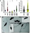
Effect of FlaG on flagellar filament lengths in C. jejuni flagellin mutants. (A) Flagellar filament lengths of WT C. jejuni and ΔflaA or ΔflaB mutants with or without flaG. Flagellar filaments were measured from micrographs of negatively stained cells acquired by TEM. Each circle represents the length of a flagellar filament on a cell. Over 100 flagellar filaments were measured from three independent cultures (n > 300). Bar represents mean filament length. Statistical significance was calculated by the Kruskal–Wallis test with Dunn’s multiple comparison post hoc test (*P < 0.05 between filament lengths of WT C. jejuni and mutants; **P < 0.05 between filament lengths of ΔflaA or ΔflaB mutants and their isogenic ΔflaG mutants). The graph on the Right is an expanded portion of the graph on the Left to show differences in the range of filament lengths of ΔflaA and ΔflaA ΔflaG. (B) Transmission electron micrographs of representative filament lengths of WT C. jejuni and mutant strains. (Scale bar, 1 μm.)
Fig. 3.
Fig. 3.
Analysis of FlaG levels in C. jejuni flagellar mutants. (A) Immunoblot analysis of flagellin, FliD, and FlaG levels in WCLs of WT C. jejuni and isogenic mutants lacking different genes. Specific antiserum to FlaA and FlaB flagellins (FlaAB), FliD (flagellar filament cap protein), and FlaG were used to detect specific proteins. Detection of RpoA served as a control to ensure equal loading of proteins across strains. (B) Levels of FlaG in WT C. jejuni and ΔfliS in WCLs and supernatants after growth in MH broth for 20 h in MH broth at 37 °C in microaerobic conditions. Strains contained vector alone (vec) or vector to express WT FliS (WT) or FliS-FLAG (WT-FLAG). WCL and supernatant proteins were analyzed by immunoblotting using specific antiserum for FlaG. Detection of RpoA served as a control to ensure equal loading of WCLs and absence of cytoplasmic proteins in supernatants.
Fig. 4.
Fig. 4.
Effect of mutating interacting residues in FlhA and FliS on flagellar filament length and FlaG levels. (A) In vivo interactions between C. jejuni FliS or FlaG and FlhA. FliS-FLAG, FlaGΔG43-E50-FLAG, or WT version of each protein without the FLAG epitope were expressed from plasmids in respective ΔfliS or ΔflaG mutants. FLAG-tagged proteins were immunoprecipitated by FLAG tag antibody resin after cross-linking cells by formaldehyde. Immunoblots to detect FlhA and RpoA were performed with specific antiserum. An antibody against the FLAG epitope was used to detect FLAG-tagged FliS and FlaG. Detection of RpoA served as a negative control for a protein that does not interact with FliS or FlaG. (B) Flagellar filament lengths of WT C. jejuni and ΔfliS with vector (vec) alone, or vector to express WT FliS-FLAG, FliSY10A-FLAG, or FliSY7A Y10A N13A-FLAG. Flagellar filaments were measured from micrographs of negatively stained cells acquired by TEM. Each circle represents the length of a flagellar filament on a cell. Over 100 flagellar filaments were measured from three independent cultures (n > 300). Bar represents mean filament length. Statistical significance was calculated by the Kruskal–Wallis test with Dunn’s multiple comparison post hoc test (*P < 0.05 between filament lengths of WT C. jejuni with vector alone and other strains; **P < 0.05 between ΔfliS complemented with WT-FliS and ΔfliS with vector alone or expressing fliS mutants). (C) Levels of FlaG, flagellins, and FliS-FLAG in WCLs or supernatants of WT C. jejuni and ΔfliS with vector (vec) alone or vector to express WT FliS-FLAG or FliSY10A-FLAG. Strains were grown for 20 h in MH broth at 37 °C in microaerobic conditions. WCL and supernatant proteins were recovered and analyzed by immunoblotting using specific antiserum for FlaG, flagellins (FlaAB), or the FLAG epitope. Detection of FlaA and FlaB flagellins are a positive control for recovery of secreted proteins in strains competent for secretion. Detection of RpoA served as a control to ensure equal loading of WCLs and absence of cytoplasmic proteins in the supernatant fraction. (D) Flagellar filament lengths of WT C. jejuni and flhA mutants with alterations of the predicted FliS binding site. Flagellar filaments were measured from micrographs of negatively stained cells acquired by TEM. Each circle represents the length of a flagellar filament on a cell. Over 100 flagellar filaments were measured from three independent cultures (n > 300). Bar represents mean filament length. Statistical significance was calculated by the Kruskal–Wallis test with Dunn’s multiple comparison post hoc test (*P < 0.05 between filament lengths of WT C. jejuni and isogenic mutants). (E) Levels of FlaG and flagellins in whole cell lysates (WCLs) and supernatants of WT C. jejuni and isogenic ΔflaG, ΔfliS and flhA mutants with alterations in the predicted FliS binding site. Strains were grown for 20 h in MH broth at 37 °C in microaerobic conditions. WCL and supernatant proteins were recovered and analyzed by immunoblotting using specific antiserum for FlaG, flagellins (FlaAB), or RpoA. Detection of FlaA and FlaB flagellins are a positive control for recovery of secreted proteins in strains competent for secretion. Detection of RpoA served as a control to ensure equal loading of WCLs and absence of cytoplasmic proteins in the supernatant fraction.
Fig. 5.
Fig. 5.
Effect of altering predicted FlhA–FlaG interactions on flagellar filament lengths and FlaG. (AC) Cartoon representation of the predicted interactions by AlphaFold 3 of (A) FliS and FlaG with the FlhA nonamer forming the torus region, (B) FliS with an FlhA torus monomer, and (C) FlaG with an FlhA torus monomer. For (AC), each C. jejuni FlhA monomer of the torus (residues 389 to 724) is represented by one of three shades of gray. The positions of the N terminus of the FlhA torus (K389) connected to the linker region of FlhA and the C terminus of the protein are indicated. The β-strand of FlhA from resides I431 through L439 (composed of IRIRDNLRL) that is the predicted interaction site for the C-terminal β-strand of FlaG (purple) is shown in yellow. Due to predicted flexibility of the N-terminal region of FlaG, only residues G40 through the C terminus of FlaG are shown. FliS is indicated in pink with teal representing Y10 that is predicted to interact with residues F485, S487, V513, and T516 (shown in red) of the FlhA torus. (D) Flagellar filament lengths of WT C. jejuni and ΔflaG containing WT flhA or flhA mutations in the predicted binding site for FlaG. Flagellar filaments were measured from micrographs of negatively stained cells acquired by TEM. Each circle represents the length of a flagellar filament on a cell. Over 100 flagellar filaments were measured from three independent cultures (n > 300). Bar represents mean filament length. Statistical significance was calculated by the Kruskal–Wallis test with Dunn’s multiple comparison post hoc test (*P < 0.05 between filament lengths of WT C. jejuni and other strains). (E) Levels of FlaG and flagellins in WCLs or supernatants of WT C. jejuni and isogenic ΔflaG and flhA mutants with alterations of the predicted FliS binding site. Strains were grown for 20 h in MH broth at 37 °C in microaerobic conditions. WCLs and supernatant proteins were recovered and analyzed by immunoblotting using specific antiserum for FlaG, flagellins (FlaAB), or RpoA. Detection of FlaA and FlaB flagellins are a positive control for recovery of secreted proteins. Detection of RpoA served as a control to ensure equal loading of WCLs and absence of cytoplasmic proteins in the supernatant fraction.
Fig. 6.
Fig. 6.
In vivo interactions of FlaG and FliS with WT FlhA and FlhA binding site mutants. (A and B) In vivo interactions between C. jejuni (A) FlaG or (B) FliS and WT FlhA or FlhA mutants with alterations at residues predicted for interactions with FliS or FlaG. C. jejuni FlaGΔG43-E50-FLAG, FliS-FLAG, or WT version of each protein without the FLAG epitope were expressed from plasmids in respective ΔflaG or ΔfliS mutants as indicated below immunoblots. FLAG-tagged proteins were immunoprecipitated by FLAG tag antibody resin after cross-linking cells by formaldehyde. Immunoblots to detect FlhA and RpoA were performed with specific antiserum. An antibody against the FLAG epitope was used to detect FLAG-tagged FliS and FlaGΔG43-E50-FLAG. Detection of RpoA served as a negative control for a protein that does not interact with FlaG or FliS. The presence of WT flhA or each flhA mutant on the chromosome is indicated above blots. FlhAF485A and FlhAV513G contain mutations predicted to disrupt interactions with FliS–flagellin complexes. FlhAR432A D435A, FlhAR434A L437A, and FlhAN436A L439A contain mutations predicted to disrupt interactions with FlaG.
Fig. 7.
Fig. 7.
Proposed model for FlaG antagonism of FliS–flagellin complex interactions with FlhA to control flagellar filament length in C. jejuni. Upon completion of the rod and hook of the C. jejuni flagellum and the start of filament polymerization (Left), a bolus of FliS–flagellin complexes (FliS, pink circles; flagellin, blue ovals) likely dominate occupancy of FlhA torus monomers (yellow wedges) in the fT3SS export gate to antagonize the ability of FlaG (purple hexagons) to bind to the FlhA torus. Binding of a FliS–flagellin complex to a FlhA monomer may antagonize FlaG to bind to the FlhA monomer in the counterclockwise position in the torus. Likewise, binding of FlaG to a FlhA monomer may antagonize FliS–flagellin complexes to bind to the FlhA monomer in the clockwise position in the torus. During this initial filament polymerization phase, the limited amount of free flagellin allows FliW (green arcs) to bind to CsrA (black rectangle) to derepress translation of the mRNA for the major flaA flagellin. During filament elongation (Right), a gradual reduction in FliS–flagellin complexes likely increases the ability of FlaG to access FlhA and bind multiple torus monomers and antagonize binding of FliS–flagellin complexes to FlhA. The resulting accumulation of cytoplasmic flagellins may contribute to a partner-switching mechanism in which binding of cytoplasmic flagellin by FliW releases CsrA to inhibit translation of flaA mRNA, blocking further production of flagellin. The combination of FlaG-dependent antagonism preventing FlhA and FliS–flagellin complex interactions and the resulting CsrA-mediated translation inhibition of flagellin may reinforce each other to allow FlaG to control flagellar filament length, resulting in shorter flagellar filaments.

Similar articles

Cited by

References

    1. Minamino T., Kinoshita M., Structure, assembly, and function of flagella responsible for bacterial locomotion. EcoSal Plus 11, eesp00112023 (2023). - PMC - PubMed
    1. Macnab R. M., How bacteria assemble flagella. Annu. Rev. Microbiol. 57, 77–100 (2003). - PubMed
    1. Kuhlen L., et al. , Structure of the core of the type III secretion system export apparatus. Nat. Struct. Mol. Biol. 25, 583–590 (2018). - PMC - PubMed
    1. Kuhlen L., et al. , The substrate specificity switch FlhB assembles onto the export gate to regulate type three secretion. Nat. Commun. 11, 1296 (2020). - PMC - PubMed
    1. Fraser G. M., et al. , Substrate specificity of type III flagellar protein export in Salmonella is controlled by subdomain interactions in FlhB. Mol. Microbiol. 48, 1043–1057 (2003). - PubMed

MeSH terms

LinkOut - more resources