. 2024 Oct 29;121(44):e2407533121.
doi: 10.1073/pnas.2407533121. Epub 2024 Oct 23.

Developing forebrain synapses are uniquely vulnerable to sleep loss

Affiliations

Developing forebrain synapses are uniquely vulnerable to sleep loss

Sean M Gay et al. Proc Natl Acad Sci U S A. .

Abstract

Sleep is an essential behavior that supports lifelong brain health and cognition. Neuronal synapses are a major target for restorative sleep function and a locus of dysfunction in response to sleep deprivation (SD). Synapse density is highly dynamic during development, becoming stabilized with maturation to adulthood, suggesting sleep exerts distinct synaptic functions between development and adulthood. Importantly, problems with sleep are common in neurodevelopmental disorders including autism spectrum disorder (ASD). Moreover, early life sleep disruption in animal models causes long-lasting changes in adult behavior. Divergent plasticity engaged during sleep necessarily implies that developing and adult synapses will show differential vulnerability to SD. To investigate distinct sleep functions and mechanisms of vulnerability to SD across development, we systematically examined the behavioral and molecular responses to acute SD between juvenile (P21 to P28), adolescent (P42 to P49), and adult (P70 to P100) mice of both sexes. Compared to adults, juveniles lack robust adaptations to SD, precipitating cognitive deficits in the novel object recognition task. Subcellular fractionation, combined with proteome and phosphoproteome analysis revealed the developing synapse is profoundly vulnerable to SD, whereas adults exhibit comparative resilience. SD in juveniles, and not older mice, aberrantly drives induction of synapse potentiation, synaptogenesis, and expression of perineuronal nets. Our analysis further reveals the developing synapse as a putative node of convergence between vulnerability to SD and ASD genetic risk. Together, our systematic analysis supports a distinct developmental function of sleep and reveals how sleep disruption impacts key aspects of brain development, providing insights for ASD susceptibility.

Keywords: development; phosphoproteome; proteome; sleep; synapse.


Conflict of interest statement

Competing interests statement:The authors declare no competing interest.

Figures
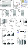
Fig. 1.
Fig. 1.
Divergent responses to SD in juveniles, adolescents, and adults. (A and B) Western blot analysis of phosphorylated GluA1 S845/S831 in forebrain PSD fractions isolated during wake (W4, ZT16) or sleep (S4, ZT4) from mice aged P21 to P70. The levels of Phospho-S845/S831 over total GluA1 at S4 are normalized to W4 (dashed line). Mice P42 and older show a significant dephosphorylation during sleep. Within ages S4 compared to W4: **P < 0.01 1-way ANOVA, Tukey’s correction. N = P21: W4(5M, 5F), S4(5M, 5F); P28: W4(3M, 5F), S4(4M, 4F); P35: W4(7M, 5F), S4(6M, 6F); P42: W4(5M, 5F), S4(5M, 5F); P70: W4(10M, 5F), S4(10M, 5F). (C) Schematic: developmental synapse dynamics predict differential responses to SD. (DH) Baseline sleep and recovery from 4 h SD treatment (SD4: ZT0-4) in juvenile (P28), adolescent (P42), or adult mice (P100). (D) 24 h sleep trace during uninterrupted baseline or during 4 h SD treatment (SD4: ZT0-4) and recovery (ZT4-24). The shaded box indicates dark-phase. (E) Sleep deficit calculated from difference in sleep amount from ZT0-4 between baseline and SD day. (F) Light-phase rebound measured as increased light-phase sleep amount during SD+recovery ZT4-12 over baseline. (G) Dark-phase sleep rebound measured as increased dark-phase sleep amount during SD+recovery ZT12-24 over baseline. (H) % Sleep deficit recovered between ages. Deficit calculated based on sleep lost compared to baseline during SD4 treatment ZT0-4; recovery calculated from increased sleep over baseline from ZT4-24. % Sleep deficit recovered is significantly greater in adults than juveniles. *P < 0.05, **P < 0.01, 1-way ANOVA, Tukey’s correction. N = juveniles (12M, 20F); adolescents (22M, 17F); adults (10M,9F). (I) Homer1a and Per2 transcripts measured using qPCR in whole forebrain from juvenile (P21), adolescent (P42), or adult mice (P100). Samples collected during the sleep phase (ZT4), wake phase (ZT16), or immediately following SD4 (ZT4). *P < 0.05, **P < 0.01, 1-way ANOVA, Tukey’s correction. N = juveniles: S4(5M, 6F), W4(6M, 7F), SD4(6M, 8F); adolescents: S4(10M, 10F), W4(10M, 10F), SD4(10M, 9F); adults: S4(6M, 5F), W4(6M, 5F), SD4(6M, 6F). (J and K) Juvenile (P28), adolescent (P42), or adult mice (P100) subjected to the novel object recognition task ±SD. Training occurs ZT0-2 followed by uninterrupted control or 4 to 6 h SD treatment, testing occurs 24 h after training. Novel object memory performance is completely impaired by SD4 in juveniles, memory impaired by SD6 in adults. *P < 0.05, **P < 0.01, ns: not significant, Student’s t test. Data include both sexes. Error bars, mean ± SEM. N = juveniles: control (5M, 6F), SD4(5M, 5F); adolescents: control (4M, 3F), SD4(5M, 4F); adults: control (8M, 12F), SD4(4M, 8F), SD6(4M, 4F).
Fig. 2.
Fig. 2.
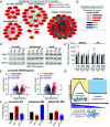
The juvenile synapse proteome is highly vulnerable to SD, transitions to wake/sleep regulation in adults. (A) Experimental design. Isolated forebrain synapse fractions (PSD) from juvenile (P21), adolescent (P42), or adult mice (P100) analyzed by quantitative mass spectrometry. Four mice pooled per sample; samples prepared in quadruplicate totaling 16 mice per condition, equal males and females. Juveniles, adolescents, and adults: W4/S4/SD4 (4 h); additional adults: W6/S6/SD6 (6 h) conditions. (B) Representative western blot from juvenile cohorts demonstrating enrichment of synapse scaffold protein PSD95 in the PSD fraction from wake, sleep, or SD conditions. (C) Venn diagram depicts high overlap in the proteins detected in the synapse fractions from juveniles, adolescents, or adults (6 h). (D) Examples of juvenile synapse proteins regulated by circadian rhythm (circadian), wake/sleep cycle (sleep), selective response to SD or combined. *P < 0.05, Student’s t test with Bonferroni correction. (EH) Volcano plots showing changes in synapse proteins in wake/sleep, SD/sleep, and SD/wake comparisons from juvenile, adolescent, or adult mice. N: number of proteins quantified; in and de: proteins significantly increased or decreased respectively (P < 0.05, Student’s t test). Total percentage of proteome altered in each comparison is indicated in purple. (IL) Pie charts indicate the significantly altered proteins assigned to regulation groups, circle scaled to number of altered proteins. Juvenile regulated proteins dominated by selective SD response, adults dominated by regulation from the wake/sleep cycle. (MO) Venn diagrams depict overlap in proteins significantly increased or decreased in each of the pairwise comparisons between juveniles, adolescents, and adults (6 h).
Fig. 3.
Fig. 3.
The developing synapse phosphoproteome is highly vulnerable to SD. (AD) Volcano plots showing changes in synapse phosphopeptides in wake/sleep, SD/sleep, and SD/wake comparisons from juvenile, adolescent, or adult (4 h and 6 h conditions) mice. N: number of phosphopeptides quantified; in and de: phosphopeptides significantly (P < 0.05, Student’s t test) increased or decreased respectively. Total percentage of phosphoproteome altered in each comparison is indicated. (EH) Pie charts indicate the significantly altered phosphopeptides assigned to regulation groups, circle scaled to number of altered phosphopeptides. Juvenile and adolescent regulated phosphopeptides dominated by selective SD response, adults (6 h) dominated by regulation from the wake/sleep cycle. (IK) Venn diagrams depict overlap in phosphopeptides significantly increased or decreased in each of the pairwise comparisons between juveniles, adolescents, and adults (6 h). Minimal overlap between ages under all conditions examined.
Fig. 4.
Fig. 4.
SD in juvenile impacts key aspects of synapse maturation, converging on nodes of ASD risk. (A) Synapse proteins up-regulated in response to SD in juveniles, related to brain development and synapse potentiation. Protein interaction network identified using GO and STRING analysis. Proteins up-regulated in one or both SD/S and SD/W comparisons are indicated. (B) KSEA indicates SD drives activation of CaMKII isoforms in juveniles. (C and D) Western blot and analysis of forebrain PSD fractions obtained from juvenile, adolescent, and adult mice. Hapln1 and Arc proteins show an increase in the PSD following SD, a pattern exclusive to juveniles. N = juveniles: S4(7M,3F), SD4(7M, 3F); adolescents: S4(3M, 3F), SD4(3M, 3F); adults: S6(3M, 3F), SD6(3M, 3F). (E) Volcano plots: SD induced changes in SFARI risk genes (bold color). Number of SFARI genes detected (n), increased (in) or decreased (de) compared to total (in brackets) are indicated. (F) Bar charts, % of total synaptic proteome represented by SFARI genes or proteins from the SD-regulation group: juveniles and adolescents, or the Adult-6 h wake-sleep regulation group (WS): * indicates SD response is significantly enriched for SFARI genes compared to expected overlap from total, ns: not significant (hypergeometric test). Proteins up-regulated by SD in juveniles or down-regulated in adolescents are significantly enriched for SFARI genes. (G) Summary model.

Update of

References

    1. Diekelmann S., Born J., The memory function of sleep. Nat. Rev. Neurosci. 11, 114–126 (2010), 10.1038/nrn2762. - DOI - PubMed
    1. Franken P., Dijk D. J., Sleep and circadian rhythmicity as entangled processes serving homeostasis. Nat. Rev. Neurosci. 25, 43–59 (2024), 10.1038/s41583-023-00764-z. - DOI - PubMed
    1. Scammell T. E., Arrigoni E., Lipton J. O., Neural circuitry of wakefulness and sleep. Neuron 93, 747–765 (2017), 10.1016/j.neuron.2017.01.014. - DOI - PMC - PubMed
    1. Weiss J. T., Blundell M. Z., Singh P., Donlea J. M., Sleep deprivation drives brain-wide changes in cholinergic presynapse abundance in Drosophila melanogaster. Proc. Natl. Acad. Sci. U.S.A. 121, e2312664121 (2024), 10.1073/pnas.2312664121. - DOI - PMC - PubMed
    1. Wang Z., et al. , Quantitative phosphoproteomic analysis of the molecular substrates of sleep need. Nature 558, 435–439 (2018), 10.1038/s41586-018-0218-8. - DOI - PMC - PubMed

LinkOut - more resources