. 2024 Oct 23;134(23):e177700.
doi: 10.1172/JCI177700.

PCDH15 dual-AAV gene therapy for deafness and blindness in Usher syndrome type 1F models

Affiliations

PCDH15 dual-AAV gene therapy for deafness and blindness in Usher syndrome type 1F models

Maryna V Ivanchenko et al. J Clin Invest. .

Abstract

Usher syndrome type 1F (USH1F), resulting from mutations in the protocadherin-15 (PCDH15) gene, is characterized by congenital lack of hearing and balance, and progressive blindness in the form of retinitis pigmentosa. In this study, we explore an approach for USH1F gene therapy, exceeding the single AAV packaging limit by employing a dual-adeno-associated virus (dual-AAV) strategy to deliver the full-length PCDH15 coding sequence. We demonstrate the efficacy of this strategy in mouse USH1F models, effectively restoring hearing and balance in these mice. Importantly, our approach also proves successful in expressing PCDH15 protein in clinically relevant retinal models, including human retinal organoids and nonhuman primate retina, showing efficient targeting of photoreceptors and proper protein expression in the calyceal processes. This research represents a major step toward advancing gene therapy for USH1F and the multiple challenges of hearing, balance, and vision impairment.

Keywords: Gene therapy; Genetic diseases; Ophthalmology; Otology.


Conflict of interest statement

Conflict of interest: The President and Fellows of Harvard College has filed US provisional application 18/025,719, “Dual-AAV vector delivery of PCDH15 and uses thereof,” with DPC, EMM, and MVI listed as inventors.

Figures

Figure 1
Figure 1. Splicing and protein production in vitro.
(A) Recombination and splicing of dual vectors to produce full-length protein. The reassembly is mediated by non-homologous end joining of the inverted terminal repeats (ITRs) and/or homologous recombination of the highly recombinogenic (AK) sequence. After transcription, splicing can occur from the splice donor site (SD) in vector 1 to the splice acceptor site (SA) in vector 2, creating a full-length Pcdh15 mRNA. (B) mRNA was obtained from HEK cells and reverse-transcribed (n = 5). A cDNA library was Sanger-sequenced around the splice junction, confirming proper recombination and splicing. (C) Dual AAVs (encoding PCDH15 with or without an N-terminal HA tag) were added to HEK293 cell cultures (n = 3 per group). Immunostaining was performed with anti-PCDH15 or anti-HA antibodies (magenta). Representative confocal images demonstrate normal trafficking of PCDH15, either with or without the N-terminal HA tag, to the cell membrane after dual-AAV delivery. Labeling was similar to that with transfection of a single plasmid encoding full-length PCDH15. No specific signal was detected in the control samples (n = 3 per group). Scale bar: 10 μm. CDS, coding sequence; CMV, cytomegalovirus; DIC, differential interference contrast; IRES, internal ribosome entry site; polyA, polyadenylation signal; WPRE, woodchuck hepatitis virus post-transcriptional regulatory element.
Figure 2
Figure 2. Dual-AAV delivery of PCDH15 successfully preserves hearing in Myo15a-Cre mouse model and alleviates vestibular deficits in constitutive knockout model of USH1F.
(A) Dual AAV vectors encoding PCDH15-CD2 protein were injected via the RWM into either conditional or constitutive knockout mice at P1. At 5 weeks, treated animals were assayed for hearing and vestibular function and processed for histology. (B) ABR click and tone thresholds for untreated Pcdh15fl/fl Cre control mice (n = 9), untreated Pcdh15fl/fl, Myo15a-Cre+/− mice (n = 10), and treated Pcdh15fl/fl, Myo15a-Cre+/− mice (n = 23). (C) Average DPOAE thresholds and amplitudes at 16 kHz for 5-week-old hearing control mice (n = 9), for untreated Pcdh15fl/fl, Myo15a-Cre+/− mice (n = 6), and treated Pcdh15fl/fl, Myo15a-Cre+/− mice (n = 22). (D) Representative open field path outlines of wild-type mice, untreated Pcdh15fl/fl, Myo15a-Cre+/− mice, Pcdh15–/– knockout mice, and treated Pcdh15–/– knockouts. (E) Summary of total rotations in wild type (n = 5), untreated Pcdh15fl/fl, Myo15a-Cre+/− mice (n = 5), Pcdh15–/– mice (n = 4), and treated Pcdh15–/– mice (n = 13). Untreated Pcdh15–/– mice exhibited severe circling behavior, while vestibular function was fully preserved at normal levels in dual AAV-treated Pcdh15–/– mice. (F) Wild-type (n = 5) and Pcdh15fl/fl, Myo15a-Cre+/− (n = 5) mice swam well. Pcdh15–/– mice (n = 4) were unable to swim normally. G) Pcdh15–/– mice showed much shorter latency to fall on the rotarod test (n = 4), while treated Pcdh15–/– mice performed at wild-type levels (n = 12). One-way ANOVA followed by Dunnett’s test was used to assess statistical significance. Data are presented as mean ± SEM. ***P < 0.001, ****P < 0.0001; n.s., not significant (P > 0.05).
Figure 3
Figure 3. Delivery of PCDH15 with dual AAVs preserves the morphology of stereocilia bundles, mechanotransduction, and tip links in the Pcdh15fl/fl, Myo15a-Cre+/− conditional knockout mouse model.
(A) Representative confocal microscopy images captured from the middle turn of 5-week-old cochleae, showing OHCs stained with phalloidin. The panels illustrate OHCs from Pcdh15fl/fl hearing control mice (n = 3), uninjected Pcdh15fl/fl, Myo15a-Cre+/− conditional knockout mice (n = 3), and knockout mice that received dual-AAV injections (n = 5). (B) Scanning electron micrographs of hair bundles of a hearing control mouse and untreated conditional knockouts. In the knockouts, the bundles were severely disrupted. However, in the OHCs of a conditional knockout treated with dual AAVs encoding PCDH15, normal bundle morphology was preserved (n = 5). (C) Representative confocal microscopy images at 5 weeks from the middle turn of the cochlea show anti-HA staining at the tips of stereocilia in knockout cochleae treated with dual AAVs encoding HA.PCDH15 at P1 (n = 4). (D) Transduction efficiency in IHCs and OHCs at 5 weeks in treated Pcdh15fl/fl, Myo15a-Cre+/− conditional knockout mice (n = 4). Data are presented as mean ± SEM. (E) Immunogold scanning electron microscopy localization of HA-tagged PCDH15 in OHC stereocilia of treated knockout mice. Multiple 12 nm gold beads (light yellow) were detected in a scanning electron microscopy image, confirming that HA-tagged PCDH15 goes to the tips of stereocilia, except the tallest (n = 3). (F) Rescue of FM1-43 uptake in a treated cochlea. (G) Average percentage of IHCs and OHCs loaded with FM1-43 in conditional knockout mice injected with dual AAVs (n = 4) and in control mice (n = 4). Data are presented as mean values ± SEM. Scale bars: 5 μm (AC and F), 100 nm (E).
Figure 4
Figure 4. Human PCDH15 exon structure and disease-associated variants.
(A) Gene schematic of PCDH15-CD2 and reported disease-causing variants. Three primary transcripts of PCDH15 (CD1, CD2, and CD3) translate 3 isoforms, which differ primarily in the 3′ end of the gene. Disease variants were gathered from the Deafness Variation Database, accessed August 2021. Disease variants are plotted based on their location on transcript NM_001142769 and color-coded based on associated disease phenotype: blue, non-syndromic hearing loss (DFNB23); red, Usher syndrome type 1F (USH1F); or green, non-syndromic retinitis pigmentosa (NSRP). Note that the variant p.P1796fs impacts the protein-coding sequence of only the CD1 isoform and has been reported to cause NSRP, suggesting that the CD1 splice form is required for retinal function but not hearing. Similarly, p.Q1576X in CD2 affects hearing but not vision. (B) Quantitative PCR evaluation of PCDH15-CD1, PCDH15-CD2, and PCDH15-CD3 expression in primate cochlea (n = 5) and retina (n = 6). One-way ANOVA followed by post hoc Tukey’s test was used to assess statistical significance. Data are presented as mean ± SEM. **P < 0.01, ***P < 0.001, ****P < 0.0001; n.s., not significant (P > 0.05).
Figure 5
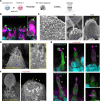
Figure 5. Expression of PCDH15 in human retinal organoids by dual AAV vectors.
(A) Organoids were transduced with dual AAV vectors, and 5 weeks later expression was analyzed by antibodies against the HA tag. (B) Representative confocal microscopy images captured from cryosections of non-transduced human retina organoids. Anti-PCDH15 labeling (green) was detected in the photoreceptors, especially at the distal ends of outer segments (OSs) (yellow arrows) (n = 3 organoids). Hoechst dye labeled nuclei, anti–cone arrestin (ARR3) labeled cones, and anti-rhodopsin (Rho) labeled rod OSs. (C) Scanning electron microscopy of human retinal organoids. The finger-like calyceal processes (yellow arrows) protrude from the apical region of the inner segments (ISs) of both rod (middle) and cone (right) photoreceptors (n = 3 organoids). (D) Immunogold scanning electron microscopy labeling of non-transduced human retinal organoids immunostained with anti-PCDH15 primary antibody and 12 nm gold-conjugated secondary antibody. Endogenous PCDH15 was located at the surfaces of ISs and of nascent calyceal processes of photoreceptors (n = 3 organoids). (E) Anti-HA fluorescent labeling of vector-delivered HA.PCDH15. Because the CMV promoter is only present on the 5′ vector, HA.PCDH15 and GFP were only expressed when both the 5′ and 3′ vectors were added to the organoids and were internalized within a cell. Anti-HA.PCDH15 is shown as magenta and GFP as green. In these images, GFP diffusely labeled doubly transduced cells. HA.PCDH15 is seen at the junction of ISs and OSs of cone photoreceptors (n = 3 organoids). (F) Immunogold scanning electron microscopy confirmed localization of HA-tagged hsPCDH15 to calyceal processes and ISs of human retinal organoids transduced with dual-AAV (n = 3). Multiple 12 nm gold beads were detected. Scale bars: 5 μm (B and E), 10 μm (C, left), 1 μm (C, right), 0.5 μm (D and F).
Figure 6
Figure 6. Localization of endogenous and dual-AAV–delivered PCDH15 in NHP retina.
(A) Dual AAV vectors were injected subretinally into the eye of a green monkey (C. sabaeus). At 9 weeks, the treated retina was assayed for expression and localization by histology. (B) Immunofluorescence labeling of endogenous PCDH15 (green) in the retina of a cynomolgus monkey (M. fascicularis). An antibody against ARR3 (magenta) marks cone photoreceptors, and an antibody against Rho (blue) marks OSs of rods. PCDH15 (green) is located at the junction between ISs and OSs and along the calyceal processes (arrows) in both cones and rods (n = 4 punches). (C) Scanning electron micrograph of the inner/outer segment junction in a cone photoreceptor (right) and rod photoreceptors (left) in a cynomolgus monkey (n = 4 punches). The calyceal processes (arrows) protrude from the apical ISs of photoreceptors to surround the OSs. (D) Immunofluorescence labeling of endogenous PCDH15 (green) in the vehicle-injected control retina of a green monkey (n = 3 punches). PCDH15 (green) is located at the junction between ISs and OSs and along the calyceal processes (arrows) in both cones and rods. White asterisk indicates the OS of a cone photoreceptor. BODIPY labels all membranes of photoreceptors. (E) Representative confocal microscopy images of cryosections from the bleb area created by the dual-AAV injection. An anti-HA signal was detected in the photoreceptors injected with dual AAVs, seen at low magnification (left) and higher magnification (right two panels). (F) Transduction efficiency in rod and cone photoreceptors injected with dual AAVs. Data are presented as mean ± SEM. (G) The HA label was not seen in the eye injected with the vehicle (n = 3 blebs). Scale bars: 5 μm (B, D, and G), 1 μm (C and E).

Update of

References

    1. Geleoc GGS, El-Amraoui A. Disease mechanisms and gene therapy for Usher syndrome. Hear Res. 2020;394:107932. doi: 10.1016/j.heares.2020.107932. - DOI - PubMed
    1. French LS, et al. A review of gene, drug and cell-based therapies for Usher syndrome. Front Cell Neurosci. 2020;14:183. doi: 10.3389/fncel.2020.00183. - DOI - PMC - PubMed
    1. Jouret G, et al. Genetics of Usher syndrome: new insights from a meta-analysis. Otol Neurotol. 2019;40(1):121–129. doi: 10.1097/MAO.0000000000002054. - DOI - PubMed
    1. Mathur P, Yang J. Usher syndrome: hearing loss, retinal degeneration and associated abnormalities. Biochim Biophys Acta. 2015;1852(3):406–420. doi: 10.1016/j.bbadis.2014.11.020. - DOI - PMC - PubMed
    1. Ahmed ZM, et al. Mutations of the protocadherin gene PCDH15 cause Usher syndrome type 1F. Am J Hum Genet. 2001;69(1):25–34. doi: 10.1086/321277. - DOI - PMC - PubMed