. 2024 Oct 25;10(43):eadr3567.
doi: 10.1126/sciadv.adr3567. Epub 2024 Oct 23.

An autonomous implantable device for the prevention of death from opioid overdose

Affiliations

An autonomous implantable device for the prevention of death from opioid overdose

Joanna L Ciatti et al. Sci Adv. .

Abstract

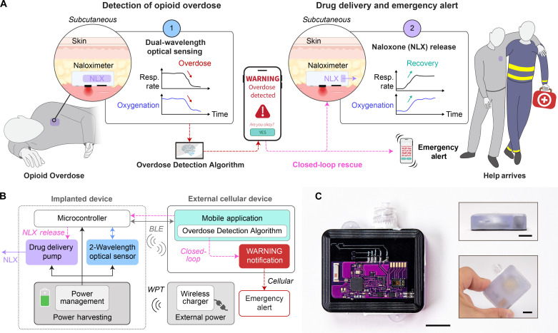
Opioid overdose accounts for nearly 75,000 deaths per year in the United States, now a leading cause of mortality among young people aged 18 to 45 years. At overdose levels, opioid-induced respiratory depression becomes fatal without the administration of naloxone within minutes. Currently, overdose survival relies on bystander intervention, requiring a nearby person to find the overdosed individual and have immediate access to naloxone to administer. To circumvent the bystander requirement, we developed the Naloximeter: a class of life-saving implantable devices that autonomously detect and treat overdose while simultaneously contacting first responders. We present three Naloximeter platforms, for fundamental research and clinical translation, all equipped with optical sensors, drug delivery mechanisms, and a supporting ecosystem of technology to counteract opioid-induced respiratory depression. In small and large animal studies, the Naloximeter rescues from otherwise fatal opioid overdose within minutes. This work introduces life-changing, clinically translatable technologies that can broadly benefit a susceptible population recovering from opioid use disorder.


Figures

Fig. 1.
Fig. 1.. Autonomous operational scheme of Naloximeter for continuous sensing, triggered drug delivery and rescue from opioid overdose.
(A) Schematic illustration of a scenario in which an implanted device (Naloximeter) detects respiratory depression leading to hypoxia, a signature of opioid overdose, delivers the rescue drug NLX, and sends an emergency alert to first responders. Created with BioRender.com. (B) Operational diagram including transdermal wireless power transfer, bidirectional communication via BLE protocols, and cellular communication that delivers an emergency alert. (C) Optical images of a Naloximeter; inset: side (top) and top view (bottom) of the device. Scale bars, 1 cm.
Fig. 2.
Fig. 2.. Device design and in-vivo validation studies.
(A) Exploded view illustration of a Naloximeter for intravenous drug delivery via catheter and electrolytic pump; inset: PCB layout containing the dual-wavelength optical sensor, mounted on the backside of the assembled pump device. (B) Cross-sectional illustration showing deformation of a flexible membrane under pressure from gas generated via electrolytic water splitting. This process drives NLX through a catheter port to a vein. (C) Drug release from the electrolytic device, N = 3 devices. (D) Exploded view illustration of a Naloximeter for drug injection via a needle and motor-driven actuator; inset: Huber needle and polytetrafluoroethylene septum at the outlet of the housing. (E) Drug release from the injector device in single- or dual-injection mode, N = 3 devices. (F) Schematic illustrations of dual-injection operation, namely: needle deployment (1–2), delivery of first dose (3), retraction (4), delivery of second dose (5), and final retraction once empty (6). (G) Validation of the dual-wavelength optical sensor for determining StO2 via sequential exposure to hypoxic gas mixtures (21, 14, 12, 10, 8, and 21% O2, denoted as colored bars) in a rodent model; SpO2 data collected using a GSD (MouseOx collar). (H) Correlation plot for SpO2 (GSD) and StO2 (Naloximeter) resulting from rodent hypoxia validation studies (R2 = 0.95). Colors signify individual animals. (I) Correlation plot for SO2 from blood gases and StO2 from the dual-wavelength optical sensor validated in porcine hypoxia studies (R2 = 0.65). Colors signify individual devices. (J) Pharmacokinetics of NLX delivered using the intravenous device in a porcine model, dose: 3 ml (2.7 mg of NLX), N = 3 animals. Inset: Optical image of a catheter tunneled subcutaneously from a device and secured in the jugular vein; scale bar, 1 cm. i.v., intravenous. s.c., subcutaneous.
Fig. 3.
Fig. 3.. Physiological signs of opioid overdose and an algorithm for overdose detection in freely moving subjects.
(A) StO2 recorded with a Naloximeter during a fentanyl overdose (OD; 10 μg/kg) in an anesthetized pig and its comparison with gold standards: SO2 (ABG) and SpO2 (pulse oximeter). (B) Decline of respiration rate, (C) elevation of end-tidal carbon dioxide (EtCO2), (D) bradycardia, and (E) reduction in blood pressure after fentanyl injection (at time = 0) represent clinical signs of overdose. (F) StO2 recorded from Naloximeters during fentanyl overdose quantify the rate of desaturation. Left, n = 9 devices and right, n = 4 devices. (G) Desaturation rates following various dosages of fentanyl. N = 29 subjects and n = 52 devices; dose-specific sample sizes are included in table S2. (H) Illustration of the analytical approach for the ODA based on the dual-wavelength optical sensor. Definition of metrics: optical absorption and its rate of change (M1), desaturation rate (M2), tissue oxygenation levels (M3), and signal power density (M4). (I) Operation of the ODA in an ambulatory pig, including (J) StO2 during opioid overdose (30 μg/kg), and the rates of change of (K) the red optical signal (λ1), (L) the difference between the red and infrared optical signals (∆λ) of the dual-wavelength optical sensor, and (M) the tissue oxygenation (∂StO2). Fentanyl was administered at time = 0. (N) Application of the ODA in a healthy, normally ambulating pig for 24 hours. Although several instances of synchronous positive metrics, i.e., EVENTs were registered, none of them led to a WARNING. Colored bars in (J) and (N) indicate time stamps where individual metrics (M1, M2, M3, and M4) or compound logical metrics (EVENT) are true.
Fig. 4.
Fig. 4.. Demonstration of autonomous rescue from life-threatening opioid overdose in rodents and freely moving porcine models.
(A) Subcutaneous implant location for a miniaturized NFC Naloximeter and corresponding fentanyl overdose experiment grouping by NLX treatments; inset: optical image of the device; scale bar, 1 cm. (B) Oxygenation in a rodent model following fentanyl overdose and manual subcutaneous administration of NLX (manual rescue), autonomous rescue enabled by the NFC Naloximeter, or self-recovery without NLX. Sample sizes are in the legend. (C) Comparison of recovery time between treatment groups (one-way ANOVA, ****P < 0.0001). Data points correspond to individual animals, and bars depict the means. (D) Experimental setup for overdose (OD) rescue in an anesthetized pig model (E) with and (F) without a Naloximeter, the latter required manually administered NLX to rescue. Oxygenation (top), respiratory vitals (middle), and pharmacokinetic data (bottom) for each fentanyl overdose. (G) Experimental setup for OD rescue in ambulatory pigs (H) with and (I) without a Naloximeter. (J) Demonstration of subcutaneous drug delivery from (K) a Naloximeter without intravenous catheter in an ambulatory pig, insufficient to recover from OD, requiring manually administered NLX to rescue. Oxygenation (top) and PK (bottom) data for each fentanyl overdose. The oxygenation in (E), (F), (H), (I), and (K) was measured with blood gases (SO2), a commercial pulse oximeter (SpO2), and the Naloximeter (StO2). (L) Longitudinal PK data from an intravenous Naloximeter at 1 to 13 days after implantation, and an infusion pump as a control (CTRL I.V.). Dose is 1.5 ml. (M) Gd pharmacokinetics from a Naloximeter (device) at 14 days after implantation in the jugular or mammary vein, or infusion pump (CTRL) in the auricular vein. Dose is 2 ml. (N) Gd PK profile when delivered by a Naloximeter intravenously (jugular vein) or subcutaneously. Dose is 3 ml. (A), (D), (G), and (J) created with BioRender.com.

Update of

References

    1. Strang J., Volkow N. D., Degenhardt L., Hickman M., Johnson K., Koob G. F., Marshall B. D. L., Tyndall M., Walsh S. L., Opioid use disorder. Nat. Rev. Dis. Primer 6, 1–28 (2020). - PubMed
    1. France C. P., Ahern G. P., Averick S., Disney A., Enright H. A., Esmaeli-Azad B., Federico A., Gerak L. R., Husbands S. M., Kolber B., Lau E. Y., Lao V., Maguire D. R., Malfatti M. A., Martinez G., Mayer B. P., Pravetoni M., Sahibzada N., Skolnick P., Snyder E. Y., Tomycz N., Valdez C. A., Zapf J., Countermeasures for preventing and treating opioid overdose. Clin. Pharmacol. Ther. 109, 578–590 (2021). - PMC - PubMed
    1. Degenhardt L., Grebely J., Stone J., Hickman M., Vickerman P., Marshall B. D. L., Bruneau J., Altice F. L., Henderson G., Rahimi-Movaghar A., Larney S., Global patterns of opioid use and dependence: Harms to populations, interventions, and future action. Lancet 394, 1560–1579 (2019). - PMC - PubMed
    1. Blanco C., Volkow N. D., Management of opioid use disorder in the USA: Present status and future directions. Lancet 393, 1760–1772 (2019). - PubMed
    1. Volkow N. D., Blanco C., The changing opioid crisis: Development, challenges and opportunities. Mol. Psychiatry 26, 218–233 (2021). - PMC - PubMed