

Choroid Plexus Volume in Pediatric-Onset Multiple Sclerosis
- PMID: 39442038
- PMCID: PMC11502105
- DOI: 10.1212/NXI.0000000000200319
Choroid Plexus Volume in Pediatric-Onset Multiple Sclerosis
Abstract
Background and objectives: Recent studies suggest that the choroid plexus (CP) may function as a site of access of inflammatory cells into the CNS in multiple sclerosis (MS). Pediatric-onset MS (POMS) is characterized by a high inflammatory burden, as evidenced by a high relapse rate and volume of T2 lesions, making patients with POMS an informative population to evaluate choroid plexus volume (CPV). The objectives of the study were (1) to evaluate CPV at symptom onset in participants with POMS compared with healthy controls (HCs); (2) to evaluate changes in CPV in the first year of disease in participants with POMS; and (3) to evaluate associations between CPV, brain volumes, relapse activity, and disability in participants with POMS.
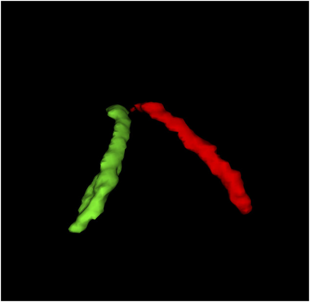
Methods: Baseline 1.5T MRI scans were acquired from 23 participants with POMS and 23 age-matched and sex-matched HCs; 18 participants with POMS also had 12-month follow-up MRI scans. The CP of the lateral ventricles was segmented manually. CP and brain structure volumes were normalized for total intracranial volume. The number of relapses, T2 and gadolinium-enhancing T1 lesion counts, and Expanded Disability Status Scale (EDSS) scores at 12 months were also analyzed. Baseline CPVs were compared between groups using the Wilcoxon exact test, and CPV change from baseline to 12 months in participants with POMS was compared using the Wilcoxon signed-rank test. The relationship between CPV and brain volumetric measures, T2 lesion volumes, lesion count, number of relapses, and EDSS scores was assessed through Spearman correlation.
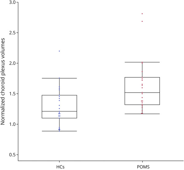
Results: The median normalized CPV was 1.51 × 10⁻³ (interquartile range [IQR]: 1.32-1.76) in POMS baseline scans and 1.21 × 10⁻³ (IQR: 1.1-1.47) in HC scans (p = 0.001). CPV did not significantly change at 12 months in the 18 participants with POMS with follow-up scans (p = 0.352). CPV in participants with POMS and HCs correlated with lateral ventricular volume (p = 0.012 for both groups) but did not correlate with brain and T2 lesion volumes or lesion count at baseline, nor with relapse activity or EDSS scores at 12 months in participants with POMS.
Discussion: CPV measured at baseline is greater in participants with POMS than in HCs. Baseline CPV did not predict higher disease activity or worse neurologic outcomes over 1 year. While higher CPV may be an early feature of inflammation in MS, its strong correlation with ventricular volumes could also reflect enlargement secondary to the mechanical attachment of CP to the ventricular wall.
Conflict of interest statement
E.A. Grasso, L. Bloy, and P. Kaplan report no disclosures relevant to the manuscript; A. Bar-Or has received personal fees for advisory board participation and/or consulting from Abata, Accure, Atara Biotherapeutics, Biogen, BMS/Celgene/Receptos, GlaxoSmithKline, Gossamer, Horizon Therapeutics, Immunic, Janssen/Actelion, Medimmune, Merck/EMD Serono, Novartis, Roche/Genentech, Sangamo, Sanofi-Genzyme, and Viracta and grant support to the University of Pennsylvania from Biogen Idec, Roche/Genentech, Merck/EMD Serono and Novartis as well as research funding from the NIH, the National MS Society (NMSS), the Juvenile Diabetes Research Foundation, the Canadian Institutes of Health Research, MS Society of Canada, MS Scientific Foundation and Melissa and Paul Anderson Chair in Neuroinflammation; E.A. Yeh received research funding from NMSS, CMSC, CIHR, NIH, OIRM, SCN, CBMH Chase an Idea, SickKids Foundation, Rare Diseases Foundation, MS Scientific Foundation, McLaughlin Centre, Leong Center, Peterson Foundation, Gary Hurvitz Centre for Brain and Mental Health, and investigator initiated research funding from Biogen. Scientific advisory: Hoffman-LaRoche, Alexion, DSMB: Pipeline Therapeutics. Speaker honoraria: Biogen, JHU, Saudi Epilepsy Society, NYU, MS-ATL, ACRS, PRIME, CNPS. Co-Editor in Chief, MSARD, Governing Council: CANTRAIN, Steering Committee: Rare-Kids CAN; D.L. Arnold reports consulting fees from Biogen, Biohaven, BMS, Eli Lilly, EMD Serono, Find Therapeutics, Frequency Therapeutics, GSK, Idorsia Pharmaceuticals, Kiniksa Pharmaceuticals, Merck, Novartis, Race to Erase MS, Roche, Sanofi-Aventis, Shionogi, and Xfacto Communications, as well as an equity interest in NeuroRx. R.A. Marrie receives research funding from CIHR, MS Canada, Crohn's and Colitis Canada, NMSS, CMSC, the Arthritis Society, Pfizer Foundation and the US Department of Defense and is a co-investigator on studies receiving funding from Biogen Idec and Roche Canada. She holds the Waugh Family Chair in MS; S. Narayanan has received research funding from the Canadian Institutes of Health Research, the International Progressive MS Alliance, the Myelin Repair Foundation, NIH (subaward), Immunotec, and F. Hoffman LaRoche; he is a consultant for Sana Biotech, has received a speaker's honorarium from Novartis Canada, and is a part-time employee of NeuroRx Research; G. Fadda has received personal compensation from Horizon therapeutics/amgen for serving as member of advisory board. B. Banwell serves as a consultant to Novartis, UCB, Sanofi-Genzyme, and Roche. Go to
Figures


References
MeSH terms
LinkOut - more resources
Full Text Sources
Medical
Miscellaneous