Case Reports
. 2024 Oct 24;391(16):1511-1518.
doi: 10.1056/NEJMoa2400718.

Neurodevelopmental Disorder Caused by Deletion of CHASERR, a lncRNA Gene

Affiliations
Case Reports

Neurodevelopmental Disorder Caused by Deletion of CHASERR, a lncRNA Gene

Vijay S Ganesh et al. N Engl J Med. .

Abstract

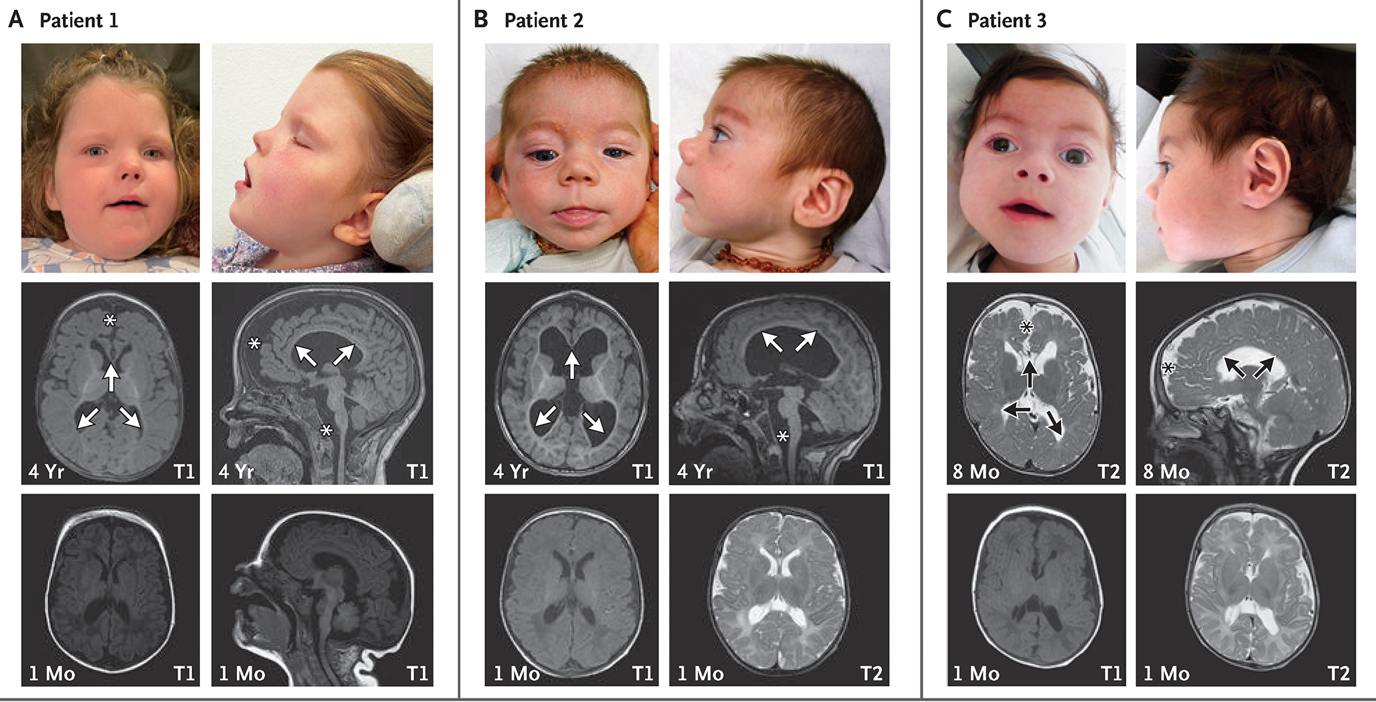
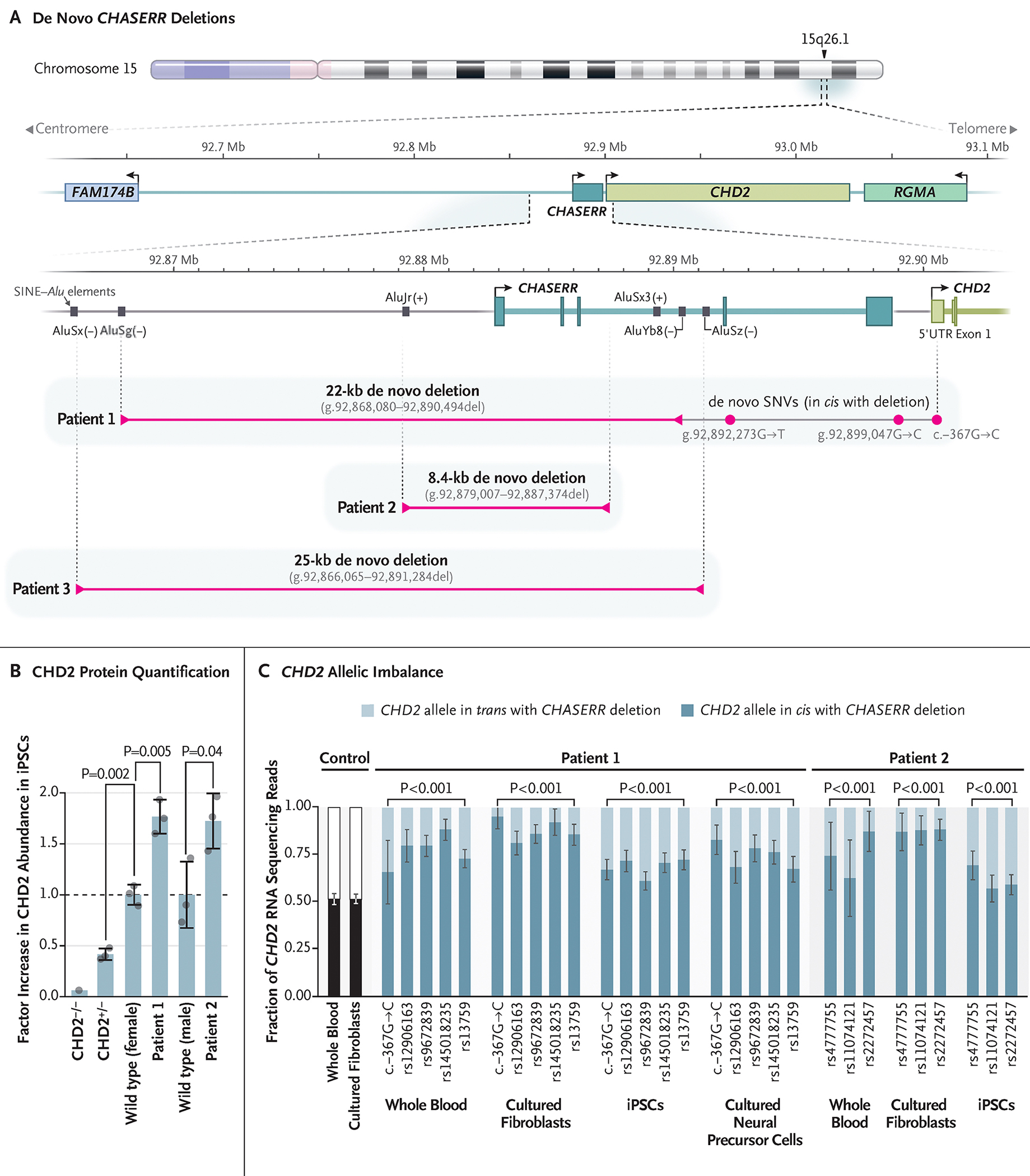
CHASERR encodes a human long noncoding RNA (lncRNA) adjacent to CHD2, a coding gene in which de novo loss-of-function variants cause developmental and epileptic encephalopathy. Here, we report our findings in three unrelated children with a syndromic, early-onset neurodevelopmental disorder, each of whom had a de novo deletion in the CHASERR locus. The children had severe encephalopathy, shared facial dysmorphisms, cortical atrophy, and cerebral hypomyelination - a phenotype that is distinct from the phenotypes of patients with CHD2 haploinsufficiency. We found that the CHASERR deletion results in increased CHD2 protein abundance in patient-derived cell lines and increased expression of the CHD2 transcript in cis. These findings indicate that CHD2 has bidirectional dosage sensitivity in human disease, and we recommend that other lncRNA-encoding genes be evaluated, particularly those upstream of genes associated with mendelian disorders. (Funded by the National Human Genome Research Institute and others.).


Figures

Figure 1.
Figure 1.. Facial Features and Findings on Magnetic Resonance Imaging of the Brain in Three Children with CHASERR Haploinsufficiency.
Shared facial dysmorphisms among the three children include wide-set eyes, anteverted nares, low-set ears, and a long philtrum. T1 and T2-weighted magnetic resonance imaging of the brain showed frontal predominant cortical atrophy with reduced volume of the brain stem (asterisks) and generalized hypomyelination of the corpus callosum and subcortical white matter (arrows) at 4 years or 8 months of age (middle row), which were all less apparent at 1 month of age (bottom row).
Figure 2.
Figure 2.. De Novo CHASERR Deletions and Characterization of CHD2 Expression.
Panel A shows human chromosome 15q26.1, with a 500-kb inset showing a gene-sparse region upstream of the long noncoding RNA (lncRNA) CHASERR (teal) and its tandem coding gene CHD2 (light green). Reads were aligned to human genome assembly GRCh38. Arrows indicate the direction of transcription. De novo deletions (bracketed pink lines) in Patient 1 (22 kb), Patient 2 (8.4 kb), and Patient 3 (25 kb) overlap the promoter and first three exons of CHASERR. Long-read DNA sequencing in Patient 1 identified three de novo single-nucleotide variants (SNVs, pink dots) in cis with the CHASERR deletion. SINE denotes short interspersed element. The signs “(+)” and “(−)” denote the strand orientation of the Alu element. Panel B shows increased CHD2 protein abundance in induced pluripotent stem cells (iPSCs) from Patients 1 and 2, as compared with iPSCs from sex-matched wild-type controls, an unrelated female with CHD2 haploinsufficiency (CHD2+/−), and CHD2−/− CRISPR (clustered regularly interspaced short palindromic repeats)–Cas9 knockout iPSCs. CHD2 protein abundance was normalized to histone deacetylase 1. I bars indicate the standard deviation. P values were calculated with a two-tailed, heteroscedastic Student’s t test. Panel C shows RNA sequencing in whole-blood samples, cultured fibroblasts, and iPSCs obtained from Patients 1 and 2 and cultured neural precursor cells obtained from Patient 1. All showed allelic imbalance toward expression of the CHD2 allele in cis with the CHASERR deletion, across all phasable variants. The control whole-blood samples and cultured fibroblasts from the Genotype-Tissue Expression (GTEx) project showed no allelic imbalance in CHD2. White and black bars represent the read fraction for each of the two CHD2 alleles. I bars indicate the 95% confidence interval (with the assumption of binomial distribution). P values were calculated with a chi-square test, with an allele balance of 1:1 as the null hypothesis.

Similar articles

Cited by

References

    1. Scheffer IE, Berkovic S, Capovilla G, et al. ILAE classification of the epilepsies: position paper of the ILAE Commission for Classification and Terminology. Epilepsia 2017;58:512–21. - PMC - PubMed
    1. Carvill GL, Heavin SB, Yendle SC, et al. Targeted resequencing in epileptic encephalopathies identifies de novo mutations in CHD2 and SYNGAP1. Nat Genet 2013;45:825–30. - PMC - PubMed
    1. Liu JC, Ferreira CG, Yusufzai T. Human CHD2 is a chromatin assembly ATPase regulated by its chromo- and DNA-binding domains. J Biol Chem 2015;290:25–34. - PMC - PubMed
    1. Shen T, Ji F, Yuan Z, Jiao J. CHD2 is required for embryonic neurogenesis in the developing cerebral cortex. Stem Cells 2015;33:1794–806. - PubMed
    1. Kim YJ, Khoshkhoo S, Frankowski JC, et al. Chd2 is necessary for neural circuit development and long-term memory. Neuron 2018;100(5):1180–1193.e6. - PMC - PubMed

Publication types