. 2024 Dec;23(12):100867.
doi: 10.1016/j.mcpro.2024.100867. Epub 2024 Oct 21.

Dehydrin Client Proteins Identified Using Phage Display Affinity Selected Libraries Processed With Paired-End Phage Sequencing

Affiliations

Dehydrin Client Proteins Identified Using Phage Display Affinity Selected Libraries Processed With Paired-End Phage Sequencing

Sandra Helena Unêda-Trevisoli et al. Mol Cell Proteomics. 2024 Dec.

Abstract

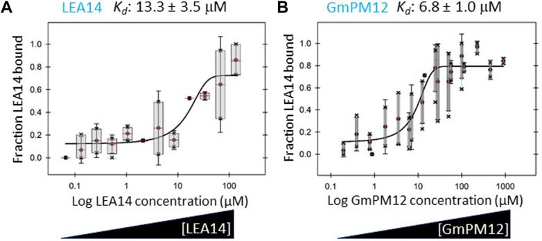
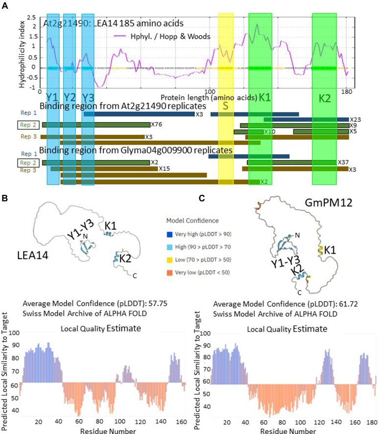
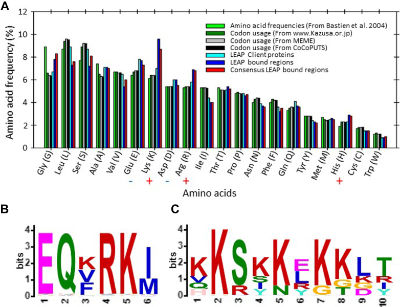
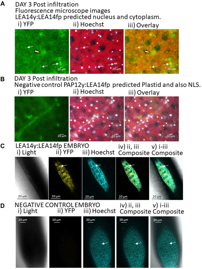
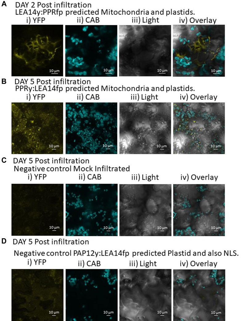
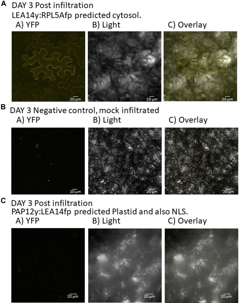
The late embryogenesis abundant proteins (LEAPs) are a class of noncatalytic, intrinsically disordered proteins with a malleable structure. Some LEAPs exhibit a protein and/or membrane binding capacity and LEAP binding to various targets has been positively correlated with abiotic stress tolerance. Regarding the LEAPs' presumptive role in protein protection, identifying client proteins (CtPs) to which LEAPs bind is one practicable means of revealing the mechanism by which they exert their function. To this end, we used phage display affinity selection to screen libraries derived from Arabidopsis thaliana seed mRNA with recombinant orthologous LEAPs from Arabidopsis and soybean (Glycine max). Subsequent high-throughput sequencing of DNA from affinity-purified phage was performed to characterize the entire subpopulation of phage retained by each LEAP ortholog. This entailed cataloging in-frame fusions, elimination of false positives, and aligning the hits on the CtP scaffold to reveal domains of respective CtPs that bound to orthologous LEAPs. This approach (paired-end phage sequencing) revealed a subpopulation of the proteome constituting the CtP repertoire in common between the two dehydrin orthologs (LEA14 and GmPm12) compared to bovine serum albumin (unrelated binding control). The veracity of LEAP:CtP binding for one of the CtPs (LEA14 and GmPM12 self-association) was independently assessed using temperature-related intensity change analysis. Moreover, LEAP:CtP interactions for four other CtPs were confirmed in planta using bimolecular fluorescence complementation assays. The results provide insights into the involvement of the dehydrin Y-segments and K-domains in protein binding.

Keywords: client proteins; late embryogenesis abundant proteins; paired-end sequencing; phage display; temperature related intensity change assay.


Conflict of interest statement

Conflict of interest The authors declare no competing interests.

Figures

None
Graphical abstract
Fig. 1
Fig. 1
Phylogenetic and sequence analysis of late embryogenesis abundant proteins.A, an alignment of the different LEAP families (6) from both soybean (79 proteins) and Arabidopsis (52 proteins). The supplemental text details the rationale for the specific sequences used. The soybean LEAPs are numbered by ascending chromosome and gene as done previously for Arabidopsis (6). B, the amino acid sequence of two orthologous LEAPs of the DHN family (AT2G21490 and Glyma.04G009900) of the type YnSKn, where n = 3 for the Y-segment and n = 2 for the K-segment (108). Both have a carboxy-terminal, hexahistidyl tag with a vector-derived “LE” amino acid linker to the ultimate carboxy-terminal amino acid in the native DHN sequence. These extraneous amino acids are in red text; as are the changes in the theoretical pI and molecular weight (Mw) that these additions made to the proteins. Clustal Omega (109) was used to generate the amino acid sequence alignment. DHN, dehydrin.
Fig. 2
Fig. 2
Recombinant protein expression and purification for use in phage display affinity selection.A, the recombinant proteins were prepared as described in Methods and analyzed by SDS-PAGE (15% gel). In A, (S1: soybean, A1: Arabidopsis) uninduced soluble fraction; (S2, A2) IPTG (0.4 mM)-induced soluble fraction; (S3, A3) boiled samples; (S4, A4) Nickel column, affinity-purified hexahistidyl-tagged recombinant protein; low range molecular weight (LMW) markers (kDa). It was determined that IPTG induction was unnecessary for GmPM12 accumulation, and this step was subsequently dropped. B, titers (means with SEM) of libraries after each round of selection with a DHN or bovine serum albumin (BSA) using an Arabidopsis seed library in the T7Select10-3b vector. Plating was conducted in triplicate dilutions of the affinity-selected library recovered from 3 independent microtiter plate wells and plaques were counted from serial dilutions. DHN, dehydrin.
Fig. 3
Fig. 3
PCR steps involved to place a three-nucleotide bar code and Illumina adaptors on either end of cDNA inserted into the T7 phage chromosome in preparation for paired-end phage sequencing.A, primers F1-T7- (red, yellow box, purple arrow) corresponding to the T7 10B coat protein 3′ end, 5′ to the cloning site (including the microtiter plate well–specific bar code and half of the forward Illumina adaptor; F Illumina adaptor; red) and R2-T7 (the reverse primer to the T7 10B coat protein 3′ end, 3′ to the cloning site) are used in a limited round PCR reaction. Template is the amalgam of T7 phage DNA extracted from the collection of clones from the terminal round of affinity selection over a specific LEAP and a specific microtiter plate well (replication). Following agarose gel electrophoresis (Supplemental Fig. S1), excision of the smear of amplicons above the primer dimers (if any), and DNA recovery, a second limited round PCR is performed. The second round uses primers PE-2F (light blue, red arrow) with PE-2R (black, green arrow) to complete the forward and reverse Illumina adaptors. B, all steps in the paired-end phage sequencing (PEPA-Seq) workflow. The width of the arrows in each step depicts the volume of reads passed from one step to the next. OOF, out of frame; LEAP, late embryogenesis abundant protein.
Fig. 4
Fig. 4
Phage sequencing scenarios.i, occasionally, cDNAs could clone into the phage genome without using the adaptors, resulting in phage “contaminating” amino acid sequence (red). If this extra phage “leader” maintained the frame between the coat protein and the plant cDNA, the clone was retained as a legitimate, in-frame CtP. ii and iii, occasionally, the recovered clone commenced in the 5′ UTR (bold black) of the Arabidopsis cDNA (yellow line). These were examined in CLC Genomics Workbench to ascertain whether there was a stop codon in-frame between the end of the phage coat protein and the commencement of the Arabidopsis clone. If a stop was in-frame, these clones were discarded. iv-vi, occasionally, the reverse primer commences in the 3′ UTR of the Arabidopsis clone. iv, PEPA-Seq allowed clones longer than the two Illumina reads (blue rectangles) to be assembled based on the forward clone, in-frame with an Arabidopsis protein being identical with the protein identified from the reverse read, and the intervening protein sequence (light blue rectangle) was hence inferred. v, some clones identified one protein from the forward read and a second, unrelated protein from the reverse read (a long clone is depicted). These chimeric proteins were cloning anomalies that occurred during library construction. Which protein that was bound by the DHN was ambiguous and so these clones were discarded. vi, there were instances where the reverse read commenced in the 3′ UTR and terminated prior to reaching the coding region, and these were retained. CtP, client protein; DHN, dehydrin.
Fig. 5
Fig. 5
TRIC assay of labeled LEA14 or GmPM12 binding to LEA14 or GmPM12, respectively. The concentration of NHS-labeled DHN (either (A) LEA14 or (B) GmPM12) was kept constant while the concentration of the non-labeled binding partner (A) LEA14 or (B) GmPM12) was varied, decreasing incrementally by 2-fold. Immediately following centrifugation, the plates containing samples were loaded into a Dianthus Pico (NanoTemper Technologies) and temperature-related intensity change (TRIC) measurements performed using 3% power. Experiments were repeated at least 3 times and the average Kd ± standared error of the mean (SE) is reported. The normalized fluorescence curves from a typical experiment were converted to fraction-bound curves for presentation. DHN, dehydrin; NHS, N-hydroxysuccinimide.
Fig. 6
Fig. 6
The LEA14 DHN can self-associate and also bind with its soybean orthologous DHN.A, LEA14 phage-displayed fragments that bound either GmPM12 or LEA4 are depicted as colored rectangles on the stylized protein portrayal and project below the figure to the rectangular depictions of the independent phage displayed CtP fragments. The bound, phage-displayed regions recovered in each of the three independent wells (replications) for LEA14 or GmPM12 are presented separately below the overlaid Hopp-Woods hydrophilicity plot for each CtP. All LEA14 fragments, whether recovered from either LEA14 or GmPM12, contained at least one partial Y-domain or one partial K-domain. B, predicted LEA14 protein topology (AlphaFold) resolved only three regions where localized amino acid alignments, in relation to nearest neighbors, could be predicted with confidence of 79% or greater (blue demarcated sequences in the model labeled Y1-Y3, K1, and K2). These corresponded to an N-terminal region encompassing the three Y domains (boxed regions highlighted in blue) and the two K-segments toward the carboxy terminus (regions highlighted in green). C, this was also the case for Y1-Y3, and the K2 domain of GmPM12 while the K1 domain was predicted with only a 55 to 61% confidence. CtP, client protein; DHN, dehydrin.
Fig. 7
Fig. 7
Estimates of the amino acid frequencies in Arabidopsis thaliana proteins were acquired and compared to the amino acid frequencies present in the DHN CtPs, in the phage-displayed fragments of these CtPs, and the consensus sequences from the CtPs (Fig. 3B).A, although the phage-displayed fragments captured by the orthologous DHNs tended to have a greater frequency of positively charged amino acids (110) than the CtPs themselves or the proteome generally, chi-square tests (critical chi-square value for 20 amino acids, 19 df: 30.1 = χ2 at p = 0.05) did not demonstrate that this was statistically significant. B and C, CtP sequences were used to acquire motifs present among them using XTREAM and the resulting WEBLOGOs (Crooks, Hon et al. 2004) were converted to peptide patterns and the Arabidopsis proteome scanned for proteins possessing the motif. There were five proteins discovered possessing each of the motifs and there were (B) three and (C) four of these captured in the phage display. CtP, client protein; DHN, dehydrin.
Fig. 8
Fig. 8
The BiFC assay, using both transient expression and stable transformation, with split YFP was used to assess LEA14–CtP interactions for five CtPs.A, transiently expressed LEA14y:LEA14fp (LATE EMBRYOGENESIS ABUNDANT PROTEIN 14, dehydrin, At2G21490) constructs resulted in a cytoplasmic- and nuclear-localized YFP signal in Nicotiana benthamiana leaves (results from 3 days following infiltration shown). Such a signal was not observed in N. benthamiana leaves (depicted are leaf disks 3 days following infiltration) with, (B) PAP12y:LEA14fp or LEA14y:PAP12fp (not shown) constructs. Embryos of Arabidopsis thaliana which were stably expressing LEA14y:LEA14fp, (C) also demonstrated a cytoplasmic- and nuclear-localized YFP signal whereas those from untransformed plants (D) did not. For both tobacco leaves and Arabidopsis embryos, tissues were vacuum infiltrated 3 times for 5 min each time with 50 μM Hoechst’s stain prior to visualization. The scale bar represents 20 μm. In A and B; i) signal from reconstituted split yellow fluorescence protein (YFP). ii, Hoechst stain signal in the nucleus. iii) overlaid image. In C and D; i, light that is bright field images. ii, reconstituted YFP signal. iii, Hoechst stain signal. iv, overlaid signal from YFP and Hoechst stain. v, overlaid signal from light, YFP, and Hoechst stain. White arrow: nucleus. Yellow broad arrow: cytoplasm. BiFC, bimolecular fluorescence complementation.
Fig. 9
Fig. 9
Split YFP assay for PPRy:LEA14fp interactions.A, LEA14y:PPRfp (PENTATRICOPEPTIDE REPEAT 596, AT1G80270), resulted in YFP signal in Nicotiana benthamiana leaves (leaf disks 2 days following infiltration depicted) outside, sometimes proximal to, the chloroplasts. B, similarly, PPRy:LEA14fp constructs (leaf disks 5 days post infiltration presented) occurred in puncta that were beside but not in, chloroplasts. The YFP signal present in (A and B) was not observed in (C) mock-infiltrated or (D) leaves infiltrated with a negative control PAP12y:LEA14fp (leaf disks 5 days following infiltration depicted). i, YFP signal; ii, chlorophyll A and B autofluorescence (CAB; cyan false color); iii) light is brightfield micrographs; and iv) overlay of all three light signals. The scale bar represents 10 μm.
Fig. 10
Fig. 10
Split YFP assay for LEA14:SES4A (AT4G27170: seed storage albumin 4) interactions.A, (LEA14y:SESA4fp (AT4G27170: SEED STORAGE ALBUMIN 4)) provided signal (3 days after infiltration shown) that is consistent with a localization in the endomembrane system, the published location of SESA4. i) YFP; ii) light; and iii) overlay. This signal was not evident in either, (B) mock-infiltrated or, (C) leaves infiltrated with the construct PAP12y:LEA14fp (images provided are 3 days after infiltration). The scale bar represents 20 μm. i, signal from reconstituted split yellow fluorescence protein (YFP). ii, bright field image (light). iii, overlaid image.
Fig. 11
Fig. 11
Split YFP assay testing a CtP LEA14y:RPL5Afp (AT5G39740:RIBOSOMAL PROTEIN LARGE SUBUNIT 5A) that was not captured using phage display but shares an amino acid motif predicted from four other CtPs.A, YFP signal using LEA14y:RPL5Afp 3 days after infiltration that was present in the cytoplasm, a predicted subcellular localization for RPL5A. This signal was not evident in either, (B) mock-infiltrated or, (C) leaves infiltrated with the construct PAP12y:LEA14fp. The scale bar represents 20 μm. CtP, client protein.

Similar articles

References

    1. Sallon S., Solowey E., Cohen Y., Korchinsky R., Egli M., Woodhatch I., et al. Germination, genetics, and growth of an ancient date seed. Science. 2008;320:1464. - PubMed
    1. Shen-Miller J., Mudgett M.B., Schopf J.W., Clarke S., Berger R. Exceptional seed longevity and robust growth: ancient sacred Lotus from China. Am. J. Bot. 1995;82:1367–1380.
    1. Tepfer D., Zalar A., Leach S. Survival of plant seeds, their UV screens, and nptII DNA for 18 months outside the International Space Station. Astrobiology. 2012;12:517–528. - PubMed
    1. Adhikari B.N., Wall D.H., Adams B.J. Effect of slow desiccation and freezing on gene transcription and stress survival of an Antarctic nematode. J. Exp. Biol. 2010;213:1803–1812. - PubMed
    1. Dinakar C., Bartels D. Desiccation tolerance in resurrection plants: new insights from transcriptome, proteome and metabolome analysis. Front. Plant Sci. 2013;4:482. - PMC - PubMed

LinkOut - more resources