Meta-Analysis
. 2025 Jun 1;97(11):1079-1090.
doi: 10.1016/j.biopsych.2024.10.013. Epub 2024 Oct 21.

Neuroimaging Meta-Analyses Reveal Convergence of Interoception, Emotion, and Social Cognition Across Neurodegenerative Diseases

Affiliations
Meta-Analysis

Neuroimaging Meta-Analyses Reveal Convergence of Interoception, Emotion, and Social Cognition Across Neurodegenerative Diseases

Jessica L Hazelton et al. Biol Psychiatry. .

Abstract

Background: Simultaneous interoceptive, emotional, and social cognition deficits are observed across neurodegenerative diseases. Indirect evidence suggests shared neurobiological bases underlying these impairments, termed the allostatic-interoceptive network (AIN). However, no study has yet explored the convergence of these deficits in neurodegenerative diseases or examined how structural and functional changes contribute to cross-domain impairments.

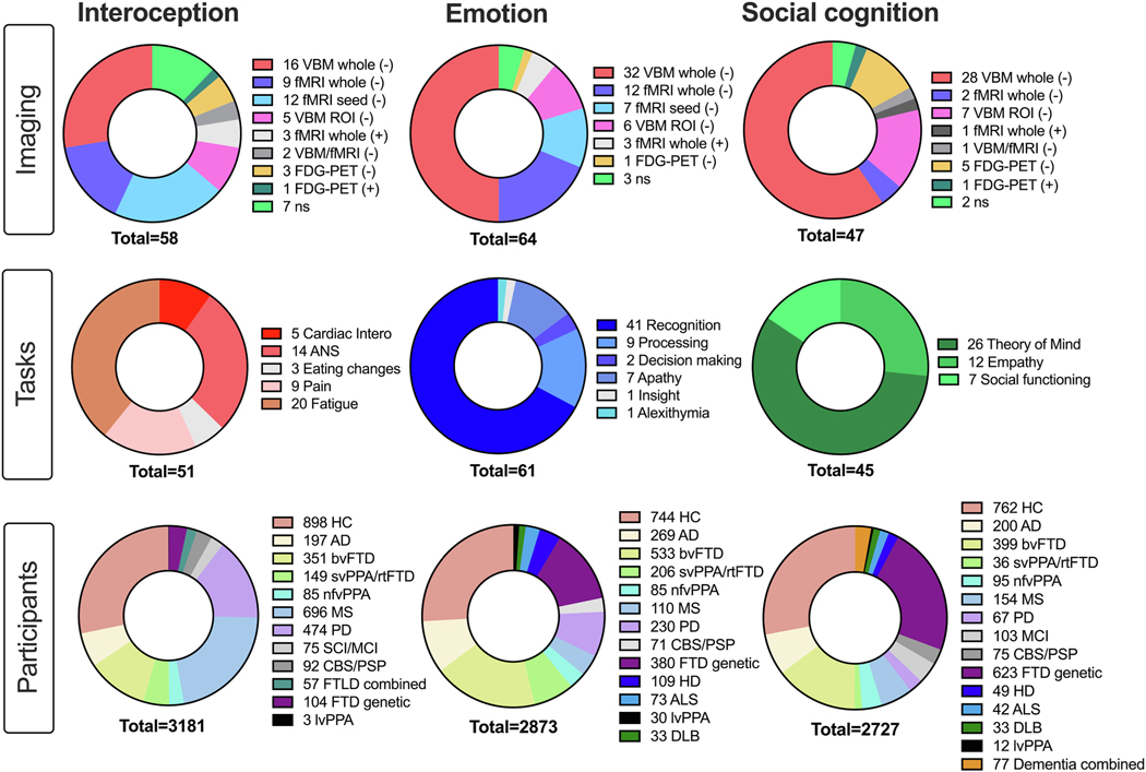
Methods: A Preferred Reporting Items for Systematic Reviews and Meta-Analyses (PRISMA) activated likelihood estimate meta-analysis encompassed studies that met the following inclusion criteria: interoception, emotion, or social cognition tasks; neurodegenerative diseases (behavioral variant frontotemporal dementia, primary progressive aphasias, Alzheimer's disease, Parkinson's disease, multiple sclerosis); and neuroimaging (structural: magnetic resonance imaging voxel-based morphometry; functional: magnetic resonance imaging and fluorodeoxyglucose-positron emission tomography).

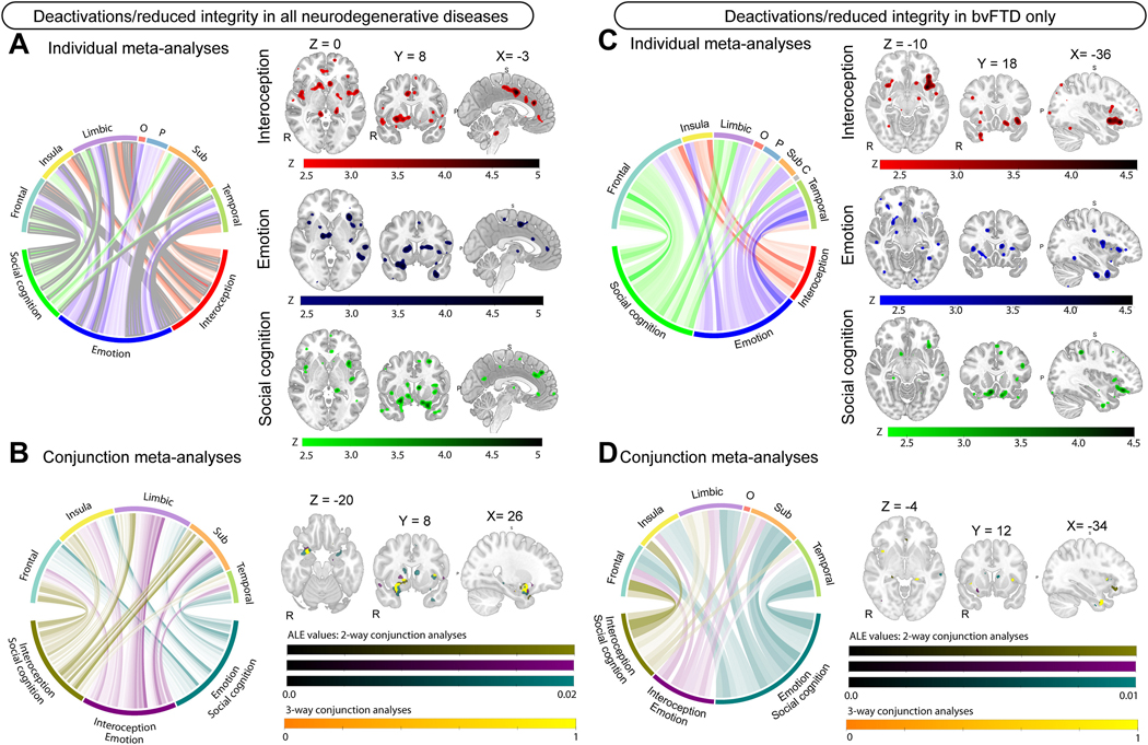
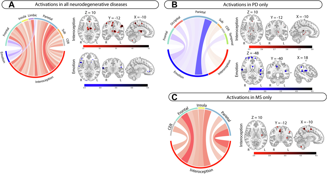
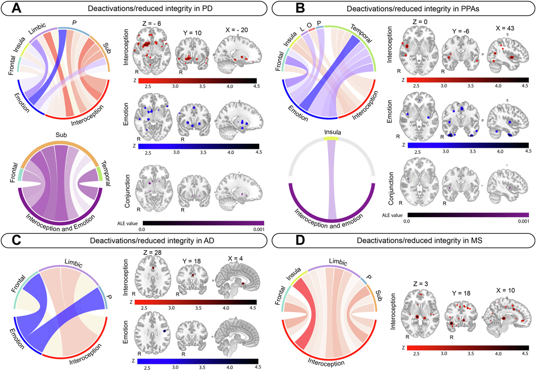
Results: Of 20,593 studies, 170 met inclusion criteria (58 interoception, 65 emotion, and 47 social cognition) involving 7032 participants (4963 patients and 2069 healthy control participants). In all participants combined, conjunction analyses revealed AIN involvement of the insula, amygdala, orbitofrontal cortex, anterior cingulate, striatum, thalamus, and hippocampus across domains. In behavioral variant frontotemporal dementia, this conjunction was replicated across domains, with further involvement of the temporal pole, temporal fusiform cortex, and angular gyrus. A convergence of interoception and emotion in the striatum, thalamus, and hippocampus in Parkinson's disease and the posterior insula in primary progressive aphasias was also observed. In Alzheimer's disease and multiple sclerosis, disruptions in the AIN were observed during interoception, but no convergence with emotion was identified.

Conclusions: Neurodegeneration induces dysfunctional AIN across atrophy, connectivity, and metabolism, more accentuated in behavioral variant frontotemporal dementia. Findings bolster the predictive coding theories of large-scale AIN, calling for more synergistic approaches to understanding interoception, emotion, and social cognition impairments in neurodegeneration.

Keywords: Allostasis; Emotion; Interoception; Meta-analysis; Neuroimaging; Social cognition.


Conflict of interest statement

Conflict of interest

The authors have no conflicts of interest to declare.

Figures

Figure 1:
Figure 1:
Studies included in the metanalysis across domains detailing imaging techniques, tasks used, and number of participants per domain. Note total N across domains > total N reported in text due to 15 studies contributing to more than 1 domain. Abbreviations: Imaging: ns = non-significant; Tasks: ANS = Autonomic nervous system; Intero = Interoception; Participants (in order): HC = Healthy control; AD = Alzheimer’s disease; bvFTD =behavioral-variant frontotemporal dementia; svPPA = semantic-variant primary progressive aphasia; rtvFTD = right temporal variant frontotemporal dementia; nfv-PPA = non-fluent variant primary progressive aphasia; MS = Multiple sclerosis; PD = Parkinson’s Disease; CBS/PSP = Corticobasal syndrome, progressive supranuclear palsy; SCI/MCI = Subjective cognitive impairment/Mild cognitive impairment; FTLD combined = frontotemporal lobar degeneration; FTD genetic = presymptomatic MAPT, GRN, C972orf mutation carriers; HD = Huntington’s Disease; ALS = Amyotrophic Lateral Sclerosis; lvPPA = logopenic-variant primary progressive aphasia; Dementia combined = AD, FTD sample.
Figure 2.
Figure 2.
Metanalyses results for deactivations/reduced structural integrity contrasts in all neuroimaging modalities and in bvFTD. Left-sided panel shows metanalyses results in all neurodegenerative diseases for A) individual metanalyses; and B) conjunction metanalyses. Right-sided panel shows metanalyses results specific to bvFTD for C) individual metanalyses; and D) conjunction metanalyses. For A and C: Circular plot displaying z-scores for brain regions in interoception (red), emotion (blue), and social cognition (green) in individual metanalyses. Larger z-scores are shown in darker colors for each metanalysis. Clusters significant at p <.05 FWE corrected are outlined in grey, with p <.001 uncorrected showing no outline. For B and D: Circular plot display ALE-scores for two-way conjunction analyses, with larger ALE-scores shown in darker colors. Interoception-social cognition (olive; color bar 1); Interoception-Emotion (purple, color bar 2); Emotion-social cognition (teal, color bar 3), Interoception-Emotion-Social cognition (yellow, color bar 4). Color bars represent ALE scores for significant clusters (color bars 1–3) and a binary outcome of the three way-analysis (color bar 4). All regions are displayed at p <.001, uncorrected. All brain slices are displayed in radiological orientation (Right = Left). MNI coordinates are displayed above brain slices. Abbreviations: C = Cerebellum; O = Occipital; P = Parietal; Sub = Subcortical grey matter.
Figure 3.
Figure 3.
Metanalyses results for activation contrasts in all neuroimaging modalities and in PD and MS. Left-sided panel shows metanalyses results in all neurodegenerative diseases for A) individual metanalyses; conjunction analyses were not significant. Right-sided panel shows metanalyses results for individual domains specific to B) PD and C) MS. All circular plot display z-scores for brain regions in interoception (red), emotion (blue) in individual metanalyses. Larger z-scores are shown in darker colors for each metanalysis. Clusters are reported at p <.001 uncorrected. All brain slices are displayed in radiological orientation (Right = Left). MNI coordinates are displayed above the first image for each panel unless different coordinates are used (e.g., 5B). Brainstem included in Sub cluster in PD. Abbreviations: CER = Cerebellum; Sub = Subcortical grey matter.
Figure 4.
Figure 4.
Disease specific metanalysis results for deactivations/reduced structural integrity for interoception (red) and emotion (blue), and conjunction analyses (purple). A) Parkinson’s disease. B) Primary progressive aphasia. C) Alzheimer’s disease. D) Multiple Sclerosis. A and B show individual metanalysis results and conjunction metanalyses results. C and D show individual metanalysis results only. All individual metanalyses results are reported at p <.001, uncorrected. Circular plots and neuroimaging panels display z-scores for each individual metanalysis, with darker colors representing larger z-scores. Conjunction metanalysis results are reported at p <.05 FWE, with 5000 permutations. Circular plots and neuroimaging panels for conjunction analyses display ALE scores, with darker colors representing larger values. MNI coordinates are displayed above the first neuroimage image for each panel. Neuroimages are displayed in radiological presentation (right = left). Brainstem included in Sub cluster in PD. Abbreviations: L = Limbic, O = Occipital, P = Parietal, Sub = Subcortical grey matter.

Similar articles

Cited by

References

    1. Berntson GG, Khalsa SS (2021): Neural circuits of interoception. Trends in neurosciences. 44:17–28. - PMC - PubMed
    1. Chen WG, Schloesser D, Arensdorf AM, Simmons JM, Cui C, Valentino R, et al. (2021): The emerging science of interoception: sensing, integrating, interpreting, and regulating signals within the self. Trends in neurosciences. 44:3–16. - PMC - PubMed
    1. Feldman M, Bliss-Moreau E, Lindquist K (2024): The neurobiology of interoception and affect. Trends in Cognitive Sciences. - PMC - PubMed
    1. Quigley KS, Kanoski S, Grill WM, Barrett LF, Tsakiris M (2021): Functions of interoception: From energy regulation to experience of the self. Trends in neurosciences. 44:29–38. - PMC - PubMed
    1. Kleckner IR, Zhang J, Touroutoglou A, Chanes L, Xia C, Simmons WK, et al. (2017): Evidence for a large-scale brain system supporting allostasis and interoception in humans. Nature human behaviour. 1:1–14. - PMC - PubMed

Publication types

MeSH terms