. 2024 Nov 6;146(44):30139-30154.
doi: 10.1021/jacs.4c08406. Epub 2024 Oct 23.

Dissecting the Conformational Heterogeneity of Stem-Loop Substructures of the Fifth Element in the 5'-Untranslated Region of SARS-CoV-2

Affiliations

Dissecting the Conformational Heterogeneity of Stem-Loop Substructures of the Fifth Element in the 5'-Untranslated Region of SARS-CoV-2

Klara R Mertinkus et al. J Am Chem Soc. .

Erratum in

Abstract

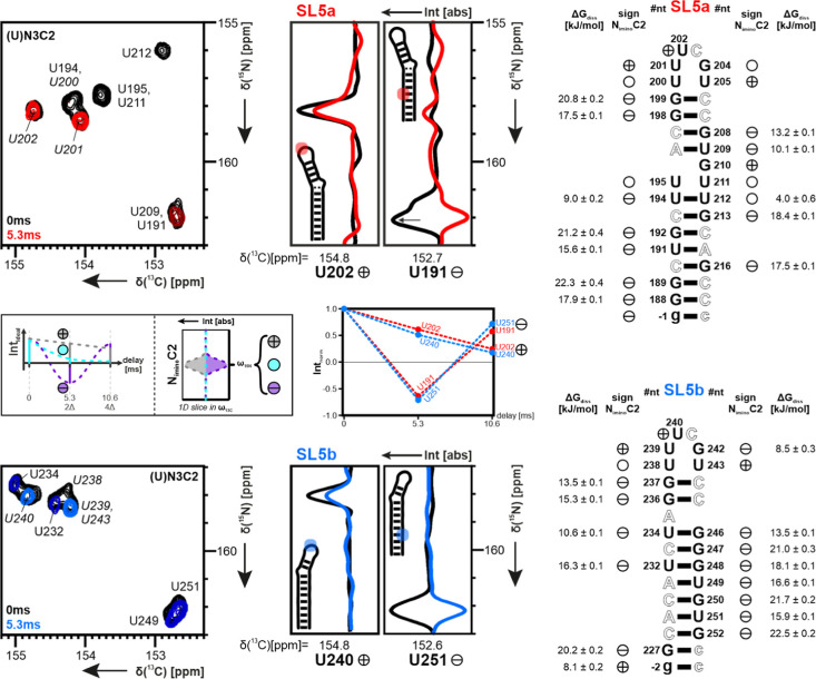
Throughout the family of coronaviruses, structured RNA elements within the 5' region of the genome are highly conserved. The fifth stem-loop element from SARS-CoV-2 (5_SL5) represents an example of an RNA structural element, repeatedly occurring in coronaviruses. It contains a conserved, repetitive fold within its substructures SL5a and SL5b. We herein report the detailed characterization of the structure and dynamics of elements SL5a and SL5b that are located immediately upstream of the SARS-CoV-2 ORF1a/b start codon. Exploiting the unique ability of solution NMR methods, we show that the structures of both apical loops are modulated by structural differences in the remote parts located in their stem regions. We further integrated our high-resolution models of SL5a/b into the context of full-length 5_SL5 structures by combining different structural biology methods. Finally, we evaluated the impact of the two most common VoC mutations within 5_SL5 with respect to individual base-pair stability.


Conflict of interest statement

The authors declare no competing financial interest.

Figures

Figure 1
Figure 1
Schematic representation of the RNA structure calculation workflow. The iterative process was repeated until the structure bundles reached convergence. Convergence was defined as an average >0.98 for back-calculated CORR R values by PALES.
Figure 2
Figure 2
Differences in 1H,13C-chemical shifts of aromatic C6H6/C8H8 (dark) and ribose C1′H1′ (light) in the sequence-identical loop regions of SL5a to SL5b. Secondary structure representations with the respective sequential numbering for comparison of SL5a (red) to SL5b (blue). Identical sequential loop region of 10 bases highlighted in the center. For an overlay of spectra, see Supporting Information Figure S4.
Figure 3
Figure 3
hetNOE values for aromatic and ribose-1′ CH bonds. (Left): SL5a (red); (right): SL5b (light blue). Measurements performed at 298 K. Sequence shown on top and bottom. Highlighted regions to identify the bulge region (color) and hexaloop (gray). A value of 1.2 was chosen as the threshold for dynamic evaluation and therefore visual orientation (dotted line). For the extended version with C2, see Supporting Information Figure S5.
Figure 4
Figure 4
Comparison of the base-pair properties for SL5a (top) and SL5b (bottom) via hydrogen-exchange-sensitive experiments. From left to right: 13C,15N-HSQC with exemplary 1D slices for U nucleotides without (black) and with a proton filter (colored), showing the impact on the phase. Scheme of analysis in small legend in-between. CN-HSQC measured at 298 K, with all corresponding peaks U N1C2 phased to a positive sign for comparison. Behavior of the signal sign in CN-HSQC with the proton filter present shown next to the sequence. See also Supporting Information Figures S6 and S7 for whole spectra. Also, next to the sequence, the stability of base pairs by ΔGdiss is calculated from kex of hydrogen exchange. Exchange rates measured in range of 278–313 K. Values of ΔGdiss highlighting the structure surfaces of SL5a (top) and SL5b (bottom). Watson–Crick (WC) base-pair partners A and C were colored the same as their U and G counterparts. The mapping shows the lower stabilities of the bonds around stem closing, bulge, and loop regions of the RNA. Color range from blue (4 kJ/mol) to red (23 kJ/mol), with transient bases colored in cyan. Structure models for the RNA are exemplary from the calculated 20 lowest-energy bundles.
Figure 5
Figure 5
(left) Representation of the final 10 best structures for SL5a. Bulge highlighted in red; loop set to be transparent due to dynamic behavior. Structure calculations with ARIA used experimental data recorded at 298 K. Shown are structures with RDC incorporated. (right) Depiction of calculated possible hydrogen bond interaction patterns for the bulge region of SL5a. Analysis performed with the RNApdbee Web server. Schematic representation and structure details colored the same for each nucleobase. Symbols of base pairs according to the Leontis-Westhof classification.
Figure 6
Figure 6
(left) Representation of the final 10 best structures for SL5b. Bulge highlighted in blue; loop set to be transparent due to dynamic behavior. Structure calculations with ARIA used experimental data recorded at 298 K with ARIA. Shown are structures with RDC incorporated. (right) Depiction of the bulge A235 (blue) of SL5b in relation to surrounding stem nucleobases (gray). Analysis for base pairs performed with the RNApdbee Web server. Symbols according to the Leontis–Westhof classification.
Figure 7
Figure 7
Comparison of 2hJ(N,N) values for the subconstructs (SL5a red, SL5b + c blue) with the full-length element (fl-SL5 gray) by assignment transfer via reporter signals (#1–10). 1H,15N–HNN–COSY experiments performed at 15N-labeled samples in a temperature range of 278–313 K. Exemplary overlay shown for 293 K. Note: Both positive and negative contour levels were colored the same for visual reasons.
Figure 8
Figure 8
Comparison of ΔGdiss values for the subconstructs SL5a (red) and SL5b + c (blue) to the full-length element SL5 (gray) by assignment transfer via reporter signals. Hydrogen exchange measured at 15N- or 13C,15N-labeled samples in a T range of 278–313 K. For exemplary spectrum overlay for reporter selection, see Supporting Information Figure S12.
Figure 9
Figure 9
Comparison of CSP of the VoC to the Wuhan sequence constructs SL5a (red) and SL5b (blue). Atoms of nucleobases colored for C6/8-H6/8 values; ribose and backbone atoms colored for C1′-H1′ values. The CSPs are mapped on exemplary structures of SL5a′s and SL5b′s final bundles of 20 lowest-energy models. The mutated nucleotides are colored in black. Secondary structure representations to show the formed base pairs. For spectra and values of CSP, see Supporting Information Figures S13 and S14.
Figure 10
Figure 10
Proposed changes in loop architecture based on experimental findings gathered by 1H,15N-TROSY (278 K) and 13C,15N-HSQC (298 K) experiments. The mutations cause changes in the chemical shifts and stability of the loops. Base-pair formation indicated by dotted lines. The prevalent C241U mutation in SL5b appears SL5a-like. Mutations are indicated by colored boxes in schemes. [Note: chemical shift difference due to temperature difference.].

Similar articles

Cited by

References

    1. Wacker A.; Weigand J. E.; Akabayov S. R.; Altincekic N.; Bains J. K.; Banijamali E.; Binas O.; Castillo-Martinez J.; Cetiner E.; Ceylan B.; Chiu L.-Y.; Davila-Calderon J.; Dhamotharan K.; Duchardt-Ferner E.; Ferner J.; Frydman L.; Fürtig B.; Gallego J.; Grün J. T.; Hacker C.; Haddad C.; Hähnke M.; Hengesbach M.; Hiller F.; Hohmann K. F.; Hymon D.; de Jesus V.; Jonker H.; Keller H.; Knezic B.; Landgraf T.; Löhr F.; Luo L.; Mertinkus K. R.; Muhs C.; Novakovic M.; Oxenfarth A.; Palomino-Schätzlein M.; Petzold K.; Peter S. A.; Pyper D. J.; Qureshi N. S.; Riad M.; Richter C.; Saxena K.; Schamber T.; Scherf T.; Schlagnitweit J.; Schlundt A.; Schnieders R.; Schwalbe H.; Simba-Lahuasi A.; Sreeramulu S.; Stirnal E.; Sudakov A.; Tants J.-N.; Tolbert B. S.; Vögele J.; Weiß L.; Wirmer-Bartoschek J.; Wirtz Martin M. A.; Wöhnert J.; Zetzsche H. Secondary structure determination of conserved SARS-CoV-2 RNA elements by NMR spectroscopy. Nucleic Acids Res. 2020, 48 (22), 12415–12435. 10.1093/nar/gkaa1013. - DOI - PMC - PubMed
    1. Rangan R.; Zheludev I. N.; Hagey R. J.; Pham E. A.; Wayment-Steele H. K.; Glenn J. S.; Das R. RNA genome conservation and secondary structure in SARS-CoV-2 and SARS-related viruses: a first look. RNA (New York, N.Y.) 2020, 26 (8), 937–959. 10.1261/rna.076141.120. - DOI - PMC - PubMed
    1. Miao Z.; Tidu A.; Eriani G.; Martin F. Secondary structure of the SARS-CoV-2 5′-UTR. RNA biology 2021, 18 (4), 447–456. 10.1080/15476286.2020.1814556. - DOI - PMC - PubMed
    1. Manfredonia I.; Nithin C.; Ponce-Salvatierra A.; Ghosh P.; Wirecki T. K.; Marinus T.; Ogando N. S.; Snijder E. J.; van Hemert M. J.; Bujnicki J. M.; Incarnato D. Genome-wide mapping of SARS-CoV-2 RNA structures identifies therapeutically-relevant elements. Nucleic Acids Res. 2020, 48 (22), 12436–12452. 10.1093/nar/gkaa1053. - DOI - PMC - PubMed
    1. Lan T. C. T.; Allan M. F.; Malsick L. E.; Woo J. Z.; Zhu C.; Zhang F.; Khandwala S.; Nyeo S. S. Y.; Sun Y.; Guo J. U.; Bathe M.; Näär A.; Griffiths A.; Rouskin S. Secondary structural ensembles of the SARS-CoV-2 RNA genome in infected cells. Nat. Commun. 2022, 13 (1), 1128.10.1038/s41467-022-28603-2. - DOI - PMC - PubMed

LinkOut - more resources