Multicenter Study
. 2025 Mar;35(2):e13303.
doi: 10.1111/bpa.13303. Epub 2024 Oct 23.

CIC/ATXN1-rearranged tumors in the central nervous system are mainly represented by sarcomas: A comprehensive clinicopathological and epigenetic series

Affiliations
Multicenter Study

CIC/ATXN1-rearranged tumors in the central nervous system are mainly represented by sarcomas: A comprehensive clinicopathological and epigenetic series

Arnault Tauziède-Espariat et al. Brain Pathol. 2025 Mar.

Abstract

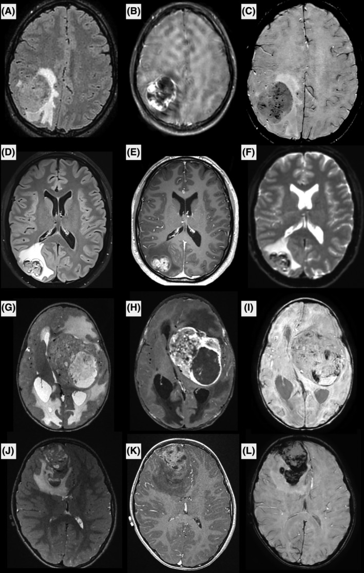
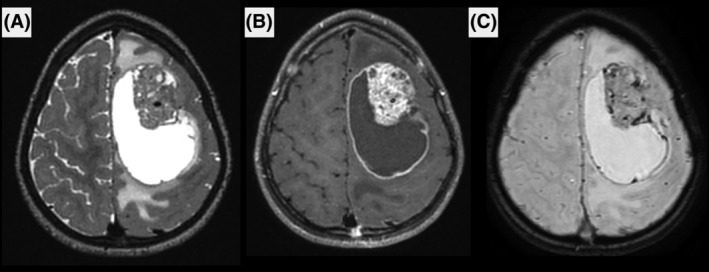
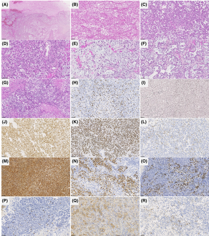
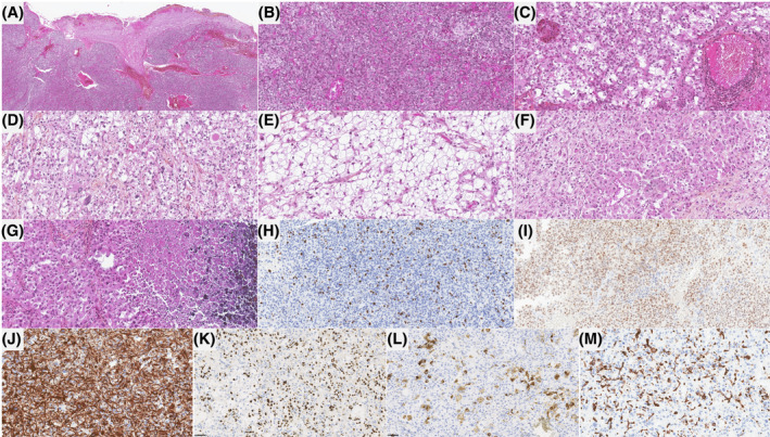
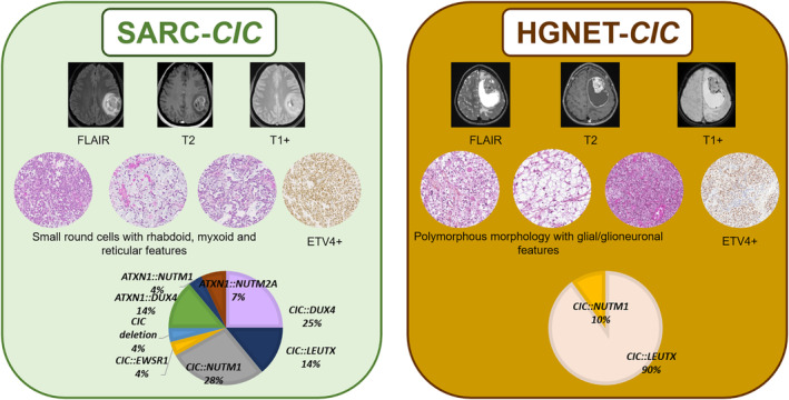
CIC fusions have been described in two different central nervous system (CNS) tumor entities. On one hand, fusions of CIC or ATXN1 genes belonging to the same complex of transcriptional repressors, were reported in the CIC-rearranged, sarcoma (SARC-CIC). The diagnosis of this tumor type, which was recently added to the World Health Organization (WHO) Classification of CNS tumors, is difficult mainly because the data concerning its histopathology (as compared to its soft tissue counterpart), immunoprofile, and clinical as well as radiological characteristics are scarce in the literature. On the other hand, a recent study, based on DNA-methylation profiling, has identified a novel high-grade neuroepithelial tumor characterized by recurrent CIC fusions (HGNET-CIC). The aim of this multicentric study was to characterize a cohort of 15 primary CNS tumors harboring a CIC or ATXN1 fusion in terms of clinical, radiological, histopathological, immunophenotypical, and epigenetic characteristics. According to the integrated diagnoses, 14/15 tumors corresponded to SARC-CIC, and only one to HGNET-CIC. The tumors showed similar clinical (mainly pediatric), radiological (mostly supratentorial, cystic, and contrast enhancing), immunophenotypical (common expression of glioneuronal markers), and genetic (similar spectrum of fusions) profiles but their histopathological appearance was clearly distinct. Moreover, we found a novel fusion transcript (CIC::EWSR1) in a SARC-CIC. Most DNA methylation profiles using the Heidelberg Brain Tumor Classifier (v12.8) annotated the samples to the methylation class "SARC-CIC" (9/14 tumors with available data). By using uniform manifold approximation and projection analysis, four other samples were classified as SARC-CIC and another clustered within the methylation class of HGNET-CIC. Our findings confirm that CNS CIC-fused tumors do not represent a single molecular tumor entity. Further analyses are needed to characterize HGNET-CIC in more detail. These results may help to refine the essential diagnostic criteria for SARC-CIC and their terminology (with a suggested consensual name of sarcoma, CIC/ATXN1-complex rearranged).

Keywords: ATXN1; CIC; DNA methylation profile; LEUTX; glioneuronal; sarcoma.


Conflict of interest statement

The authors declare no conflicts of interest.

Figures

FIGURE 1
FIGURE 1
Radiological features of CIC‐rearranged sarcomas. Axial FLAIR (A, D), T2‐weighted (G, J), postcontrast T1‐weighted (B, E, H, K), and Susceptibility‐weighted (C, I, L) or echo‐planar (F) images of cases #1 (A, B, C), #3 (D, E, F), #5 (G, H, I) and #6 (J, K, L). The tumors were located supratentorially, and had partially cystic content, intense contrast enhancement, intra‐tumoral hemorrhage, and large peritumoral edema. Most of the tumors had pachymeningeal contact, which is clearly seen in patient #6.
FIGURE 2
FIGURE 2
Radiological features of the CIC‐rearranged high‐grade neuroepithelial tumor. Images show a large left fontal tumor, with cystic content and peritumoral edema on T2‐weighted images (A), intense contrast enhancement (B) and intra‐tumoral microbleeds (C).
FIGURE 3
FIGURE 3
Histopathological features of CIC‐rearranged sarcomas. (A) Well‐defined delineation of the tumor from the brain parenchyma (HPS, magnification ×80). (B) Sheets of cells with a lobular appearance and epithelioid differentiation (HPS, magnification ×400). (C) Sheets of small round cells (HPS, magnification ×400). (D) Rhabdoid features (HPS, magnification ×600). (E) Myxoid modifications (HPS, magnification ×400). (F) A reticular pattern was present in a subset of cases (HPS, magnification ×400). (G) Areas of necrosis (HPS, magnification ×400). (H) High MIB1 labeling index (magnification ×400). (I) Reticulin deposition (magnification ×400). (J) Diffuse immunopositivity for ETV4 (magnification ×400). (K) Diffuse immunoreactivity for NUT in a tumor with a CIC::NUTM1 fusion (magnification ×400). (L) A focal membranous staining for CD99 (magnification ×400). (M) Diffuse expression of vimentin (magnification ×400). (N) Expression of GFAP by a subset of tumor cells (magnification ×400). (o) Expression of OLIG2 in some tumor cells (magnification ×400). (P) Expression of neurofilament by some tumor cells (magnification ×400). (Q) Expression of CD56 (magnification ×400). (R) Expression of synaptophysin by a subset of tumor cells (magnification ×400). Black scale bars represent 250 μm (A) and 50 μm (B–R). HPS, Hematoxylin Phloxin Saffron.
FIGURE 4
FIGURE 4
Histopathological features of the CIC‐rearranged high‐grade neuroepithelial tumor. (A) Well‐defined delineation of the tumor from the brain parenchyma (HPS, magnification ×80). (B) Sheets of small round cells (HPS, magnification ×400). (C, D) Glioneuronal features with perivascular lymphocytic infiltrates and eosinophilic granular bodies (HPS, magnification ×400). (E) A microcystic pattern (HPS, magnification ×400). (F) A glial gemistocytic pattern (HPS, magnification ×400). (G) Area of necrosis (HPS, magnification ×400). (H) High MIB1 labeling index (magnification ×400). (I) Diffuse immunopositivity for ETV4 (magnification ×400). (J) Diffuse expression of GFAP (magnification ×400). (K) Expression of OLIG2 in some tumor cells (magnification ×400). (L) Expression of synaptophysin by some tumor cells (magnification ×400). (M) An extravascular cellular expression of CD34 (magnification ×400). HPS, Hematoxylin Phloxin Saffron.
FIGURE 5
FIGURE 5
Summary of CIC‐rearranged sarcomas and CIC‐rearranged high‐grade neuroepithelial tumors main characteristics extracted from the literature and the current series. Both SARC‐CIC and HGNET‐CIC were large tumors with marked peritumoral edema and intense contrast enhancement. Hemorrhage was seen in all tumors. SARC‐CIC present densely cellular sheets of small round cells with rhabdoid differentiation, myxoid modifications and reticular pattern whereas HGNET‐CIC showed obvious signs of neuronal differentiation (eosinophilic granular bodies, perivascular lymphocytic infiltrates and ganglion cells), and other components (clear cells and small round cells). The two entities share the expression of ETV4. SARC‐CIC present a wide variety of alterations (mostly fusions) implicating CIC or ATXN1 genes whereas HGNET‐CIC are associated to date with CIC::LEUTX or CIC::NUTM1 fusions. HGNET‐CIC, high‐grade neuroepithelial tumor, CIC‐fused; SARC‐CIC, CIC‐rearranged, sarcoma.

Similar articles

Cited by

References

    1. Louis DN, Perry A, Wesseling P, Brat DJ, Cree IA, Figarella‐Branger D, et al. The 2021 WHO classification of tumors of the central nervous system: a summary. Neuro‐Oncol. 2021;23:1231–1251. - PMC - PubMed
    1. WHO Classification of Tumours Editorial Board . WHO classification of tumours series. Central nervous system tumours. Volume 6. 5th ed. Lyon, France: International Agency for Research on cancer; 2021.
    1. Watson S, Perrin V, Guillemot D, Reynaud S, Coindre J‐M, Karanian M, et al. Transcriptomic definition of molecular subgroups of small round cell sarcomas. J Pathol. 2018;245:29–40. - PubMed
    1. Kallen ME, Hornick JL. The 2020 WHO classification: What's new in soft tissue tumor pathology? Am J Surg Pathol. 2021;45:e1–e23. - PubMed
    1. Sturm D, Orr BA, Toprak UH, Hovestadt V, Jones DTW, Capper D, et al. New brain tumor entities emerge from molecular classification of CNS‐PNETs. Cell. 2016;164:1060–1072. - PMC - PubMed

Publication types