Review
. 2024 Oct 24;14(1):163.
doi: 10.1186/s13613-024-01395-y.

Methodologies and clinical applications of lower limb muscle ultrasound in critically ill patients: a systematic review and meta-analysis

Affiliations
Review

Methodologies and clinical applications of lower limb muscle ultrasound in critically ill patients: a systematic review and meta-analysis

Roberto Venco et al. Ann Intensive Care. .

Abstract

Background: Reduced muscle mass upon admission and development of muscle wasting are frequent in critically ill patients, and linked to unfavorable outcomes. Muscle ultrasound is a promising instrument for evaluating muscle mass. We summarized the findings of lower limb muscle ultrasound values and investigated how the muscle ultrasound parameters of the examination or the patient characteristics influence the results.

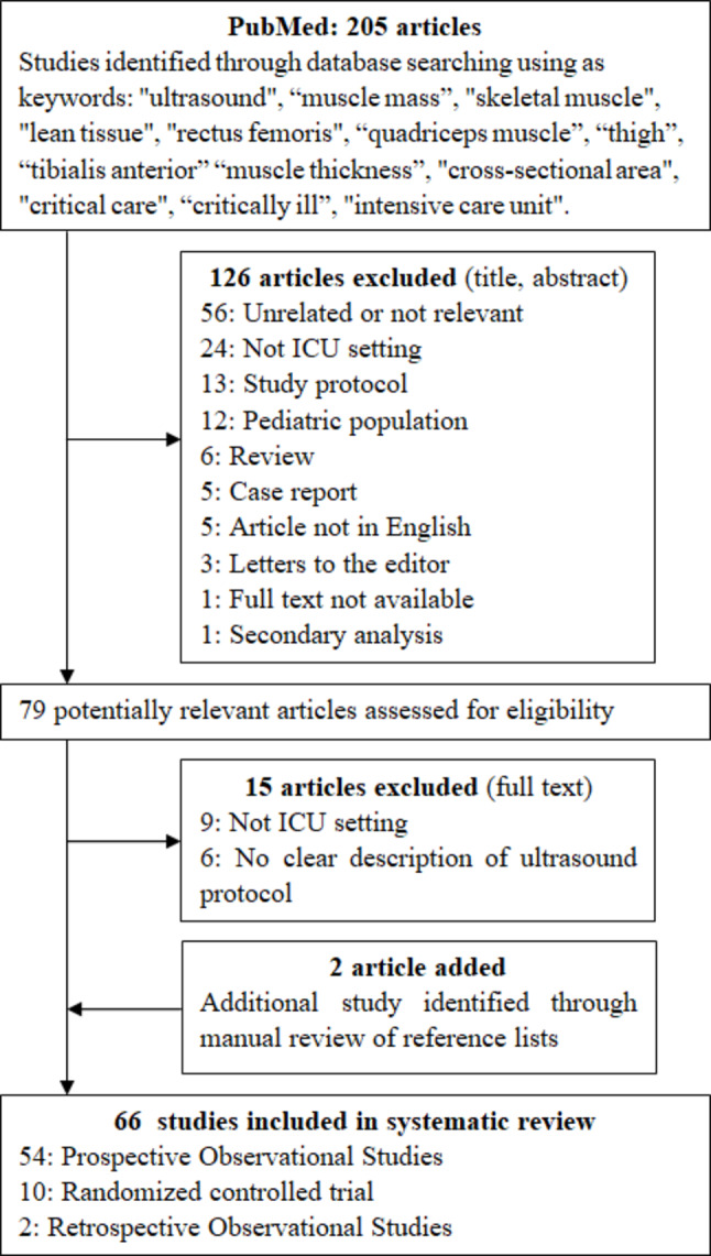
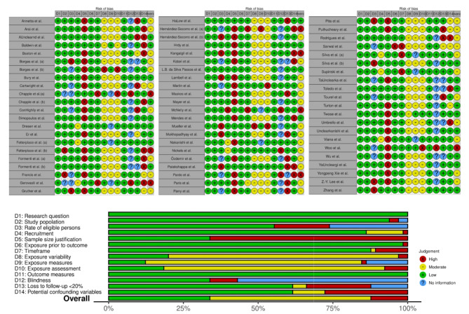
Methods: Systematic review and meta-analysis of studies of lower limb ultrasound critically ill adults. PubMed, CINAHL, Embase, PEDro and Web of Science were searched. PRISMA guidelines were followed, and studies evaluated with the appropriate NIH quality assessment tool. A meta-analysis was conducted to compare the values at admission, short and long follow-up during ICU stay, and the association between baseline values and patient characteristics or ultrasound parameters was investigated with a meta-regression.

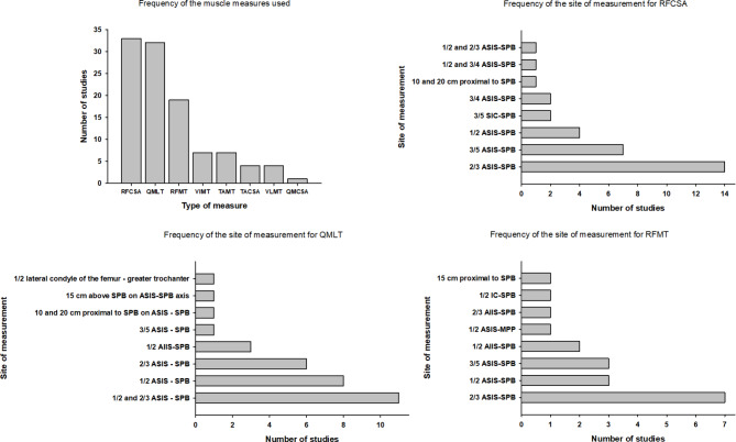
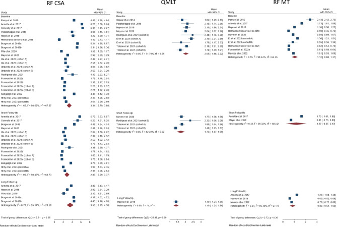
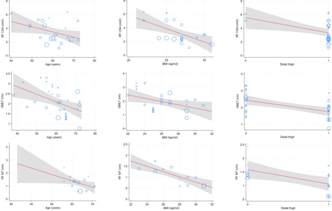
Results: Sixty-six studies (3839 patients) were included. The main muscles investigated were rectus femoris cross-sectional area (RF-CSA, n = 33/66), quadriceps muscle layer thickness (n = 32/66), and rectus femoris thickness (n = 19/66). Significant differences were found in the anatomical landmark and ultrasound settings. At ICU admission, RF-CSA ranged from 1.1 [0.73-1.47] to 6.36 [5.45-7.27] cm2 (pooled average 2.83 [2.29-3.37] cm2) with high heterogeneity among studies (I2 = 98.43%). Higher age, higher BMI, more distal landmark and the use of probe compression were associated with lower baseline muscle mass.

Conclusions: Measurements of muscle mass using ultrasound varied with reference to patient characteristics, patient position, anatomical landmarks used for measurement, and the level of compression applied by the probe; this constrains the external validity of the results and highlights the need for standardization.

Study registration: PROSPERO CRD42023420376.

Keywords: Critically ill; Lean body mass; Meta-analysis; Muscle ultrasound; Muscle wasting; Systematic review; Ultrasound.


Conflict of interest statement

MU has received speaker fees from Baxter, Fresenius Kabi and Nestlè. The other authors declare that they have no conflicts of interest.

Figures

Fig. 1
Fig. 1
Flow-chart of the study selection process
Fig. 2
Fig. 2
Type and relative frequencies of the ultrasound measurements of muscle mass at the thigh and the landmarks used. The figure shows the different muscle measures used in the studies included in the current systematic review (upper left panel), as well as the anatomical landmarks and sites of measurements selected in the different studies for each of the most frequently used muscle measurements. RFCSA: Rectus Femoris cross sectional area; QMLT: Quadriceps layer muscle thickness; RFMT: Rectus Femoris muscle thickness; VIMT: VastusIntermedius muscle thickness; TAMT: Tibialis Anterior muscle thickness; TACSA: Tibialis Anterior cross sectional area; VLMT: VastusLateralis muscle thickness; QMCSA: Quadriceps muscle cross sectional area; ASIS: anterior superior iliac spine; SIC: superior iliac crest; SPB: superior border patella; SIC: superior iliac crest; MPP: middle point of the patella
Fig. 3
Fig. 3
Forest plot of the average value of muscle mass for the three most frequently reported ultrasound methods (Left panel: Rectus femoris CSA, Middle panel: Quadriceps muscle layer thickness, Right panel: Rectus femoris MT) at baseline and at the shortest and longest follow-up. The figure reports the value of muscle mass assessment using no or minimal compression and a distal thigh landmark
Fig. 4
Fig. 4
Summary of the risk of bias of the included studies using the appropriate NIH quality assessment tool
Fig. 5
Fig. 5
Meta-regression for the crude effects of age (left panel), body-mass index (middle panel) or the use of a distal thigh landmark of subjects included in the studies on baseline RF CSA (upper row), QMLT (middle row) or RF MT (lower row). The point sizes are inversely proportional to the standard error of the mean of the individual studies (i.e., larger/more precise studies are shown as larger circles). Regression line and its prediction intervals (95% CIs) were presented in the figure

Similar articles

Cited by

References

    1. Hermans G, Van Den Berghe G. Clinical review: intensive care unit acquired weakness. Crit Care. 2015;19(1):274. - PMC - PubMed
    1. Jolley SE, Bunnell AE, Hough CL. ICU-acquired weakness. Chest. 2016;150(5):1129–40. - PMC - PubMed
    1. Batt J, Dos Santos CC, Cameron JI, Herridge MS. Intensive care unit–acquired weakness: clinical phenotypes and molecular mechanisms. Am J Respir Crit Care Med. 2013;187(3):238–46. - PubMed
    1. Weijs PJM, Looijaard WGPM, Dekker IM, et al. Low skeletal muscle area is a risk factor for mortality in mechanically ventilated critically ill patients. Crit Care. 2014;18(2):R12. - PMC - PubMed
    1. Jaitovich A, Khan MMHS, Itty R, et al. ICU admission muscle and Fat Mass, Survival, and disability at Discharge. Chest. 2019;155(2):322–30. - PMC - PubMed