. 2024 Oct 23;28(11):609.
doi: 10.1007/s00784-024-06000-x.

Evaluation of clinical and radiographic warning signs for prediction of oroantral communication following tooth extractions

Affiliations

Evaluation of clinical and radiographic warning signs for prediction of oroantral communication following tooth extractions

Alexandra Jurásek et al. Clin Oral Investig. .

Abstract

Objectives: Oroantral communication (OAC) is a relatively common and mild complication of maxillary tooth extractions. Preoperative prediction of OAC can reduce treatment duration and prepare both operators and patients for the procedure. This study aims to identify alarming radiographic and clinical indicators that can predict OAC therefore assisting clinical decision making to practicing general dentists.

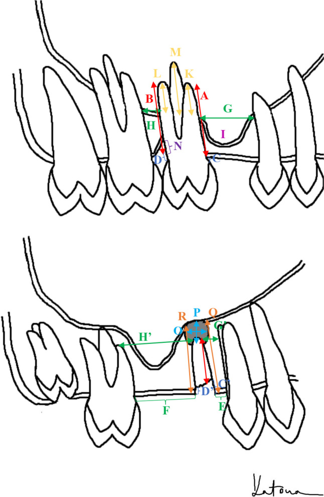
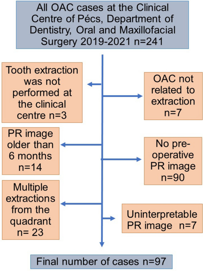
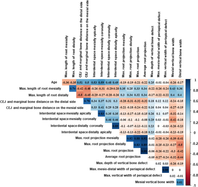
Methods: In this retrospective case-control study the OAC group consisting of 97 cases and a control group twice the size was established. Clinical data were collected, and measurements were conducted separately by two blinded observers on digital panoramic radiographs. Inter-rater reliability was assessed. In case of disagreement a third observer's results were utilized. The correlation between OAC and demographic data (age, sex), as well as various factors assessed on panoramic radiographs (including, but not limited to, the length of the root, root projection into the sinus, bone width, presence of mesial and distal adjacent teeth), was statistically evaluated.

Results: Inter-rater reliability was found to be excellent. Several factors were identified as potential predictors of OAC. According to our model, the strongest predictors were the distance between the cemento-enamel junction and marginal bone, extent of root projection into the sinus, presence of sinus recess around the roots, angulation, and absence of the mesial adjacent tooth.

Conclusions: Well-defined measurements on panoramic radiographs may aid in predicting OAC. Further prospective investigations are necessary to confirm these indicators and address factors related to clinical examination and operation.

Clinical relevance: We present several clinical and radiographic warning signs of OAC that can facilitate pre-extraction decision-making.

Keywords: Intraoperative Complications; Maxillary Sinus; Oroantral Fistula; Orthopantomography; Panoramic radiograph; Tooth Extraction.


Conflict of interest statement

We report no competing interests.

Figures

Fig. 1
Fig. 1
Schematic representation of the measurements conducted on the panoramic radiographical images: A and B- length of the root measured mesially and distally from the coronal level of the bone to the apex; C and D- distance between the cemento-enamel junction (CEJ) and the coronal bone level on the mesial (C) and distal (D) sides. In cases of crown restoration, the most apical point of the crown was used instead of the CEJ. For severely destructed teeth (root remnant) below the bone level (C', D') or in cases of impacted teeth, this value is negative; E and F- interdental space measured coronally on the mesial and distal sides, at the coronal bone level or, in the case of an impacted tooth, at the most coronal point of the crown. If no more tooth was present in the quadrant, the distal measurement point was designated at the distal side of the maxillary tuber and the mesial at the median sagittal suture; G and H- interdental space measured apically on the mesial and distal sides at the level of the sinus base, or if the root did not reach this, then at the most apical point of the root (G' & H'). In cases of completely missing distal or mesial teeth in the quadrant, the same approach was used as previously described; I-sinus recess on the mesial side (or the distal side); K and L-root projection into the sinus, on the mesial and/or distal side: distance between the level of the sinus base and the apex on the mesial and distal side, note that if the apex does not reach the level of the sinus base this value is negative; M- maximal root projection- the length between the base of the sinus and the apex of the root with the most protrusion into the sinus. If the root(s) do not reach the base of the sinus, the distance between the base and the closest root was measured and indicated by a negative value; N-depth of vertical bone loss: distance between coronal level of bone and most apical point of vertical defect; O and P- mesiodistal (O) and vertical (P) diameter of periapical defect; Q and R- vertical bone width on the mesial and distal sides, measuring the distance between the coronal bone level and the base of the sinus on the mesial and distal sides of the analyzed tooth, respectively
Fig. 2
Fig. 2
Flow chart of case selection, OAC-oroantral communication, PR-panoramic radiographic (image)
Fig. 3
Fig. 3
Results of random forest analysis. Each column represents a variable (1–39), factors above the line (1–13) were identified as potential predictors of oroantral communication: 1- maximal and 2-average root projection, 3- distance between cementoenamel junction and marginal bone on the distal and 4- mesial side, 5-vertical bone width distally, 6- maximal root projection mesially, 7- maximal root projection distally, 8- vertical bone width mesially, 9-presence of distal adjacent tooth, 10- extent of caries lesion, 11- presence of mesial adjacent tooth, 12-length of root mesially, 13-presence of caries, 14-distal sinus recess present, 15-angulation, 16-presence of sinus recess, 17-root canal treatment (yes/no), 18-interdental space distally coronally, 19-age of patient (years), 20-interdental space mesially apically, 21- relation of periapical lesion to the base of the sinus, 22- interruption in the basal line of maxillary sinus, 23-interdental space mesially coronally, 24-presence of periapical radiolucency, 25- interdental space distally apically, 26- maximal mesio-distal width of periapical defect, 27-single or multiple roots, 28-presence of mesial sinus recess, 29-impaction, 30-root length distally, 31-depth of vertical bone defect, 32-type of tooth, 33-presence of restoration, 34- extent of restoration, 35-relation of apex(es) to base of maxillary sinus, 36-sex, 37-presence of vertical bone defect, 38-maximal vertical width of periapical defect, 39-notation of tooth (FDI)
Fig. 4
Fig. 4
Results of variable correlation: values closer to 0 indicate no correlation while values closer to 1 or -1 indicate strong positive and negative correlation respectively. CEJ-cemento-enamel junction, max.-maximal
Fig. 5
Fig. 5
Decision tree for oroantral communication occurrence

Similar articles

References

    1. Punwutikorn J, Waikakul A, Pairuchvej V (1994) Clinically significant oroantral communications - a study of incidence and site. Int J Oral Maxillofac Surg 23:19–21. 10.1016/S0901-5027(05)80320-0 - PubMed
    1. Miclotte I, Agbaje JO, Spaey Y et al (2018) Incidence and treatment of complications in patients who had third molars or other teeth extracted. Br J Oral Maxillofac Surg 56:388–393. 10.1016/j.bjoms.2018.02.001 - PubMed
    1. Kiencało A, Jamka-Kasprzyk M, Panaś M, Wyszyńska-Pawelec G (2021) Analysis of complications after the removal of 339 third molars. Dent Med Probl 58:75–60. 10.17219/dmp/127028 - PubMed
    1. Shahrou R, Sha P, Withana T et al (2021) Oroantral communication, its causes, complications treatments and radiographic features: A pictorial review. Imaging Sci Dent 51:1–5. 10.5624/ISD.20210035 - PMC - PubMed
    1. Belmehdi A, El Harti K (2019) Management of oroantral communication using buccal advanced flap. Pan Afr Med J 34:. 10.11604/pamj.2019.34.69.19959 - PMC - PubMed

LinkOut - more resources