. 2024 Oct 23;15(1):9138.
doi: 10.1038/s41467-024-53364-5.

Characterization of Medusavirus encoded histones reveals nucleosome-like structures and a unique linker histone

Affiliations

Characterization of Medusavirus encoded histones reveals nucleosome-like structures and a unique linker histone

Chelsea M Toner et al. Nat Commun. .

Abstract

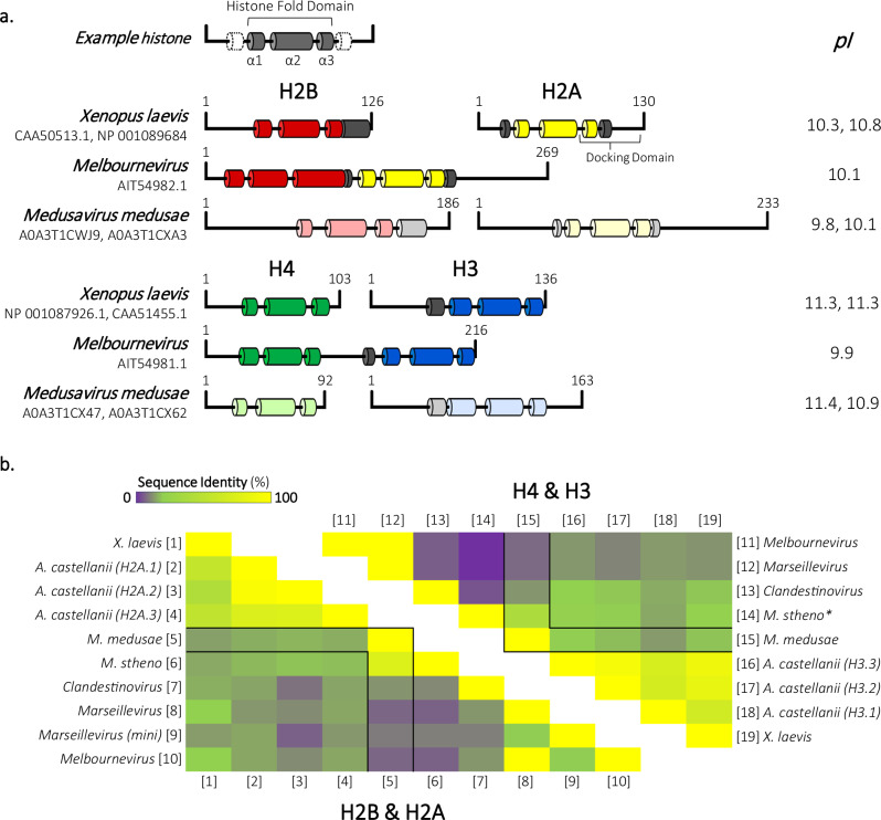
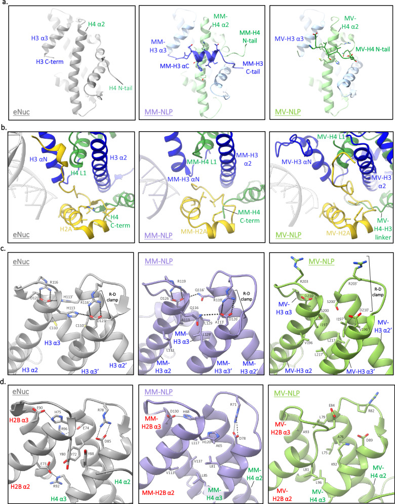
The organization of DNA into nucleosomes is a ubiquitous and ancestral feature that was once thought to be exclusive to the eukaryotic domain of life. Intriguingly, several representatives of the Nucleocytoplasmic Large DNA Viruses (NCLDV) encode histone-like proteins that in Melbournevirus were shown to form nucleosome-like particles. Medusavirus medusae (MM), a distantly related giant virus, encodes all four core histone proteins and, unique amongst most giant viruses, a putative acidic protein with two domains resembling eukaryotic linker histone H1. Here, we report the structure of nucleosomes assembled with MM histones and highlight similarities and differences with eukaryotic and Melbournevirus nucleosomes. Our structure provides insight into how variations in histone tail and loop lengths are accommodated within the context of the nucleosome. We show that MM-histones assemble into tri-nucleosome arrays, and that the putative linker histone H1 does not function in chromatin compaction. These findings expand our limited understanding of chromatin organization by virus-encoded histones.


Conflict of interest statement

The authors declare no competing interests.

Figures

Fig. 1
Fig. 1. Secondary structure prediction and sequence alignment of Medusavirus medusae histones reveals conservation of key eukaryotic residues.
a Schematic of Eukarya (Xenopus laevis) and Nucleocytoviricota histones from Melbournevirus (MV) and Medusavirus (MM). Known X. laevis and MV α helices representative of the histone fold domain are represented in dark-colored tubes (H2B, red; H2A, yellow; H4, green; H3, blue; and additional helices, gray). α helices in MM histones were predicted using HHpred’s Quick 2D prediction webserver (shown in lighter designated colors). Isoelectric points (pI) of each histone are shown to the right. b Heat map comparing percent identity of Eukarya and Nucleocytoviricota H2B-H2A histone sequences (left triangle) to each other. Equivalent H2B-H2A histone sequences are represented along the bottom side [1–10]. Heat map (right triangle) of percent identity of Eukarya and Nucleocytoviricota H4-H3 sequences to each other. Equivalent H4-H3 histone sequences are represented along the top side [11–19]. MM histones are outlined in black within both triangles. A comparison of different dimer pairs (H2B-H2A to H4–H3) sequence identity is not displayed. *M. stheno H4-H3 alignment values determined from H3–H4 alignment shown in Supplementary Fig. 1C. Source data are provided as a Source Data file.
Fig. 2
Fig. 2. Medusavirus medusae histones and DNA assemble into stable mono nucleosome-like particles (NLP) and tri-NLP in vitro.
a SDS-PAGE of refolded Medusavirus medusae (MM) core histone octamer (H2A, H2B, H3, and H4). b MM-NLP, eNuc, and Melbournevirus NLP (MV-NLP) reconstituted with Widom 601–207 bp DNA and analyzed by 5% Native-PAGE stained with SYBRGold (DNA visualization). c SV-AUC of reconstituted NLP and histone-DNA complexes. Van Holde–Weischet plot of eNuc on 207 bp DNA, histone-DNA complexes with H2A-H2B and (H3–H4)2, and MM-NLP207W. Quantitative evaluation is given in Table 1. d 5% Native-PAGE of reconstituted MM-NLP and eNuc on 150 bp ‘random sequence DNA’ (50% G/C DNA (150 R)), and on Widom 601–207 bp DNA. e Thermal stability of MM-NLP and eNuc shown in d including MV207W from b. All points are the mean (SEM as error bars, n = 3). Tm values of each MM-NLP and eNuc are shown in the inset. f Mass photometry analysis of eNuc-tri (gray) and MM-tri-NLP (purple). The solid lines represent the Gaussian function fit the main species observed on particle counts versus molecular mass distribution histograms, with the estimated molecular weight (in kDa) corresponding to the respective mass at the centre of each peak (i.e., mean ± SD (μ ± σ)). Theoretical and measured molecular masses are shown in the inset. This is one representative data set of triplicate MP measurements. g Representative AFM topography images of eNuc-tri and MM-tri-NLP. Scale bar = 50 nm. Unless otherwise noted, all experiments have been repeated independently more than three times with similar results. Source data are provided as a Source Data file.
Fig. 3
Fig. 3. Medusavirus medusae NLP (MM-NLP207W) resemble eukaryotic nucleosomes.
a Overview of MM-NLP207W structure. Individual histones H2A, H2B, H3, and H4 and their surrounding density are shown in yellow, red, blue, and green, respectively. DNA is shown in purple. b Overlay of MM-NLP207W with eNuc (gray) with only 71 bp of DNA, one H2A-H2B dimer, H3–H3′ four-helix bundle, H3′ N-terminal helix, and a single H4 displayed for clarity. Superhelix locations (SHLs) are numbered from 0 to 6 starting at the nucleosome dyad (ɸ). c Comparison of native (light blue) and GraFix (purple) MM-NLP207W electron densities.
Fig. 4
Fig. 4. Comparison to eNuc reveals unique roles for longer tails and loops.
a Comparison of crosslinked and native MM-NLP. Superposition of GraFix MM-H3 density and native MM-H3 density. The MM-H3 tail from H135 to G153 is displayed (inset), with each density shown as purple and blue surface, respectively. b Superposition of MM-(H3–H4) with eukaryotic (H3–H4) and 30 bp of associated DNA. Close-ups (inset) are provided of (Top) MM residues in DNA minor groove with residue density shown as gray surface, (Bottom) MM-H3 C-tail (blue) orientation in relation to the eukaryotic H3 C-terminus (gray), the eukaryotic (gray), and MM H4 N-tail (green). Corresponding residues of each H3 C-tail are denoted below. c Superposition of MM-H2A-H2B dimer aligned with eukaryotic H2A-H2B dimer (gray) and 40 bp of associated DNA. Close-ups (inset) are provided of (left) eH2B (gray) and MM-H2B loop 1, highlighting MM loop extension, (Right) MM-H2B loop 1 and H2A residues form hydrophobic core with density shown as gray surface.
Fig. 5
Fig. 5. Medusavirus NLPs have unique structural features.
a The H4 α2 helix is stabilized by different structural elements in Medusavirus (MM) and Melbournevirus (MV) NLPs. H3–H4 heterodimer for eNuc and MM-NLP, and H3–H4 doublet for MV-NLP shown. b H2A docking domain (yellow) in eNuc, MM-NLP, MV-NLP with key residues shown as sticks. c Residues contributing to the H3–H3′ four-helix bundle interactions in eNuc, MM-NLP, and MV-NLP. d Residues contributing to the H2B–H4 four-helix bundle interactions in eNuc, MM-NLP, and MV-NLP.
Fig. 6
Fig. 6. Medusavirus medusae linker histone H1 contains a second winged-helix domain and lacks canonical compaction residues.
a Acanthamoeba castellanii (A. castellanii) H1.1, Xenopus laevis (X. laevis) H1.0, and Medusavirus medusae (MM) linker histones were aligned using HHpred’s multiple sequence alignment tool (ClustalΩ). Predicted A. castellanii α helices and beta strands in Mamonoviridae histones were generated using HHpred’s Quick 2D prediction webserver (shown as tubes and arrows, respectively). Known X. laevis H1.0 and A. castellanii α helices and beta strands representative of the winged-helix (WH) domain shown as tubes and arrows, respectively. Corresponding isoelectric points (pI) of each linker histone are provided to the right. b Superposition of AlphaFold MM-H1 (dark pink) and X. laevis H1.0 (dark gray; 5NL0) within the charged surface representation of MM-H1. c Overlay of MM-H1 (shown in dark pink) and X. laevis H1 (dark gray; 5NL0). Inset: X. laevis H1 residues involved in chromatin compaction and corresponding residues in MM-H1.
Fig. 7
Fig. 7. Medusavirus medusae linker histone H1 does not compact tri-nucleosomes.
a Representative AFM topography image of eNuc-tri:eH1.0 sample, imaged in air (scale bar = 50 nm). More than three technical replicates of this representative data were observed with same result. b Height profile through particles (indicated by white box in a) along the lines as depicted in graphical inset. c Representative AFM topography image of eNuc-tri:MM-H1 sample, imaged in air (scale bar = 50 nm). More than three technical replicates of this representative data were observed with same result. d Height profile through particles (indicated by white box in c) along the lines as depicted in graphical inset. Dashed lines at 3 and 5 nm shown for reference. e Histogram and Gaussian fitting (data are shown as mean ± SD (μ ± σ)) of particle height for eNuc-tri alone (2.9 ± 0.9 nm, N = 774, gray) and after incubation with either eH1.0 (3.8 ± 0.9 nm, N = 784, blue) or MM-H1 (3.0 ± 0.9, N = 770, pink). f AFM compaction analysis for eNuc-tri alone (99 ± 22 nm, N = 260, gray) and after incubation with either eH1.0 (87 ± 16 nm, N = 245, blue) or MM-H1 (95 ± 22 nm, N = 249, pink). These values represent mean ± SD and statistical significance was determined by unpaired two-tailed Student’s t test, **** represents P = 4.69 × 10−11, ns represents P = 0.0703. Center line, median; dashed lines, upper and lower quartiles. Source data are provided as a Source Data file.

References

    1. Luger, K. et al. Crystal structure of the nucleosome core particle at 2.8 A˚ resolution. Nature389, 251–260 (1997). - PubMed
    1. Malik, H. S. & Henikoff, S. Phylogenomics of the nucleosome. Nat. Struct. Mol. Biol.10, 882–891 (2003). - PubMed
    1. Luger, K. & Richmond, T. J. DNA binding within the nucleosome core. Curr. Opin. Struct. Biol.8, 33–40 (1998). - PubMed
    1. Zhou, K., Gaullier, G. & Luger, K. Nucleosome structure and dynamics are coming of age. Nat. Struct. Mol. Biol.26, 3–13 (2019). - PMC - PubMed
    1. Koonin, E. V. Origin of eukaryotes from within archaea, archaeal eukaryome and bursts of gene gain: eukaryogenesis just made easier? Philos. Trans. R. Soc. B Biol. Sci.370, 20140333 (2015). - PMC - PubMed

Publication types