. 2024 Oct 23;14(1):25081.
doi: 10.1038/s41598-024-75757-8.

Sacubitril/valsartan improves diastolic left ventricular stiffness with increased titin phosphorylation via cGMP-PKG activation in diabetic mice

Affiliations

Sacubitril/valsartan improves diastolic left ventricular stiffness with increased titin phosphorylation via cGMP-PKG activation in diabetic mice

Nozomi Furukawa et al. Sci Rep. .

Abstract

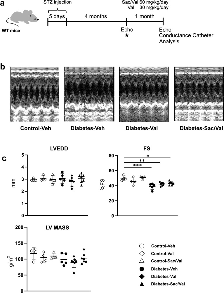
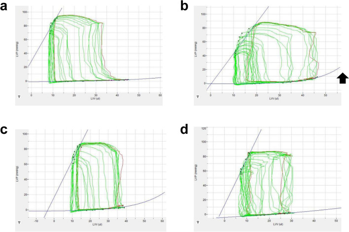
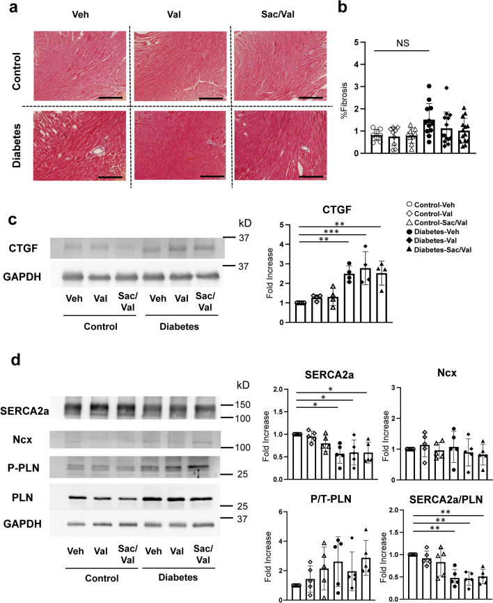
Titin, a giant sarcomeric protein, regulates diastolic left ventricular (LV) passive stiffness as a molecular spring and could be a therapeutic target for diastolic dysfunction. Sacubitril/valsartan (Sac/Val), an angiotensin receptor neprilysin inhibitor, has been shown to benefit patients with heart failure with preserved ejection fraction. The effect of Sac/Val is thought to be due to the enhancement of the cGMP/PKG pathway via natriuretic peptide. In this study, the effects of Sac/Val on LV diastolic dysfunction are demonstrated in a mouse diabetic cardiomyopathy model focusing on titin phosphorylation. Sac/Val-treated diabetic mice showed a greater increase in myocardial levels of cGMP-PKG than Val-treated and control mice. Conductance catheter analysis showed a significant reduction in LV stiffness in diabetic mice, but not in non-diabetic mice. Notably, diastolic LV stiffness was significantly reduced in Sac/Val-treated diabetic hearts compared with Val-treated or vehicle-treated diabetic mice. The phosphorylation level of titin (N2B), which determines passive stiffness and modulates active contraction, was higher in Sac/Val-treated hearts compared with Val-treated hearts in diabetic mice. Given that alteration of titin phosphorylation through PKG contributes to myocardial stiffness, the beneficial effects of Sac/Val in heart failure might be partly attributed to the induction of titin phosphorylation.

Keywords: Cardiomyopathy; Diabetes; Diastolic dysfunction; HFpEF; Myocardial stiffness; Titin.


Conflict of interest statement

N.K. was supported by a joint research fund from Novartis Pharma, which provided the sacubitril/valsartan and valsartan used in the present study.

Figures

Fig. 1
Fig. 1
Experimental schedule and echocardiographic parameters after treatment. Experimental schedule (a). Preliminary conductance catheter measurements are performed four months after STZ injection, shown in Supplementary table S1★; Control, diabetes, diabetes with valsartan treatment, and diabetes with sacubitril/valsartan (Sac/Val). Representative M-mode echocardiography images of Control with Veh (vehicle), Diabetes with Veh, Diabetes with Val, and Diabetes with Sac/Val (b). Summarized graphs of echocardiographic parameters (c), Control with Veh, N = 4; Control with Val, N = 4; Control with Sac/Val, N = 4; Diabetes with Veh, N = 5; Diabetes with Val, N = 7; Diabetes with Sac/Val, N = 7 STZ, streptozotocin; LVEDD, left ventricular end-diastolic diameter; FS, left ventricular fractional shortening; LV mass, calculated left ventricular mass. *p < 0.05, **p < 0.01, and ***p < 0.001 by two-way ANOVA followed by Tukey’s post hoc test. All data are shown as mean and SD values.
Fig. 2
Fig. 2
Representative pressure–volume loops. The arrow indicates the increase in stiffness estimated by the end-diastolic pressure–volume relationship. (a). control with vehicle; (b). diabetes with vehicle; (c). diabetes with Val; (d). diabetes with Sac/Val. The arrow in B show the increase in diastolic LV stiffness.
Fig. 3
Fig. 3
Effect of Sac/Val or Val treatment on the fibrosis area and calcium handling proteins. Representative Masson’s trichrome staining of mouse heart (a). Scale bar: 100 μm. The myocardial fibrosis area is quantified by Masson’s trichrome staining (b) Tissue fibrosis is shown as the ratio (%) of the stained area to the total tissue area. (Control with vehicle, N = 8; Control with Val, N = 9; Control with Sac/Val, N = 9; Diabetes with vehicle, N = 12; Diabetes with Val, N = 14; Diabetes with Sac/Val N = 14). (c). Western blotting for connective tissue growth factor (CTGF). Total protein was quantified, GAPDH was used as an internal standard, and the percent change is presented as a graph for each group. (Control with vehicle, N = 4; Control with Val, N = 4; Control with Sac/Val, N = 4; Diabetes with vehicle, N = 4; Diabetes with Val, N = 4; Diabetes with Sac/Val, N = 4); (d). Western blotting for SERCA2a (sarco/endoplasmic reticulum Ca2+-ATPase 2a), Ncx (sodium-calcium exchanger), and PLN (Phospholamban). Total protein was quantified, GAPDH was used as an internal standard, and the percent change is presented as a graph for each group. For PLN, phosphorylation and total protein intensity (P/T-PLN) are quantified as ratios and presented graphically for each group. For SERCA2a/PLN are quantified as ratios and presented graphically for each group. (Control with vehicle, N = 5; Control with Val, N = 5; Control with Sac/Val, N = 5; Diabetes with vehicle, N = 5; Diabetes with Val, N = 5; Diabetes with Sac/Val, N = 5) *p < 0.05, **p < 0.01, and ***p < 0.001 by two-way ANOVA followed by Tukey’s post hoc test. All data are shown as mean and SD values.
Fig. 4
Fig. 4
Sac/Val treatment increases the phosphorylation levels of titin in diabetic mouse heart. Titin phosphorylation and total titin detection were performed in control and diabetic mice hearts. Pro-Q Diamond staining for phosphorylated titin and Sypro-Ruby staining for total titin detection (a) For titin, phosphorylation and total protein intensity are quantified as ratios and presented graphically for each group. (Control with vehicle, N = 5; Control with Val, N = 5; Control with Sac/Val, N = 5; Diabetes with vehicle, N = 5; Diabetes with Val, N = 5; Diabetes with Sac/Val, N = 5). Western blotting for Phospho/Total-VASP, sGC, pGC, and GAPDH (b). For VASP, phosphorylation and total protein intensity are quantified as ratios and presented graphically for each group. The quantified data for sGC and pGC proteins and corrected for GAPDH are presented as a graph of the ratio of increase to the Control-Veh group. (Control with vehicle, N = 4; Control with Val, N = 4; Control with Sac/Val, N = 4; Diabetes with vehicle, N = 4; Diabetes with Val, N = 4; Diabetes with Sac/Val, N = 4); PKG activity (c) and PKC activity (d). (Control with vehicle, N = 6; Control with Val, N = 4; Control with Sac/Val, N = 4; Diabetes with vehicle, N = 5; Diabetes with Val, N = 7; Diabetes with Sac/Val, N = 6) *p < 0.05, **p < 0.01, ***p < 0.001, and ****p < 0.0001 by two-way ANOVA followed by Tukey’s post hoc test. All data are shown as mean and SD values.

Similar articles

Cited by

References

    1. Dunlay, S. M., Roger, V. L. & Redfield, M. M. Epidemiology of heart failure with preserved ejection fraction. Nat. Rev. Cardiol. 14, 591–602. 10.1038/nrcardio.2017.65nrcardio.2017.65[pii] (2017). - PubMed
    1. Kawaguchi, M., Hay, I., Fetics, B. & Kass, D. A. Combined ventricular systolic and arterial stiffening in patients with heart failure and preserved ejection fraction: implications for systolic and diastolic reserve limitations. Circulation 107, 714–720. 10.1161/01.cir.0000048123.22359.a0 (2003). - PubMed
    1. Zile, M. R., Baicu, C. F. & Gaasch, W. H. Diastolic heart failure–abnormalities in active relaxation and passive stiffness of the left ventricle. N. Engl. J. Med. 350, 1953–1959. 10.1056/NEJMoa032566 (2004). - PubMed
    1. Burkhoff, D., Mirsky, I. & Suga, H. Assessment of systolic and diastolic ventricular properties via pressure-volume analysis: a guide for clinical, translational, and basic researchers. Am. J. Physiol. Heart Circ. Physiol. 289, H501-512. 10.1152/ajpheart.00138.2005 (2005). - PubMed
    1. Zile, M. R. et al. Myocardial stiffness in patients with heart failure and a preserved ejection fraction: contributions of collagen and titin. Circulation 131, 1247–1259. 10.1161/CIRCULATIONAHA.114.013215 (2015). - PMC - PubMed

MeSH terms

LinkOut - more resources