. 2024 Oct 23;14(1):25001.
doi: 10.1038/s41598-024-75747-w.

Effects of coagulation factors on bone cells and consequences of their absence in haemophilia a patients

Affiliations

Effects of coagulation factors on bone cells and consequences of their absence in haemophilia a patients

Giulia Battafarano et al. Sci Rep. .

Abstract

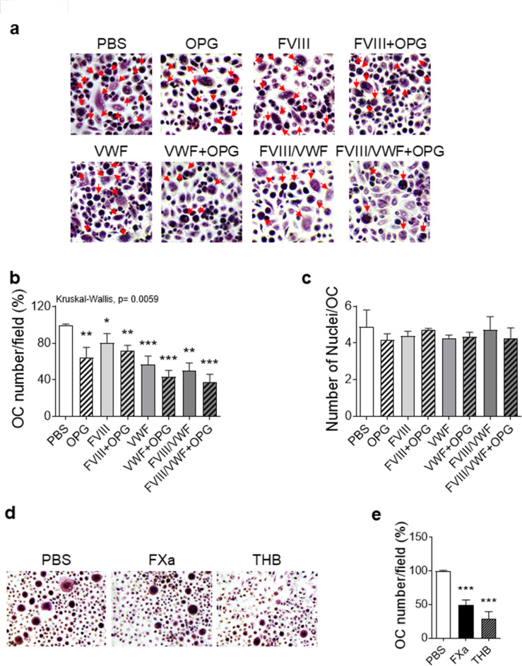
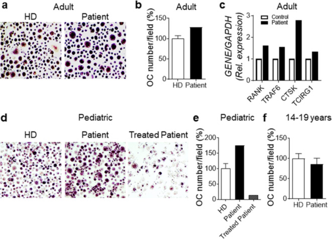
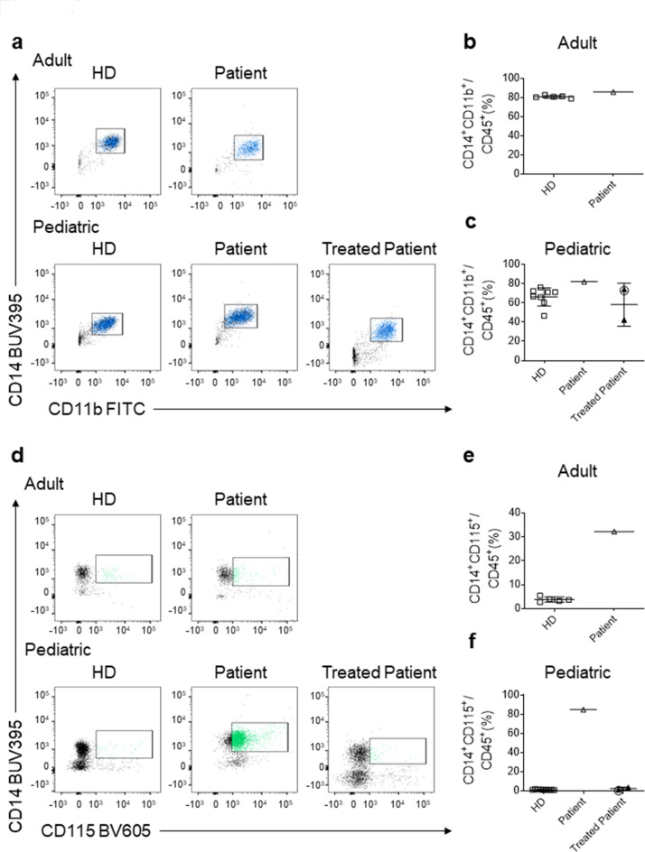
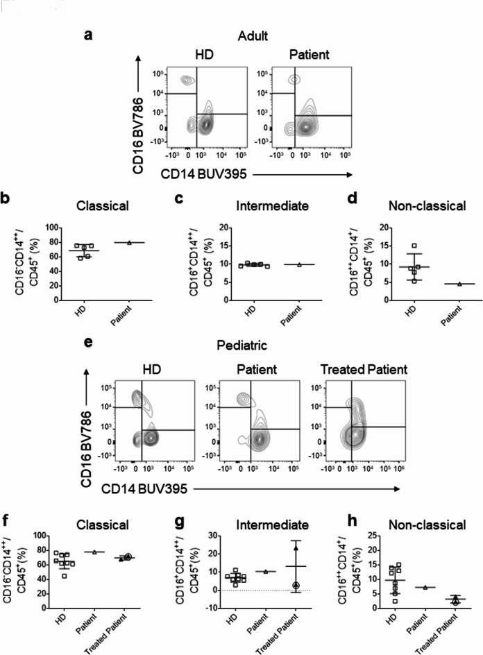
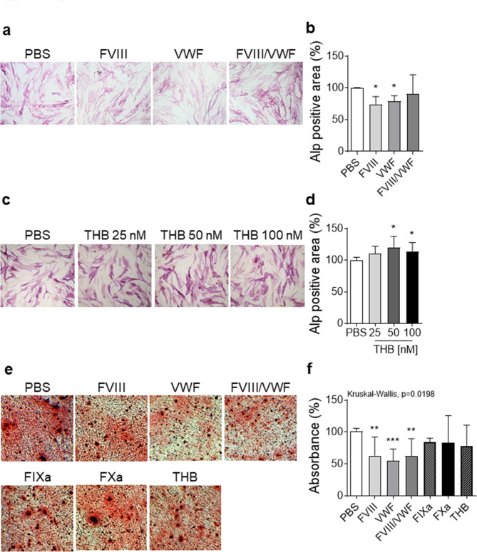
Haemophilia is associated with reduced bone mass and mineral density. Due to the rarity of the disease and the heterogeneity among the studies, the pathogenesis of bone loss is still under investigation. We studied the effects of coagulation factors on bone cells and characterized in a pilot study the osteoclastogenic potential of patients' osteoclast precursors. To evaluate the effect of coagulation factors on osteoclasts, we treated Healthy Donor-Peripheral Blood Mononuclear Cells (HD-PBMC) with Factor VIII (FVIII), von Willebrand Factor (VWF), FVIII/VWF complex, activated Factor IX (FIXa), activated Factor X (FXa) and Thrombin (THB). FVIII, VWF, FVIII/VWF, FXa and THB treatments reduced osteoclast differentiation of HD-PBMC and VWF affected also bone resorption. Interestingly, PBMC isolated from patients with moderate/severe haemophilia showed an increased osteoclastogenic potential due to the alteration of osteoclast precursors. Moreover, increased expression of genes involved in osteoclast differentiation/activity was revealed in osteoclasts of an adult patient with moderate haemophilia. Control osteoblasts treated with the coagulation factors showed that FVIII and VWF reduced ALP positivity; the opposite effect was observed following THB treatment. Moreover, FVIII, VWF and FVIII/VWF reduced mineralization ability. These results could be important to understand how coagulation factors deficiency influences bone remodeling activity in haemophilia.

Keywords: Bone diseases; Coagulation factors; Haemophilia A; Inherited coagulation disorders; Rare disease.


Conflict of interest statement

The authors declare no competing interests.

Figures

Fig. 1
Fig. 1
In vitro effects of coagulation factors on osteoclastogenesis. (ac) Peripheral Blood Mononuclear Cells isolated from 3 Healthy Donors (HD-PBMC) were cultured for 14 days in presence of M-CSF (20 ng/ml)/RANK-L (30 ng/ml) and PBS or OPG (25 ng/ml), FVIII (2 U/ml), VWF (20 µg/ml ≅ 200 U/dl), FVIII/VWF complex (2 U/ml). (a) Representative pictures of osteoclast cultures stained for TRAcP. Original magnification: 10×. Red arrows indicate multinucleated (≥ 3 nuclei) TRAcP positive osteoclasts. (b) Number of osteoclasts (OC) per field and (c) number of nuclei per osteoclast. (d) TRAcP staining of HD-PBMC cultured for 14 days in presence of M-CSF (20 ng/ml)/RANK-L (30 ng/ml) and PBS or activated factor X (FXa, 10 nM) and thrombin (THB, 50 nM). Original magnification: 10×. (e) Number of osteoclasts (OC) per field. Results are expressed as percentage of osteoclasts vs. PBS-treated cells and as mean ± SD of at least three independent experiments. p-values are reported as each treatment compared to PBS. *p ≤ 0.05, **p ≤ 0.01 and ***p ≤ 0.001.
Fig. 2
Fig. 2
Increased osteoclastogenesis in moderate and severe PWH. (a) Representative pictures of TRAcP staining of osteoclast cultures obtained by the differentiation of PBMC isolated from one adult moderate PWH (patient #1) and three age-matched healthy donors (HD). (b) Number of osteoclasts (OC) per field of (a). (c) Gene expression analysis of RANK, TRAF6, CTSK and TCIRG1 in osteoclasts of adult patient with moderate haemophilia (patient #1) and healthy control. (d) Representative pictures of TRAcP staining of osteoclast cultures obtained by the differentiation of PBMC isolated from three HD and one pediatric severe patient before and after treatment with emicizumab. (e) Number of osteoclasts per field of (d). (f) Number of osteoclasts per field of ten HD and three adolescent patients with mild haemophilia. Results are expressed as mean ± SD.
Fig. 3
Fig. 3
Evaluation of osteoclast precursors in monocytes CD45+ cell population of adult and pediatric patients with haemophilia. (a) Representative FACS plots of CD14+CD11b+ monocytes in the gate of CD45+ cells in adult moderate (upper panels) and pediatric severe untreated or treated patients with haemophilia (lower panels) and age-matched HD. (b,c) Percentage of CD14+CD11b+ cells in the gate of CD45+ population, in (b) adult moderate, and (c) pediatric severe patients. (d) Representative FACS plots of CD14+CD115+ population in the monocyte gate of adult moderate (upper panels) and pediatric severe patients (lower panels). (e,f) Percentage of CD14+CD115+ in the gate of CD45+ cells, in (e) adult moderate and (f) pediatric severe patients. Adult moderate: patient #1, Table 1; pediatric severe patients: patients #2 and #6, Table 1. In (c) and (f) empty and filled triangles indicate patient #2 before and after emicizumab treatment, respectively; circled triangle represents patient #6 treated with denecimig. Results are expressed as mean ± SD.
Fig. 4
Fig. 4
Monocyte subsets distribution in PWH. (a,e) FACS plots showing the monocytes subpopulations in adult moderate and pediatric severe untreated or treated patients and age-matched healthy donors (HD). In monocyte gate, (b,f) the classical (CD16CD14++), (c,g) the intermediate (CD16+CD14++), and (d,h) the non-classical (CD16++CD14+) populations were identified. Adult moderate: patient #1, Table 1; pediatric severe patients: patients #2 and #6, Table 1. In (fh) empty and filled triangles indicate patient #2 before and after emicizumab treatment, respectively; circled triangle represents patient #6 treated with denecimig. Results are expressed as mean ± SD.
Fig. 5
Fig. 5
In vitro effects of coagulation factors on osteoblasts. Human control osteoblasts were cultured for 48 h with FVIII (2 U/ml), VWF (20 µg/ml), FVIII/VWF complex (2 U/ml) and THB. (a) Representative pictures of ALP positive osteoblasts. Original magnification: 10×. (b) Densitometric analysis of ALP positive staining of osteoblasts. (c) Representative pictures of ALP positive osteoblasts treated with different concentrations of THB. (d) Densitometric analysis of ALP staining. Results are expressed as a percentage of ALP-positive area in cultures of coagulation factors-treated osteoblasts vs. PBS-treated cells and as mean ± SD. (e) Alizarin red staining of osteoblast treated with FVIII (2 U/ml), VWF (20 µg/ml), FVIII/VWF complex (2 U/ml), FIXa (10 nM), FXa (10 nM), THB (50 nM) or PBS. Original magnification: 4×. (f) Absorbance analysis of Alizarin red staining. Results are expressed as a percentage of absorbance of coagulation factors-treated cultures vs. PBS-treated cells and as mean ± SD. p-values are reported as each treatment compared to PBS. *p ≤ 0.05, **p ≤ 0.01 and ***p ≤ 0.001.

Similar articles

Cited by

References

    1. Escobar, M., Sallah, S. & Hemophilia A and hemophilia B: focus on arthropathy and variables affecting bleeding severity and prophylaxis. J. Thromb. Haemost. 11, 1449–1453. 10.1111/jth.12317 (2013). - PubMed
    1. Rodriguez-Merchan, E. C. et al. Joint protection in haemophilia. Haemophilia. 17 (Suppl 2), 1–23. 10.1111/j.1365-2516.2011.02615.x (2011). - PubMed
    1. Stephensen, D. & Rodriguez-Merchan, E. C. Orthopaedic co-morbidities in the elderly haemophilia population: a review. Haemophilia. 19, 166–173. 10.1111/hae.12006 (2013). - PubMed
    1. Rodriguez-Merchan, E. C. Hemophilic arthropathy: a teaching approach devoted to hemophilia treaters in under-development countries. Expert Rev. Hematol.14, 887–896. 10.1080/17474086.2021.1977118 (2021). - PubMed
    1. Samuelson Bannow, B. et al. Long-established role in haemophilia A and emerging evidence beyond haemostasis. Blood Rev.35, 43–50. 10.1016/j.blre.2019.03.002 (2019). Factor VIII. - PubMed