. 2024 Oct 23;14(1):25080.
doi: 10.1038/s41598-024-76851-7.

Anti-IL-1RAP scFv-mSA-S19-TAT fusion carrier as a multifunctional platform for versatile delivery of biotinylated payloads to myeloid leukemia cells

Affiliations

Anti-IL-1RAP scFv-mSA-S19-TAT fusion carrier as a multifunctional platform for versatile delivery of biotinylated payloads to myeloid leukemia cells

Aref Farokhi-Fard et al. Sci Rep. .

Abstract

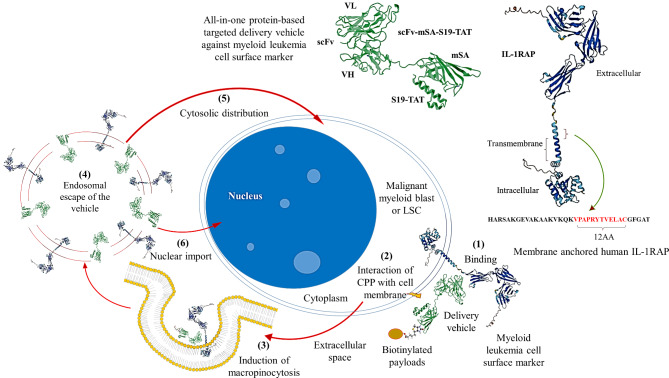
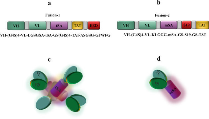
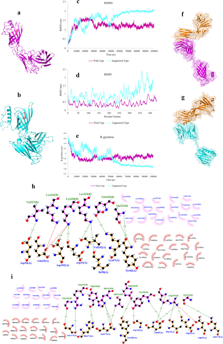
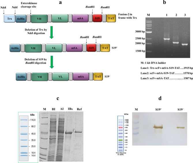
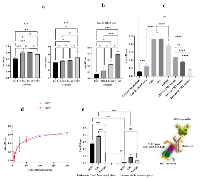
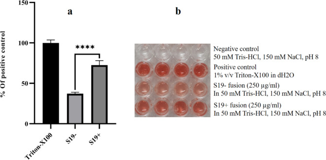
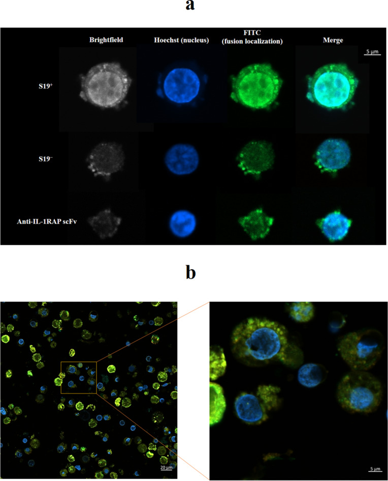
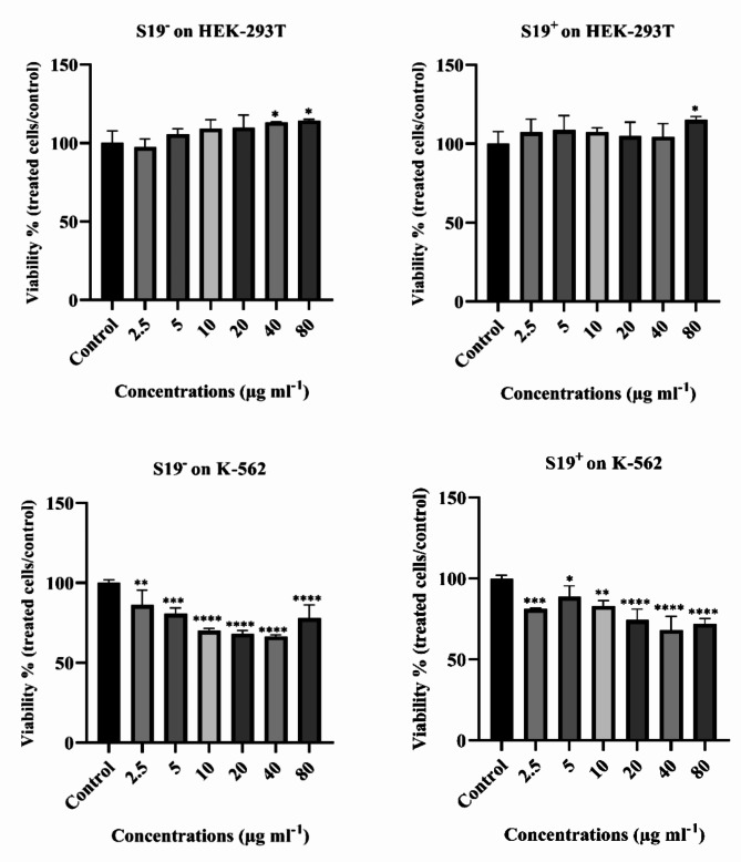
Acute myeloid leukemia (AML) is an aggressive blood cancer with frequently poor clinical outcomes. This heterogeneous malignancy encompasses genetically, molecularly, and even clinically different subgroups. This makes it difficult to develop therapeutic agents that are effective for all subtypes of the disease. Therefore, a selective, universal, and adaptable delivery platform capable of carrying various types of anti-neoplastic agents is an unmet requirement in this area. Two multifunctional fusion proteins were designed for the delivery of biotinylated cargoes to human myeloid leukemia cells by fusing an anti-IL-1RAP single-chain antibody with streptavidin (tetramer or monomer), a cell-penetrating peptide (CPP), and an endosomolytic peptide in a single biomacromolecule. The designed fusions were analyzed primarily in silico, and the biofunctionality of the selected fusion was fully characterized via several binding assays, hemolysis assay, confocal microscopy and cell cytotoxicity assay after production via the Escherichia coli (E. coli) system. The refolded protein exhibited desirable binding activity to leukemic cells, pure antigen and biotinylated BSA. Further analyses revealed efficient cellular uptake, endosomolytic activity, and nuclear penetration without any detectable cytotoxicity toward normal epithelial cells. The described platform seems to have great potential for targeted delivery of different therapeutics to malignant myeloid cells.

Keywords: Bispecific fusion protein; Cell-penetrating peptide; Endosomal escape; Myeloid leukemia; Protein-based delivery vehicle.


Conflict of interest statement

All benefits, profits, and intellectual rights associated with the possible patenting of this research belong exclusively to the first author, the corresponding author, and Pasteur Institute of Iran. There is no conflict of interest by the other authors.

Figures

Fig. 1
Fig. 1
The concept of the study. The targeted vehicle composed of monomeric streptavidin (mSA), single chain variable fragment (scFv), and S19-TAT specifically binds to the myeloid leukemia cell surface marker (membrane isoform of human IL-1RAP, hmIL-1RAP) via recognition of a hmIL-1RAP-specific 12-amino acid sequence (red). The binding of the fusion, causes TAT sequence to be placed in the vicinity to cell membrane. The interaction of TAT with the target cell membrane leads to internalization of the fusion and its biotinylated cargo into the target cell through the induction of macropinocytosis. The vehicle-cargo complex then escapes from the endocytic vesicle and distributed in the cytoplasm with the help of fusogenic properties of S19-TAT. Some of the escaped complexes can then penetrate into the cell nucleus through the nuclear localization functioning of TAT peptide. 3D structure of the full-length hmIL-1RAP has been predicted by AlphaFold (AlphaFold ID: AF-Q9NPH3-F1).
Fig. 2
Fig. 2
Design of anti-hmIL-1RAP fusion proteins. (a, b) Arrangement of functional fragments of tetrameric (a) and monomeric (b) fusions; VH: variable domain of the heavy chain; VL: variable domain of the light chain; tSA: tetrameric core streptavidin; TAT: 9-aa peptide (residues 49-57) from the HIV-1 transactivator; EED: endosomal escape domain pentapeptide; mSA: engineered monomeric streptavidin; S19: 19-aa peptide (residues 322–340) from the human syncytin-1 protein. The gray fragments are flexible linkers. (c, d) Graphic configuration of tetrameric (c) and monomeric (d) fusions.
Fig. 3
Fig. 3
In silico analysis of designed fusions. (a, b) 3D models (biotin-bound state) of fusion-1 (a) and fusion-2 (b) predicted by Galaxyweb. (c-e) RMSD (c), RMSF (d) and Rg (e) plots obtained from MDS on the designed fusions. (f, g) Docking complexes of fusion-1:hmIL-1RAP (f) and fusion-2:hmIL-1RAP (g). (h, i) LigPlot analysis of docking complexes to determine the molecular interactions in the fusion-1hm: IL-1RAP (h) and fusion-2:hmIL-1RAP (i) complexes.
Fig. 4
Fig. 4
Construction and bacterial production of S19+ and S19 fusions. (a) Creation of S19+ and S19 fusion constructs via enzymatic deletions. The NdeI deletion of the Trx-encoding fragment in the fusion-2 construct subcloned in pET32a resulted in the formation of the scFv-mSA-S19-TAT construct (S19+). Subsequent deletion of the S19-encoding fragment from the S19+ construct via BamHI resulted in the formation of the scFv-mSA-TAT construct (S19). (b) Confirmation PCR of the three constructs shown in part A via T7 universal primers (with the T7 promoter as the forward primer and the T7 terminator as the reverse primer). The reactions were performed using Taq DNA Polymerase Master Mix RED (Ampliqon, Denmark) and an Eppendorf thermocycler with an annealing temperature of 60°C. (c) SDS‒PAGE of S19+ expression/refolding samples. For expression, a single colony of the transformant (BL21(DE3) harboring the S19+ construct) was cultured in LB medium and induced at 37°C (4 h induction with 0.1 mM IPTG). M: unstained protein marker, BI: before induction sample, AI: after induction sample, IBs: denatured sample of isolated washed inclusion bodies, Ref: refolded S19+ protein. The SDS‒PAGE pattern of the S19 construct was not presented due to similarity. (d) Confirmation of the proteins by western blotting. Denatured inclusion bodies were subjected to SDS‒PAGE, and the transferred bands were detected using HRP-conjugated anti-6xHis-tag antibody. 3,3’-Diaminobenzidine (DAB) was utilized as a substrate. M: prestained protein marker. The protein quantities are not equal in the lanes.
Fig. 5
Fig. 5
Evaluation of the binding ability of the produced fusions. (a) Cell-based ELISA in triplicate using produced fusions on different myeloid leukemia cell lines. (b) Cell-based ELISA using anti-hmIL-1RAP scFv under nearly similar conditions. (c) Cell-based ELISA on K-562 cells in the absence and presence of AML serum. Three different fusion proteins (S19, S19+, and the isotype molecule Trx-mSA-S19-TAT (40 µg/ml) were added to the test wells in triplicate. The fusion protein solutions were prepared either in HBSS or in 50:50 diluted AML serum. (d) Evaluation of the biotin-binding activity of the refolded proteins. The fusion proteins were added at different concentrations (3–200 µg/ml, in triplicate) to the wells precoated with biotinylated BSA. An HRP-conjugated anti-6xHis tag antibody was utilized for the detection of bound molecules. (e) ELISA on the wells coated with purified antigenic fusions proteins (consisting of Trx and the 12-aa epitope from hmIL-1RAP). Trx or Trx-12aa fusion proteins used for coating the wells. S19+, S19 or an isotype molecule (Trx-mSA-S19-TAT fusion) were added to the coated wells in quadruplicate. Biotinylated IgG and HRP-streptavidin were used for detection. The data are displayed as the mean ± standard deviation (SD). The graphs were plotted, and statistical analyses (one-way ANOVA) were performed via GraphPad Prism version 8. (ns: not significant, *P < 0.05, **P < 0.01, ***P < 0.001, and ****P < 0.0001).
Fig. 6
Fig. 6
Evaluation of the endosomal escape potential of fusions via a hemolysis assay. The diluted RBC suspension (2% v/v in PBS) was added to the fusion proteins (250 µg/ml) in quadruplicate. Protein-free buffer and Triton X-100 (1% v/v in dH2O) were used as negative and positive controls, respectively. The supernatants of the wells were transferred to a flat-bottom 96-well plate after 2 h of incubation at 37 °C. The absorbance of each well was measured spectrophotometrically (Epoch microplate reader) at a wavelength of 405 nm. The data are reported as percentages of the positive control and are presented as the means ± standard deviations (SDs) from four independent tests. The graphs were plotted, and statistical analyses (one-way ANOVA) were performed via GraphPad Prism version 8. (****P ˂ 0.0001).
Fig. 7
Fig. 7
Confocal microscopy of K-562 cells. (a) Evaluation of the cellular internalization and localization of different fusion proteins via confocal microscopy. K-562 cells were treated with ~500 picomoles of Trx-scFv, S19, or S19+ fusion proteins for 3 h. After washing, fixation and permeabilization, the cells were blocked and incubated with a FITC-labeled anti-6xHis-tag monoclonal antibody and stained with Hoechst before imaging with a Zeiss LSM 800 confocal laser scanning microscope. (b) Investigation of S19+ fusion-assisted delivery of macromolecular cargo to K-562 cells. Confocal microscopy was performed on living K-562 cells coincubated (37 °C for 2 h) with 46 pmol of S19+ fusion agent and a FITC-labeled anti-6xHis-tag monoclonal antibody. The treated cells were thoroughly washed after treatment and stained with Hoechst and rhodamine B before imaging.
Fig. 8
Fig. 8
Cytotoxicity evaluation of the produced fusions. An MTT assay was performed on normal (HEK293T) and leukemic (K-562) cells. The cells were seeded at a density of ~5,000 cells/well in 96-well plates and treated with 2.5–80 µg/ml fusion agent in quadruplicate for 48 h. Untreated wells were considered negative controls, and the cells treated with 20% DMSO were considered positive controls (not shown). The data are reported as percentages of the negative control and are presented as the means ± standard deviations (SDs) from four independent tests. The outlier data were removed via Grubbs’ method. The graphs were plotted, and statistical analyses (one-way ANOVA) were performed via GraphPad Prism version 8; *P < 0.05, **P < 0.01, ***P < 0.001, and ****P < 0.0001.

Similar articles

References

    1. 1 Deschler, B. & Lübbert, M. Acute myeloid leukemia: epidemiology and etiology. Cancer Interdiscip. Int. J. Am. Cancer Soc. 107, 2099–2107. 10.1002/cncr.22233 (2006). - PubMed
    1. Maksimovic, N. et al. Incidence and mortality patterns of Acute myeloid leukemia in Belgrade, Serbia (1999–2013). Med. 54, 5. 10.3390/medicina54010005 (2018). - PMC - PubMed
    1. Shallis, R. M., Wang, R., Davidoff, A., Ma, X. & Zeidan, A. M. Epidemiology of acute myeloid leukemia: recent progress and enduring challenges. Blood Rev. 36, 70–87. 10.1016/j.blre.2019.04.005 (2019). - PubMed
    1. Southam, C. M., Craver, L. F., Dargeon, H. W. & Burchenal, J. H. A study of the natural history of acute leukemia with special reference to the duration of the disease and the occurrence of remissions. Cancer. 4, 39–59. 10.1002/1097-0142(195101)4 (1951). :1%3C39::AID-CNCR2820040105%3E3.0.CO;2-G. - PubMed
    1. Cheung, E. et al. The leukemia strikes back: a review of pathogenesis and treatment of secondary AML. Ann. Hematol. 98, 541–559. 10.1200/JCO.2014.60.0890 (2019). - PubMed

MeSH terms

LinkOut - more resources