Multicenter Study
. 2024 Oct 23;14(1):25103.
doi: 10.1038/s41598-024-76933-6.

Differentiating MYCN-amplified RB1 wild-type retinoblastoma from biallelic RB1 mutant retinoblastoma using MR-based radiomics: a retrospective multicenter case-control study

Affiliations
Multicenter Study

Differentiating MYCN-amplified RB1 wild-type retinoblastoma from biallelic RB1 mutant retinoblastoma using MR-based radiomics: a retrospective multicenter case-control study

Christiaan M de Bloeme et al. Sci Rep. .

Abstract

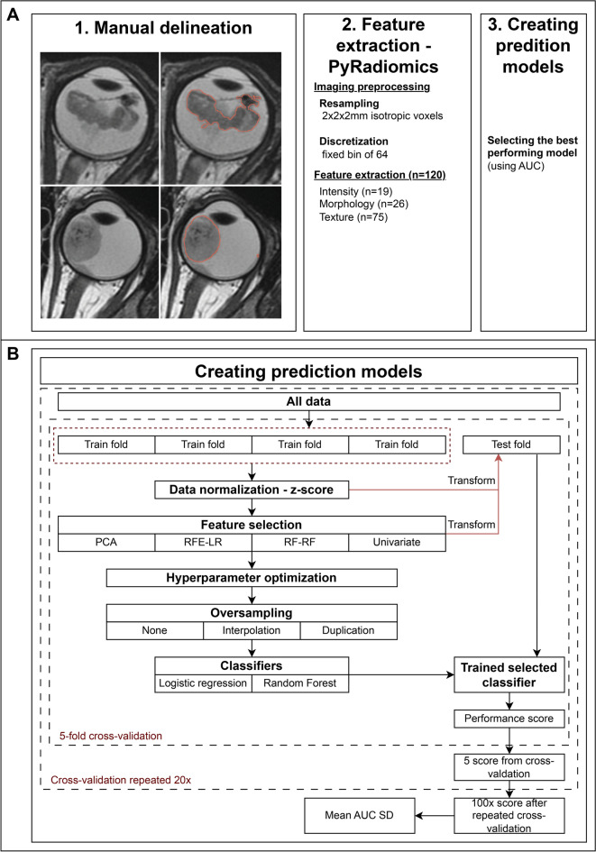
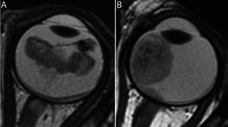
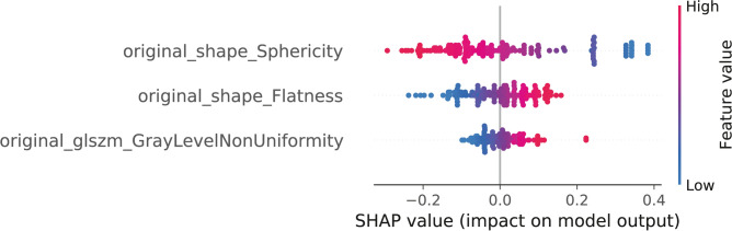
MYCN-amplified RB1 wild-type (MYCNampRB1+/+) retinoblastoma is a rare and aggressive subtype, often resistant to standard therapies. Identifying unique MRI features is crucial for diagnosing this subtype, as biopsy is not recommended. This study aimed to differentiate MYCNampRB1+/+ from the most prevalent RB1-/- retinoblastoma using pretreatment MRI and radiomics. Ninety-eight unilateral retinoblastoma patients (19 MYCN cases and 79 matched controls) were included. Tumors on T2-weighted MR images were manually delineated and validated by experienced radiologists. Radiomics analysis extracted 120 features per tumor. Several combinations of feature selection methods, oversampling techniques and machine learning (ML) classifiers were evaluated in a repeated fivefold cross-validation machine learning pipeline to yield the best-performing prediction model for MYCN. The best model used univariate feature selection, data oversampling (duplicating MYCN cases), and logistic regression classifier, achieving a mean AUC of 0.78 (SD 0.12). SHAP analysis highlighted lower sphericity, higher flatness, and greater gray-level heterogeneity as predictive for MYCNampRB1+/+ status, yielding an AUC of 0.81 (SD 0.11). This study shows the potential of MRI-based radiomics to distinguish MYCNampRB1+/+ and RB1-/- retinoblastoma subtypes.

Keywords: MYCN-amplification; MRI; radiomics; Retinoblastoma.


Conflict of interest statement

C.M.d.B. No relevant relationships. R.W.J. No relevant relationships. L.C. No relevant relationships. S.G. No relevant relationships. S.v.E. No relevant relationships. J.L.J. No relevant relationships. A.R. No relevant relationships. A.H.S. No relevant relationships. A.K.M. No relevant relationships. P.M. No relevant relationships. O.E.U. No relevant relationships. G.B.H. No relevant relationships. H.G. No relevant relationships. H.C.B. No relevant relationships. K.E.N. No relevant relationships. R.C.B. Consultant for Aileron Therapeutics. S. Sen No relevant relationships. M.K. No relevant relationships. S. Sirin No relevant relationships. H.J.B. No relevant relationships. P.G. No relevant relationships. C.J.D. No relevant relationships. M.C. No relevant relationships. R.B. No relevant relationships. J.D. No relevant relationships. A.C.M. No relevant relationships. M.C.d.J. No relevant relationships. P.d.G. No relevant relationships.

Figures

Fig. 1
Fig. 1
Work flowchart of the study. (1) Manual delineation of the whole-tumor and validated by expert radiologists; (2) Processing data through PyRadiomics (version 3.1.0). (2.1) Resampling of MR imaging to 2×2×2 mm isotropic voxels and discretization by a 64 fixed bin. (2.2) Extracting 120 radiomics features divided into three categories: intensity (n = 19), morphology (n = 26), texture (n = 76); 3) Schematic framework of the creation of prediction models for MYCN-status utilizing different combinations of feature selection methods, oversampling techniques, and classifiers; (4) Selection and evaluation of the best-performing model. (4.1) Selection of the best-performing model by the highest mean AUC after the fivefold cross-validation which was 20 times repeated. (4.2) Evaluating the highest selected model by “random guessing” in which the original mean AUC was compared to permuted mean AUC. The permuted mean AUC was calculated by randomly shuffling the MYCN status and using it as input in the fivefold cross-validation which was repeated 20 times. This shuffling and 20 times repeated fivefold cross-validation was repeated 1000 times to obtain the permuted mean AUC.
Fig. 2
Fig. 2
MRI phenotype MYCN-amplified RB1 wild-type (MYCNampRB1+/+) retinoblastoma versus RB1 pathogenic variation (RB1−/−) retinoblastoma. (A) Axial 2D T2-weighted MR image of a 42-month-old patient with MYCNampRB1+/+ retinoblastoma. The tumor has a diffuse growth pattern and is plaque shaped. Also, the intensity in the tumor varies greatly. (B) Axial 2D T2-weighted MR image of a 35-month-old patient with RB1−/− retinoblastoma. The tumor has an endophytic growth pattern and is dome shaped.
Fig. 3
Fig. 3
All features in the best-performing model with univariate feature selecting, duplicate scaling, and a logistic regression as classifier. Features in three categories were assessed: Intensity features, texture features and morphology features. Intensity features comprised of first order statistics [19 features]. Morphology features comprised of shaped-based 2D features [10 features] and 3D features [16 features]. Texture features comprised on features based on gray-level co-occurrence matrices (GLCM) [24 features], gray-level run length matrices (GLRLM) [16 features], gray-level size zone matrices (GLSZM) [16 features], neighborhood gray-tone difference matrices (NGTDM) [5 features], and gray-level dependence matrices (GLDM) [14 features]. Original_shape_Sphericity = original shape sphericity; original_shape_Flatness = original shape flatness; original_glszm_GrayLevelNonUniformity = Original glszm gray level non uniformity.

References

    1. Moll, A. C. et al. Incidence and survival of retinoblastoma in The Netherlands: a register based study 1862–1995. Br J Ophthalmol 81(7), 559–562 (1997). - PMC - PubMed
    1. Pritchard, E. M., Dyer, M. A. & Guy, R. K. Progress in Small Molecule Therapeutics for the Treatment of Retinoblastoma. Mini Rev Med Chem 16(6), 430–454 (2016). - PMC - PubMed
    1. Aubry, A., Yu, T. & Bremner, R. Preclinical studies reveal MLN4924 is a promising new retinoblastoma therapy. Cell Death Discov 6, 2 (2020). - PMC - PubMed
    1. Kooi, I. E. et al. Loss of photoreceptorness and gain of genomic alterations in retinoblastoma reveal tumor progression. Ebiomedicine 2(7), 660–670 (2015). - PMC - PubMed
    1. Afshar, A. R. et al. Next-Generation Sequencing of Retinoblastoma Identifies Pathogenic Alterations beyond RB1 Inactivation That Correlate with Aggressive Histopathologic Features. Ophthalmology 127(6), 804–813 (2020). - PMC - PubMed

Publication types

MeSH terms