. 2024 Oct 24;21(1):60.
doi: 10.1186/s12014-024-09505-5.

SPOT: spatial proteomics through on-site tissue-protein-labeling

Affiliations

SPOT: spatial proteomics through on-site tissue-protein-labeling

Yuanwei Xu et al. Clin Proteomics. .

Abstract

Background: Spatial proteomics seeks to understand the spatial organization of proteins in tissues or at different subcellular localization in their native environment. However, capturing the spatial organization of proteins is challenging. Here, we present an innovative approach termed Spatial Proteomics through On-site Tissue-protein-labeling (SPOT), which combines the direct labeling of tissue proteins in situ on a slide and quantitative mass spectrometry for the profiling of spatially-resolved proteomics.

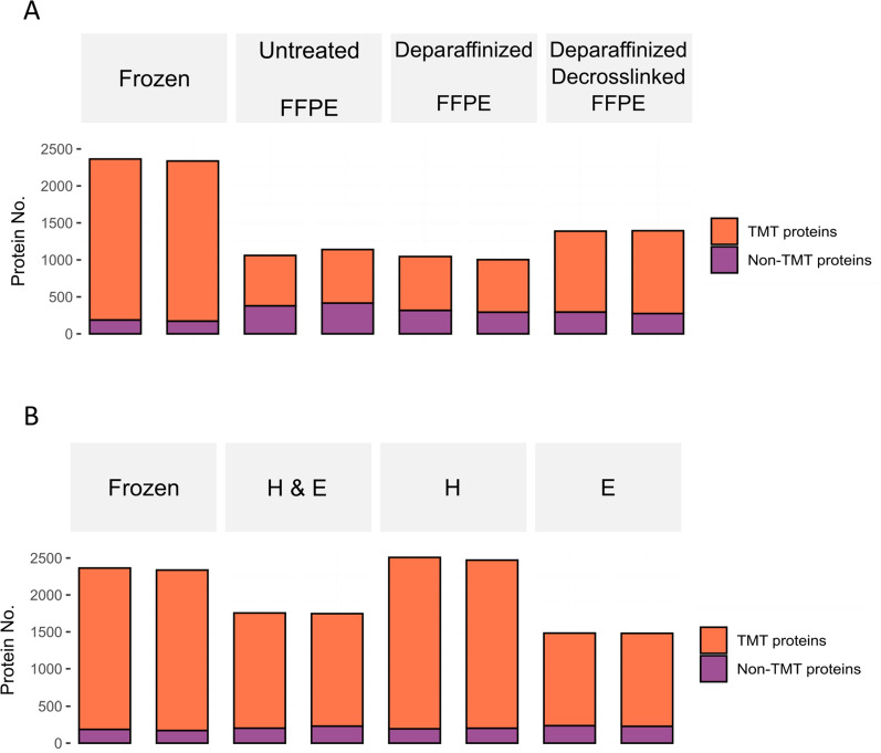
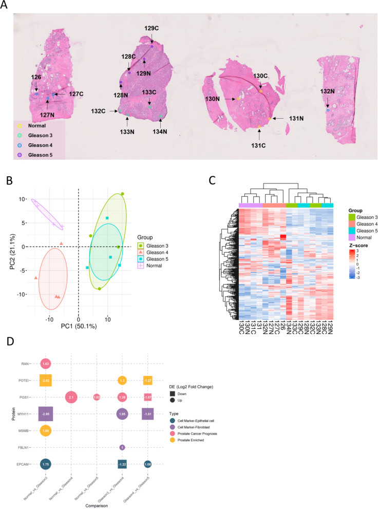
Materials and methods: Efficacy of direct TMT labeling was investigated using seven types of sagittal mouse brain slides, including frozen tissues without staining, formalin-fixed paraffin-embedded (FFPE) tissues without staining, deparaffinized FFPE tissues, deparaffinized and decrosslinked FFPE tissues, and tissues with hematoxylin & eosin (H&E) staining, hematoxylin (H) staining, eosin (E) staining. The ability of SPOT to profile proteomes at a spatial resolution was further evaluated on a horizontal mouse brain slide with direct TMT labeling at eight different mouse brain regions. Finally, SPOT was applied to human prostate cancer tissues as well as a tissue microarray (TMA), where TMT tags were meticulously applied to confined regions based on the pathological annotations. After on-site direct tissue-protein-labeling, tissues were scraped off the slides and subject to standard TMT-based quantitative proteomics analysis.

Results: Tissue proteins on different types of mouse brain slides could be directly labeled with TMT tags. Moreover, the versatility of our direct-labeling approach extended to discerning specific mouse brain regions based on quantitative outcomes. The SPOT was further applied on both frozen tissues on slides and FFPE tissues on TMAs from prostate cancer tissues, where a distinct proteomic profile was observed among the regions with different Gleason scores.

Conclusions: SPOT is a robust and versatile technique that allows comprehensive profiling of spatially-resolved proteomics across diverse types of tissue slides to advance our understanding of intricate molecular landscapes.

Keywords: Mass spectrometry; Prostate cancer; Spatial proteomics; Tissue-protein-labeling.


Conflict of interest statement

The authors declare no competing interests.

Figures

Fig. 1
Fig. 1
Overview of the SPOT workflow. Tissue slides are first annotated by cell types, histological patterns, or pathological states, followed by applying TMT tags directly onto regions of interest. After on-slide labeling and quenching of TMT, the tissue would be lysed, digested, and cleaned up for the downstream proteomic analysis using a mass spectrometer
Fig. 2
Fig. 2
Total identified proteins from each type of sagittal mouse brain slide. A Protein identifications of frozen, untreated FFPE, deparaffinized FFPE, and deparaffinized/decrosslinked slides (all were unstained). B Protein identifications of frozen, H&E stained, H stained, and E stained slides
Fig. 3
Fig. 3
A Mouse brain slide in horizontal view. Eight different regions are color-coded as shown and a scale bar to show the size of the brain slide. The scanning image was augmented using the filter “Hematoxylin” and brain regions were marked using QuPath52 [49]. B Hierarchical clustering illustrating the proteomic quantification results across 8 brain regions. Protein expressions could be clustered into 8 clusters, each revealing a distinctive spatial trend displayed on the left side of the heatmap
Fig. 4
Fig. 4
On-site TMT labeled frozen prostate cancer tissue slide. A Bright-field scanning of the adjacent prostate cancer H&E slide annotated with normal (yellow), Gleason 3 (cyan), Gleason 4 (blue), and Gleason 5 (purple) regions. B Principal component analysis of Gleason score regions based on the protein expression profiles. C Hierarchical clustering based on the expression profiles of 289 proteins across different Gleason score regions. D Significantly changed proteins (absolute log2 fold change > 1, p-value < 0.05) from pairwise comparison of two different Gleason score regions
Fig. 5
Fig. 5
On-site TMT labeled prostate cancer TMA slide with paraffin. A Bright-field scanning image of selected cores from the adjacent H&E TMA of prostate cancer. Three normal cores, five Gleason score 3 cores, five Gleason score 4 cores and five Gleason score 5 cores were represented. B PCA analysis based on the protein expression profiles in different Gleason score regions. C Hierarchical clustering using the expression profiles of 265 proteins across different Gleason score regions. D Significantly changed proteins (absolute log2 fold change > 1, p-value < 0.05) from pairwise comparison of two different Gleason score cores

References

    1. Christopher JA, et al. Subcellular proteomics. Nat Rev Methods Prim. 2021;1(1):1–24. - PMC - PubMed
    1. Thul PJ, et al. A subcellular map of the human proteome. Science. 1979;356:1. - PubMed
    1. Bridges RJ, Bradbury NA. Cystic fibrosis, cystic fibrosis transmembrane conductance regulator and drugs: Insights from cellular trafficking. Handb Exp Pharmacol. 2018;245:385–425. - PubMed
    1. Meyer K, et al. Mutations in disordered regions can cause disease by creating dileucine motifs. Cell. 2018;175:239-253.e17. - PubMed
    1. Banworth MJ, Li G. Consequences of Rab GTPase dysfunction in genetic or acquired human diseases. Small GTPases. 2017;9:158–81. 10.1080/21541248.2017.13978339. - PMC - PubMed

LinkOut - more resources