. 2024 Oct 23;22(1):242.
doi: 10.1186/s12915-024-02042-5.

MdSVWC1, a new pattern recognition receptor triggers multiple defense mechanisms against invading bacteria in Musca domestica

Affiliations

MdSVWC1, a new pattern recognition receptor triggers multiple defense mechanisms against invading bacteria in Musca domestica

Ting Tang et al. BMC Biol. .

Abstract

Background: Single-domain von Willebrand factor type C (SVWC) constitute a protein family predominantly identified in arthropods, characterized by a SVWC domain and involved in diverse physiological processes such as host defense, stress resistance, and nutrient metabolism. Nevertheless, the physiological mechanisms underlying these functions remain inadequately comprehended.

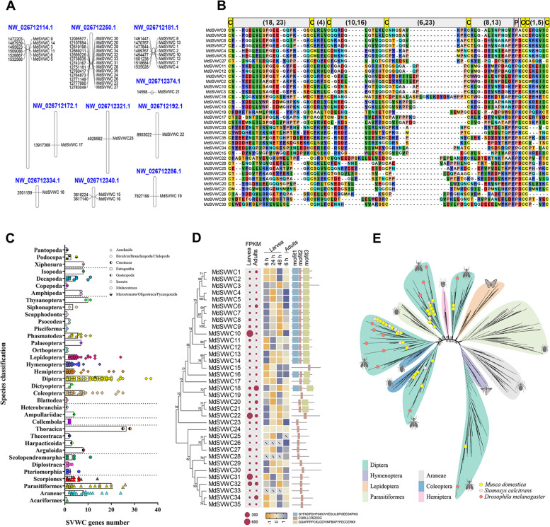
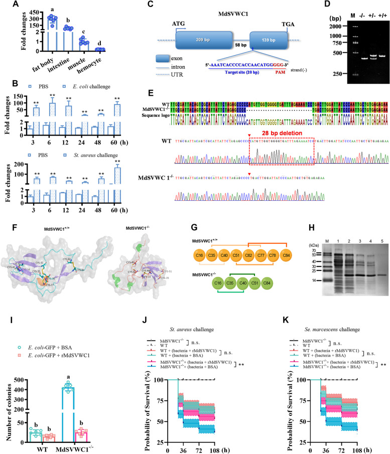
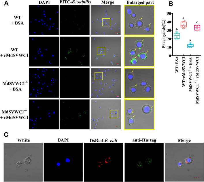
Results: A massive expansion of the SVWC gene family in Musca domestica (MdSVWC) was discovered, with a count of 35. MdSVWC1 was selected as the representative of the SVWC family for functional analysis, which led to the identification of the immune function of MdSVWC1 as a novel pattern recognition receptor. MdSVWC1 is highly expressed in both the fat body and intestines and displays acute induction upon bacterial infection. Recombinant MdSVWC1 binds to surfaces of both bacteria and yeast through the recognition of multiple pathogen-associated molecular patterns and exhibits Ca2+-dependent agglutination activity. MdSVWC1 mutant flies exhibited elevated mortality and hindered bacterial elimination following bacterial infection as a result of reduced hemocyte phagocytic capability and weakened expression of antimicrobial peptide (AMP) genes. In contrast, administration of recombinant MdSVWC1 provided protection to flies from bacterial challenges by promoting phagocytosis and AMP genes expression, thereby preventing bacterial colonization. MdSPN16, a serine protease inhibitor, was identified as a target protein of MdSVWC1. It was postulated that the interaction of MdSVWC1 with MdSPN16 would result in the activation of an extracellular proteolytic cascade, which would then initiate the Toll signaling pathway and facilitate the expression of AMP genes.

Conclusions: MdSVWC1 displays activity as a soluble pattern recognition receptor that regulates cellular and humoral immunity by recognizing microbial components and facilitating host defense.

Keywords: Musca domestica; Cellular immunity; Humoral immunity; Pattern recognition receptor; Single von Willebrand factor C-domain protein.


Conflict of interest statement

The authors declare that they have no competing interests.

Figures

Fig. 1
Fig. 1
Bioinformatics analysis of single-domain von Willebrand factor type C (SVWC) genes. A Chromosomal maps of the MdSVWCs in Musca domestica. B Multiple sequence alignments of the SVWC domain of MdSVWCs. Conservation cysteine residues are shadowed in yellow. C The copy number of the SVWC gene varies across different species’ genomes. D Phylogenetic relationships, expression profiles, and protein motif analyses of SVWC from M. domestica. The basal expression of the MdSVWC genes in both house fly larvae and adults, as determined by RNA-seq analysis, is indicated as fragments per kilobase million (FPKM). The presented heat map illustrates the variation in MdSVWC expression following mixed Escherichia coli and Staphylococcus aureus stimulation. The slash (\) characters indicate that no expression was detected. E Phylogenetic tree comprising 2869 SVWC protein sequences derived from 407 species across 36 orders. The maximum-likelihood method was employed to construct the phylogenetic tree with a bootstrap value of 1000 using iqtree. Please refer to the supporting information section for the respective gene IDs and protein sequences analyzed in this study (Additional file 1: SI-1 and SI-2)
Fig. 2
Fig. 2
The MdSVWC1-deficient flies (MdSVWC1−/−) are hypersensitive to bacterial infections. A Expression of MdSVWC1 was evaluated in varied tissues of healthy larvae through qRT-PCR analysis (n = 6, one-way ANOVA, Dunnett’s test). B The expression of MdSVWC1 over time after challenge with E. coli or St. aureus was examined by qRT-PCR in the fat body. The control group received PBS (n = 6, two-way ANOVA, Sidak’s test). C A schematic diagram of the M. domestica SVWC1 gene structure. The exons are indicated by boxes and introns are indicated by lines (the number presents the length of exons and introns). The gRNA-target site, located on the second exon of the antisense strand, consists of a 20-nucleotide guide sequence and a PAM site. D The homozygous mutant was detected using a 6% DNA-PAGE gel. The PCR product derived from the heterozygous (+ / −) contained two bands, whereas the wild type (+ / +) and homozygous (− / −) had one band each, which could be easily distinguished on an acrylamide:bis (29:1) gel. The gel was electrophoresed at 150 V for 2.5 h in 1 × TBE buffer. Lane M: DNA marker. E Mutations at the MdSVWC1 locus were identified through DNA sequencing. The peaks in Sanger sequencing of wild type (WT) and MdSVWC1−/− flies indicate the deletion site within the region marked by the dotted box. F Predicted protein structure resulting from both the WT allele and the mutation allele. G The mutation results in the disruption of two disulfide bonds within the MdSVWC1 protein molecule. H Recombinant expression and purification of MdSVWC1. The proteins underwent analysis using SDS-PAGE and were stained with Coomassie blue. Lane M: protein marker; lane 1: normal E. coli BL21 (DE3)—containing recombinant vector; lane 2: E. coli BL21 (DE3)—containing recombinant vector after induction by 1 mM IPTG; lane 3: precipitate of recombinant MdSVWC1 (rMdSVWC1); lane 4: supernatant of rMdSVWC1; lane 5: purified rMdSVWC1. I The removal of invasive bacteria by larvae was promoted by MdSVWC. Bacterial counts (CFU) were assessed in hemolymph samples collected from both MdSVWC1−/− and WT larvae 30 min following injection with GFP-expressing E. coli coated with rMdSVWC1. GFP-expressing E. coli coated with BSA used as a control (n = 6, one-way ANOVA, Dunnett’s test). J, K The survival rate of adult MdSVWC1 mutants after infection with St. aureus and Se. marcescens, respectively (n = 100, log-rank (Mantel-Cox) test). The error bar represents the mean ± SEM of three biological replicates. Different letters indicate significant differences (p < 0.05). Statistical significance is indicated by asterisks in the figures (**p < 0.01). n.s., no significance
Fig. 3
Fig. 3
rMdSVWC1 binds to the surfaces of microbes and hemocytes. A rMdSVWC1 lacks direct bactericidal activity in vitro. The rMdSVWC1 was incubated with E. coli, Pseudomonas aeruginosa, St. aureus, or Bacillus subtilis for 12 h. Bacterial growth was evaluated by measuring the absorbance at 600 nm (n = 6). TBS and ampicillin were used as negative and positive controls, respectively. The error bar represents the mean ± SEM of three biological replicates. B The microbial binding of rMdSVWC1 was assessed using a western blot. Various microbes were incubated with rMdSVWC1, then washed four times with PBS. The resulting bacterial precipitate was analyzed via western blot using anti-His antibody. C Carbohydrate-binding assay of rMdSVWC1 by ELISA. Seven sugars were used for ELISA analysis (n = 3), including LPS, DAP-PGN, Lys-PGN, D-galactose, D-mannan, and β-1,3-glucan, while sucrose served as the negative control for ligands. D rMdSVWC1 was observed to agglutinate various microbes when Ca2+ was present. BSA was used as a negative control instead of rMdSVWC1. The size of the bar is 50 μm
Fig. 4
Fig. 4
The enhancement of hemocyte phagocytosis activity towards bacteria is achieved by the coating of bacteria with rMdSVWC1. A Hemocyte phagocytosis was observed under a fluorescence microscope, where FITC-labeled B. subtilis (green) had been treated with rMdSVWC1 or BSA before injection into larvae. The bar represents 10 μm. The right panel shows a closer view of a particular section of the image. B The rate at which hemocytes phagocytose bacteria coated with rMdSVWC1 was measured. The mean ± SEM of the results (n = 6, one-way ANOVA, Dunnett’s test) was calculated based on three independent repeats. Statistical significance was indicated by different letters (p < 0.05). C The cellular localization of MdSVWC1 following phagocytosis by hemocytes in the presence of E. coli. A fluorescent immunocytochemical assay was conducted to investigate the localization of MdSVWC1 and bacteria expressing red fluorescence in the hemocytes of larvae. The red-fluorescing E. coli were injected into the larvae, and hemocytes were collected from 50 larvae at 30 min post-injection, then incubated with anti-His antibody (green). Nuclei were counterstained with DAPI (blue). The scale bar is 5 μm
Fig. 5
Fig. 5
MdSVWC1 regulates the transcription of antibacterial peptide (AMP) genes by binding with MdSPN16. A Visualization of RNA-seq results with a heat map displaying gene expression of AMPs and Toll target transcription factors in WT, MdSVWC1−/−, and MdSPN16−/− groups following a mixed infection with E. coli and St. aureus for 6 h. Three biological replicate samples were collected for each genotype. B The interaction between MdSVWC1 and MdSPN16 was examined via co-immunoprecipitation. HEK293T cells were utilized to generate the proteins. The symbols ( +) or ( −) indicate the presence or absence of recombinant proteins in the system. The symbol (IP: HA) indicates that the HA antibody was used as the bait protein antibody for the IP experiment. The symbol (Input) is used as a positive control in the IP experiment. The symbols (IB: Myc) or (IB: HA) indicate the western blot detection of the samples with Myc or HA antibody. If the bait MdSVWC1-HA protein was present in the system, the MdSPN16-Myc protein was pulled down. C Schematic diagram of the gRNA’s targeting of the MdSPN16 locus. The gRNA was specifically designed to target exon I of the open reading frame. The red highlighted area indicates the PAM site. D Mutant and WT alleles of exon I from the MdSPN16 gene were separated using a 6% DNA-PAGE with acrylamide:bis (29:1) and run in 1 × TBE buffer at 150 V for 2.5 h. The gel contained lane M, a DNA marker. Lane (+ / +) showed the WT fragment while lane (+ / −) contained both the mutant and WT fragments. Finally, lane (− / −) contained the mutant fragment exclusively. E DNA sequencing of the MdSPN16 mutant revealed a deletion of 58 base pairs. F Venn diagram depicting the AMPs that demonstrated notable alterations in expression in the MdSVWC1−/− and MdSPN16−/− flies as a result of E. coli and St. aureus stimulation. G Expression profiles of AMPs in WT, MdSVWC1−/−, and MdSPN16.−/− flies through qRT-PCR following a mixed infection with E. coli and St. aureus for 6 h. The AMP genes and their respective IDs are as follows: diptericin (LOC101896897), defensin (LOC105261620), attacin (LOC109612355), cecropin (LOC101889972). Graphs show mean ± SEM. All trials were performed at least thrice. The statistical significance was determined utilizing a two-way ANOVA and Sidak’s test (n = 6) (*p < 0.05, **p < 0.01)
Fig. 6
Fig. 6
A schematic representation illustrates the roles of MdSVWC1 in regulating the cellular and humoral response of M. domestica to microbial pathogens. MdSVWC1 functions as a pattern recognition receptor that detects pathogen-associated molecular patterns of invading microorganisms and mediates Ca2+-dependent agglutination reactions. MdSVWC1 acts as an opsonin, inhibiting infection by marking pathogens for destruction by hemocytes. Phagocytic hemocytes utilize unknown receptors to bind to MdSVWC1-opsonized pathogens, facilitating the first step of attachment and enhancing subsequent phagocytosis. Additionally, MdSVWC1 regulates the production of AMPs by targeting SPN16, a negative regulator of the Spätzle activation process in the Toll receptor-mediated signaling pathway. Upon detection of invading microorganisms, the interaction between MdSVWC1 and MdSPN16 leads to the release of the Spätzle-processing enzyme. This, in turn, triggers the hydrolytic activation of proSpätzle. Processed Spätzle subsequently initiates the Toll intracellular signaling pathway, ultimately facilitating the expression of AMPs

Similar articles

Cited by

References

    1. Nunes C, Sucena E, Koyama T. Endocrine regulation of immunity in insects. FEBS J. 2021;288:3928–47. - PubMed
    1. Hoffmann JA. The immune response of Drosophila. Nature. 2003;426(6962):33–8. - PubMed
    1. Brubaker SW, Bonham KS, Zanoni I, Kagan JC. Innate immune pattern recognition: a cell biological perspective. Annu Rev Immunol. 2015;33:257–90. - PMC - PubMed
    1. Stokes BA, Yadav S, Shokal U, Smith LC, Eleftherianos I. Bacterial and fungal pattern recognition receptors in homologous innate signaling pathways of insects and mammals. Front Microbiol. 2015;6:19. - PMC - PubMed
    1. Akira S, Uematsu S, Takeuchi O. Pathogen recognition and innate immunity. Cell. 2006;124(4):783–801. - PubMed

MeSH terms

LinkOut - more resources