. 2024 Oct 23;22(1):652.
doi: 10.1186/s12951-024-02920-8.

Copper hydrogen phosphate nanosheets functionalized hydrogel with tissue adhesive, antibacterial, and angiogenic capabilities for tracheal mucosal regeneration

Affiliations

Copper hydrogen phosphate nanosheets functionalized hydrogel with tissue adhesive, antibacterial, and angiogenic capabilities for tracheal mucosal regeneration

Pengli Wang et al. J Nanobiotechnology. .

Abstract

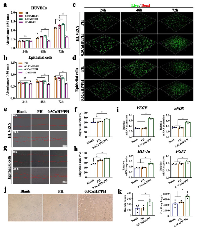
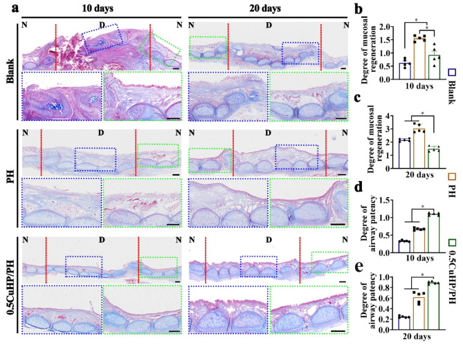
Timely and effective interventions after tracheal mucosal injury are lack in clinical practices, which elevate the risks of airway infection, tracheal cartilage deterioration, and even asphyxiated death. Herein, we proposed a biomaterial-based strategy for the repair of injured tracheal mucosal based on a copper hydrogen phosphate nanosheets (CuHP NSs) functionalized commercial hydrogel (polyethylene glycol disuccinimidyl succinate-human serum albumin, PH). Such CuHP/PH hydrogel achieved favorable injectability, stable gelation, and excellent adhesiveness within the tracheal lumen. Moreover, CuHP NSs within the CuHP/PH hydrogel effectively stimulate the proliferation and migration of endothelial/epithelial cells, enhancing angiogenesis and demonstrating excellent tissue regenerative potential. Additionally, it exhibited significant inhibitory effects on both bacteria and bacterial biofilms. More importantly, when injected injured site of tracheal mucosa under fiberoptic bronchoscopy guidance, our results demonstrated CuHP/PH hydrogel adhered tightly to the tracheal mucosa. The therapeutic effects of the CuHP/PH hydrogel were further confirmed, which significantly improved survival rates, vascular and mucosal regeneration, reduced occurrences of intraluminal infections, tracheal stenosis, and cartilage damage complications. This research presents an initial proposition outlining a strategy employing biomaterials to mitigate tracheal mucosal injury, offering novel perspectives on the treatment of mucosal injuries and other tracheal diseases.

Keywords: Angiogenesis; Antibacteria; Copper hydrogen phosphate nanosheets, Hydrogel; Tracheal mucosa repair.


Conflict of interest statement

The authors declare no competing interests.

Figures

Fig. 1
Fig. 1
Display of overall experiments. CuHP NSs, PEG-(SS)2 and HAS were evenly mixed and immediately injected to the region of tracheal mucosal injury, which quickly attached onto the native tissue and transformed to a composite hydrogel with antibacterial and angiogenic capabilities due to the released Cu ions and acid-sensitive POD-like catalytic activity, thereby facilitating tracheal mucosal regeneration, preserving airway patency, and safeguarding cartilage
Fig. 2
Fig. 2
Synthesis and characterization of CuHP nanosheets and CuHP/PH hydrogel SEM (a), XRD analysis (b), and AFM imaging (c) were conducted on CuHP nanosheets. The MB degradation by CuHP nanosheets under varying concentrations (d), durations (e), and pH levels (f). Gross observation (g) and determination of gelation time (h) were performed for CuHP/PH hydrogel. In vitro gelation studies for both PH and CuHP/PH hydrogel were conducted (i). Adhesion assessment of CuHP/PH hydrogel was carried out in situ (j). Intratracheal injection of CuHP/PH hydrogel under fiberoptic bronchoscopy guidance (k)
Fig. 3
Fig. 3
Biocompatibility of CuHP/PH hydrogel on HUVECs and TECs. (a-b) Impact of varied concentrations of CuHP/PH (0.25, 0.5, 1 wt%) hydrogel on the proliferation of HUVECs and TECs; (c-d) Live/dead staining for PH and 0.5CuHP/PH hydrogel on HUVEC and TECs; Cell scratch assay (e) and quantitative outcomes (f) regarding HUVEC cell migration ability; Cell scratch assay (g) and quantitative outcomes (h) regarding epithelial cell migration ability; (i) Expression levels of key angiogenesis-related genes in HUVECs; Tube formation (j) and quantitative outcomes regarding branch points and capillary length (k) of HUVECs. (h: hours; *, p < 0.05)
Fig. 4
Fig. 4
Antibacterial activity of CuHP/PH hydrogel against S. aureus and E. coli. (a) S. aureus and (b) E. coli was subjected to live/dead staining using green fluorescence (SYTO9) and red fluorescence (PI) after treatment with PBS, PH, and 0.5CuHP/PH hydrogel, respectively. (c) Images depict E. coli and S. aureus growth on agar plates under different treatments. (d) Quantitative assessment of the inhibition percentage of E. coli and S. aureus. (e) Visualization of E. coli and S. aureus biofilms stained with crystal violet. (f) OD values at 570 nm of biofilms stained with crystal violet. (*, p < 0.05)
Fig. 5
Fig. 5
Treatment of injured tracheal mucosa in a rabbit model. (a) Elucidation of the surgical methodology for treating injured tracheal mucosa with CuHP/PH hydrogel; (b) Evaluation of the probability of survival in experimental rabbits post-surgery (Sample 1 to 8 for Blank group; Sample 9–16 for PH group; Sample 17–24 for CuHP group). Endoscopic images (c) and gross view (d) showcase the tracheal lumen condition after treatment with Blank (Samples 2 at 10 days and Sample 8 at 20 days), PH (Samples 10 at 10 days and Sample 14 at 20 days), and 0.5CuHP/PH hydrogels (Samples 19 at 10 days and Sample 21 at 20 days). (Purulent secretion is indicated by black arrows; granulation tissue is denoted by red arrows; D: defected tracheal mucosa; N: normal mucosa; *, p < 0.05)
Fig. 6
Fig. 6
The evaluation of mucosal regeneration and the status of airway lumen following treatment with various hydrogel at 10 and 20 days. (a) Masson’s trichrome staining was conducted on samples from Blank (Samples 2 at 10 days and Sample 8 at 20 days), PH (Samples 10 at 10 days and Sample 14 at 20 days), and 0.5CuHP/PH groups (Samples 19 at 10 days and Sample 21 at 20 days); (b-e) Quantitative analyses of mucosal regeneration and airway patency were performed. (Blue box: regenerated epithelium; Green box: natural epithelium; Scale bar = 500 μm; *, p < 0.05)
Fig. 7
Fig. 7
Development of tracheal cartilage following treatment with distinct hydrogel for 10 and 20 days. (a) HE, Collagen II, Safranin-O, and Masson’s trichrome staining were performed on samples from the Blank (Samples 3 at 10 days and Sample 7 at 20 days), PH (Samples 11 at 10 days and Sample 16 at 20 days), and 0.5CuHP/PH (Samples 18 at 10 days and Sample 22 at 20 days) groups; (b) Quantitative analysis of GAG content, (c) total collagen content, and (d) Young’s modulus was carried out. (N: natural tracheal cartilage; Scale bar = 200 μm; *, p < 0.05)
Fig. 8
Fig. 8
Assessment of in vivo epithelial regeneration, revascularization, infection, and inflammatory reactions after treatment with various hydrogel at 10 and 20 days. Immunofluorescence staining for cytokeratin (a1-a6); Immunohistochemical staining for CD31 (b1-b6); Immunofluorescence staining for bacteria (c1-c6), TNF-α (d1-d6), and IL-1β (e1-e6) were performed on samples from the Blank (Samples 2 at 10 days and Sample 6 at 20 days), PH (Samples 10 at 10 days and Sample 15 at 20 days), and 0.5CuHP/PH (Samples 20 at 10 days and Sample 24 at 20 days) groups. Quantitative analyses were conducted for neonatal blood vessels (f), bacteria (g), TNF-α (h), and IL-1β density (i). (Scale bar = 20 μm; *, p < 0.05)

References

    1. Lei D, Luo B, Guo YF, Wang D, Yang H, Wang SF, Xuan HX, Shen A, Zhang Y, Liu ZH, He CL, Qing FL, Xu Y, Zhou GD. You, 4-Axis printing microfibrous tubular scaffold and tracheal cartilage application. Sci China Mater. 2019;62(12):1910–20.
    1. Jahshan F, Ertracht O, Eisenbach N, Daoud A, Sela E, Atar S, Abu Ammar A, Gruber M. A novel rat model for tracheal mucosal damage assessment of following long term intubation. Int J Pediatr Otorhinolaryngol. 2020;128:109738. - PubMed
    1. Deshmukh A, Jadhav S, Wadgoankar V, Takalkar U, Deshmukh H, Apsingkar P, Sonwatikar P, Antony P. Airway Management and Bronchoscopic Treatment of Subglottic and Tracheal Stenosis using Holmium laser with balloon dilatation. Indian J Otolaryngol Head Neck Surg. 2019;71(Suppl 1):453–8. - PMC - PubMed
    1. Gao W, Chen K, He W, Zhao S, Cui D, Tao C, Xu Y, Xiao X, Feng Q, Xia H. Synergistic chondrogenesis promotion and arthroscopic articular cartilage restoration via injectable dual-drug-loaded sulfated hyaluronic acid hydrogel for stem cell therapy. Compos Part B: Eng 263 (2023).
    1. Xu Y, Dai J, Zhu XS, Cao RF, Song N, Liu M, Liu XG, Zhu JJ, Pan F, Qin LL, Jiang GN, Wang HF, Yang Y. Biomimetic Trachea Engineering via a modular Ring Strategy based on bone-marrow stem cells and Atelocollagen for use in extensive Tracheal Reconstruction. Adv Mater 34(6) (2022). - PubMed

MeSH terms