Review
. 2024 Jul-Sep;34(3):99-113.
doi: 10.4103/jcecho.jcecho_37_24. Epub 2024 Sep 21.

Clinical Applications of Myocardial Work in Echocardiography: A Comprehensive Review

Affiliations
Review

Clinical Applications of Myocardial Work in Echocardiography: A Comprehensive Review

Giancarlo Trimarchi et al. J Cardiovasc Echogr. 2024 Jul-Sep.

Abstract

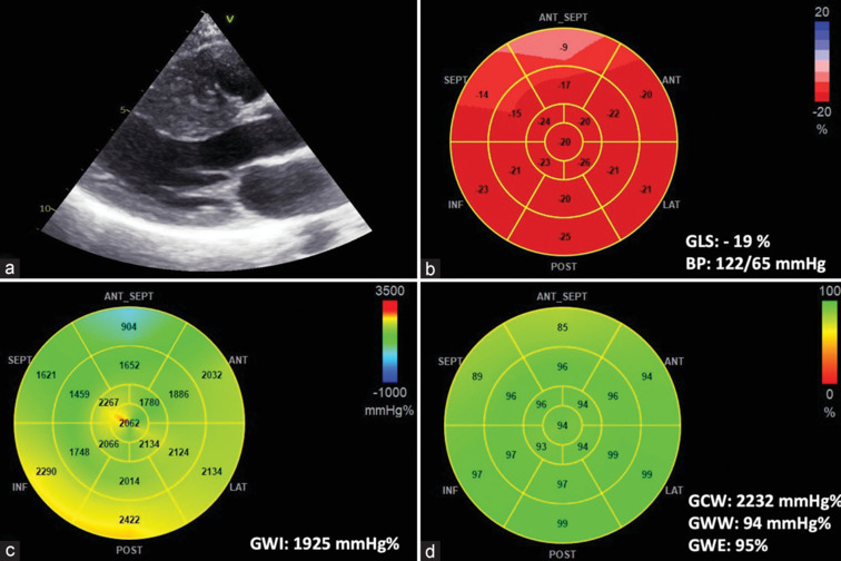
Left ventricular (LV) global longitudinal strain (GLS) has recently garnered attention as a reliable and objective method for evaluating LV systolic function. One of the key advantages of GLS is its ability to detect subtle abnormalities even when the ejection fraction (EF) appears to be preserved. However, it is important to note that GLS, much like LVEF, is significantly influenced by load conditions. In recent years, researchers and clinicians have been exploring noninvasive myocardial work (MW) quantification as an innovative tool for assessing myocardial function. This method integrates measurements of strain and LV pressure, providing a comprehensive evaluation of the heart's performance. Notably, MW offers an advantage over GLS and LVEF because it provides a load-independent assessment of myocardial performance. The implementation of commercial echocardiographic software that facilitates the noninvasive calculation of MW has significantly broadened the scope of its application. This advanced technology is now being utilized in multiple clinical settings, including ischemic heart disease, valvular diseases, cardiomyopathies, cardio-oncology, and hypertension. One of the fundamental aspects of MW is its correlation with myocardial oxygen consumption, which allows for the assessment of work efficiency. Understanding this relationship is crucial for diagnosing and managing various cardiac conditions. The aim of this review is to provide an overview of the noninvasive assessment of myocardial by echocardiography, from basic principles and methodology to current clinical applications.

Keywords: Cardiomyopathies; echocardiography; global longitudinal strain; heart failure; myocardial work; speckle-tracking echocardiography; valvular diseases.


Conflict of interest statement

There are no conflicts of interest.

Figures

Figure 1
Figure 1
The left ventricular (LV) global longitudinal strain (GLS) data, measured using the R-wave onset in the electrocardiogram as a common time reference (left panel), are combined with peak arterial pressure, measured using a cuff manometer (central panel). The right panel displays a representative trace showing the LV pressure–strain loop and a 17-segment bull’s-eye representation of myocardial work index (top), along with 17-segment bull’s-eye representations of GLS and myocardial work efficiency (bottom)
Figure 2
Figure 2
Hypertension. Blood pressure at the time of the echocardiogram was 185/90 mmHg. Left ventricular ejection fraction (63%) and global longitudinal strain (-19%) are both preserved (a), global work index is elevated (2393 mmH%) with multiple, red-shaded segments, especially in the apex (b), pressure-strain loop is normal (c) and global work efficiency is relatively unaffected (92%), as demonstrated by all green-shaded segments, except for the basal septum that is yellow-shaded which indicates reduced efficiency (d). GWE = Global work efficiency, GWW = Global wasted work, GCW = Global constructive work, GWI = Global work index, GLS = Global longitudinal strain, BP = Blood pressure
Figure 3
Figure 3
Coronary artery disease. A patient presenting with an anterior ST elevation myocardial infarction caused by occlusion at the mid-segment of the left anterior descending artery. Left ventricular ejection fraction is mildly reduced (48%). (a) The global longitudinal strain (GLS) bull’s eye shows impaired longitudinal deformation of apical and mid-anteroseptal segments with coronary distribution and reduced GLS value (−7%), (b) Global work index bull’s eye shows the regional impairment of myocardial work index in the same segments that show reduced longitudinal deformation, (c) The pressure–strain loop shows a reduced area under the curve, and it is shifted toward right, which means a significant impairment of myocardial work, (d) Global work efficiency (GWE) bull’s eye shows impairment of myocardial work efficiency especially in apical segment, that are red shaded, with a GWE significantly reduced (72%). GWE = Global work efficiency, GWW = Global wasted work, GCW = Global constructive work, GWI = Global work index, GLS = Global longitudinal strain, BP = Blood pressure
Figure 4
Figure 4
Hypertrophic cardiomyopathy. (a) Parasternal long axis view showing severe hypertrophy of the interventricular septum in a patient affected by sarcomeric hypertrophic cardiomyopathy, (b) Global longitudinal strain’s bull’s eye showing impairment of longitudinal deformation in anterior-septum segments, interested by hypertrophy, (c) Myocardial work index bull’s eye showing a decreased amount of myocardial work performed by basal anteroseptal segment, light-blue shaded, (d) Myocardial work efficiency’s bull’s eye showing preserved global work efficiency of 95% and decreased regional work efficiency in segments interested by hypertrophy. GWE = Global work efficiency, GWW = Global wasted work, GCW = Global constructive work, GWI = Global work index, GLS = Global longitudinal strain, BP = Blood pressure
Figure 5
Figure 5
Dilatative cardiomyopathy. (a) Four-chamber view demonstrates severely dilated left ventricle in a patient with nonischemic dilatative cardiomyopathy and left ventricular ejection fraction equal to 30%, (b) A bull’s-eye plot for the same patient correlates with a severely reduced global longitudinal strain (−4%), (c) A bull’s-eye plot demonstrates severely decreased global work index (397 mmHg%), (d) Myocardial efficiency bull’s eye showing diffuse impairment of work efficiency with global work efficiency severely impaired and equal to 66%. GWE = Global work efficiency, GWW = Global wasted work, GCW = Global constructive work, GWI = Global work index, GLS = Global longitudinal strain, BP = Blood pressure
Figure 6
Figure 6
Transthyretin cardiac amyloidosis. (a) Four-chamber view demonstrates severely thickened left ventricular walls and granular sparkling aspect of the interventricular septum, (b) A bull’s-eye plot of strain from the same patient demonstrates the typical apical sparing seen in amyloid heart disease, (c) A bull’s-eye plot of myocardial work index in the same patient shows decreased global work index (global work index; 824 mmHg%) with normal work index at the apex and negative work at the base, (d) Work efficiency bull’s eye showing preserved myocardial work efficiency at the apical segments. GWE = Global work efficiency, GWW = Global wasted work, GCW = Global constructive work, GWI = Global work index, GLS = Global longitudinal strain, BP = Blood pressure

References

    1. Suga H. Total mechanical energy of a ventricle model and cardiac oxygen consumption. Am J Physiol. 1979;236:H498–505. - PubMed
    1. Suga H, Hayashi T, Shirahata M. Ventricular systolic pressure-volume area as predictor of cardiac oxygen consumption. Am J Physiol. 1981;240:H39–44. - PubMed
    1. Marwick TH. Ejection fraction pros and cons: JACC state-of-the-art review. J Am Coll Cardiol. 2018;72:2360–79. - PubMed
    1. Zito C, Longobardo L, Citro R, Galderisi M, Oreto L, Carerj ML, et al. Ten years of 2D longitudinal strain for early myocardial dysfunction detection: A clinical overview. Biomed Res Int. 2018;2018:8979407.. - PMC - PubMed
    1. Cameli M, Mandoli GE, Sciaccaluga C, Mondillo S. More than 10 years of speckle tracking echocardiography: Still a novel technique or a definite tool for clinical practice? Echocardiography. 2019;36:958–70. - PubMed

LinkOut - more resources