. 2024 Oct 4;10(19):e38936.
doi: 10.1016/j.heliyon.2024.e38936. eCollection 2024 Oct 15.

Rosmarinic acid promotes cartilage regeneration through Sox9 induction via NF-κB pathway inhibition in mouse osteoarthritis progression

Affiliations

Rosmarinic acid promotes cartilage regeneration through Sox9 induction via NF-κB pathway inhibition in mouse osteoarthritis progression

Ye Eun Sim et al. Heliyon. .

Abstract

Background: The natural polyphenolic compound known as Rosmarinic acid (RosA) can be found in various plants. Although its potential health benefits have been extensively studied, its effect on osteoarthritis (OA) progression and cartilage regeneration function still needs to be fully elucidated in OA animal models. This study elucidated the effect of RosA on OA progression and cartilage regeneration.

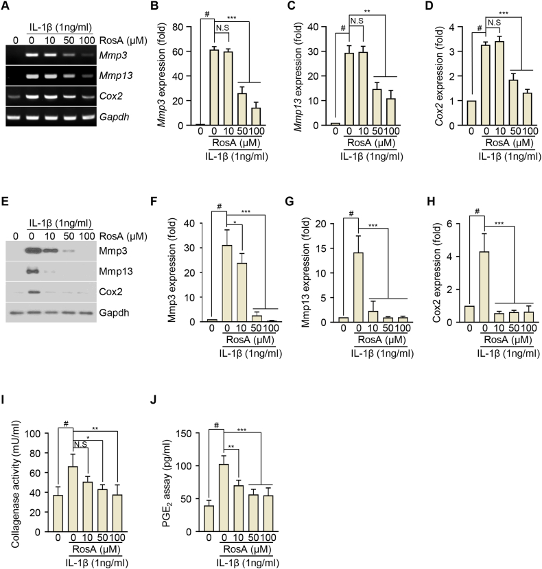
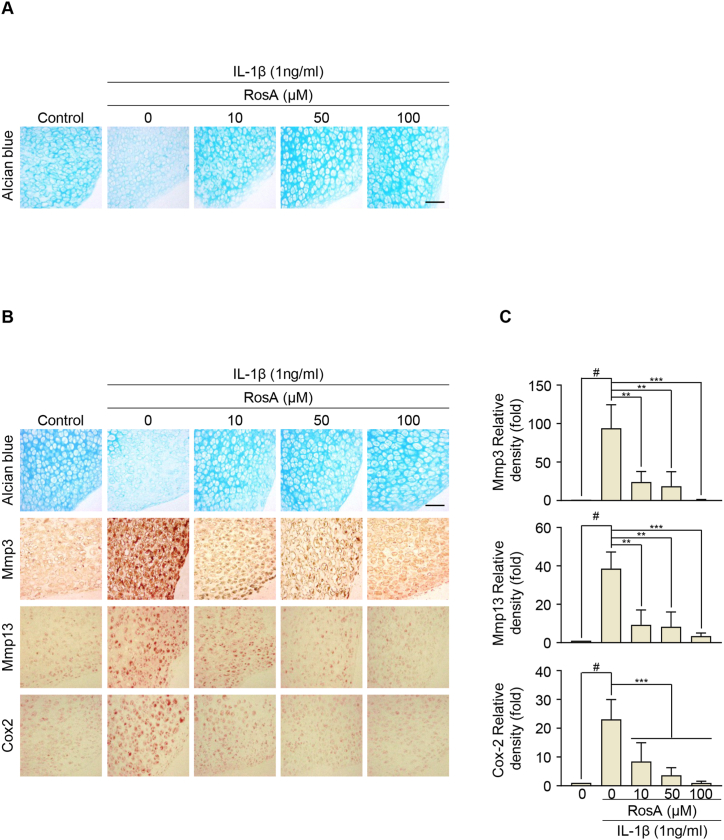
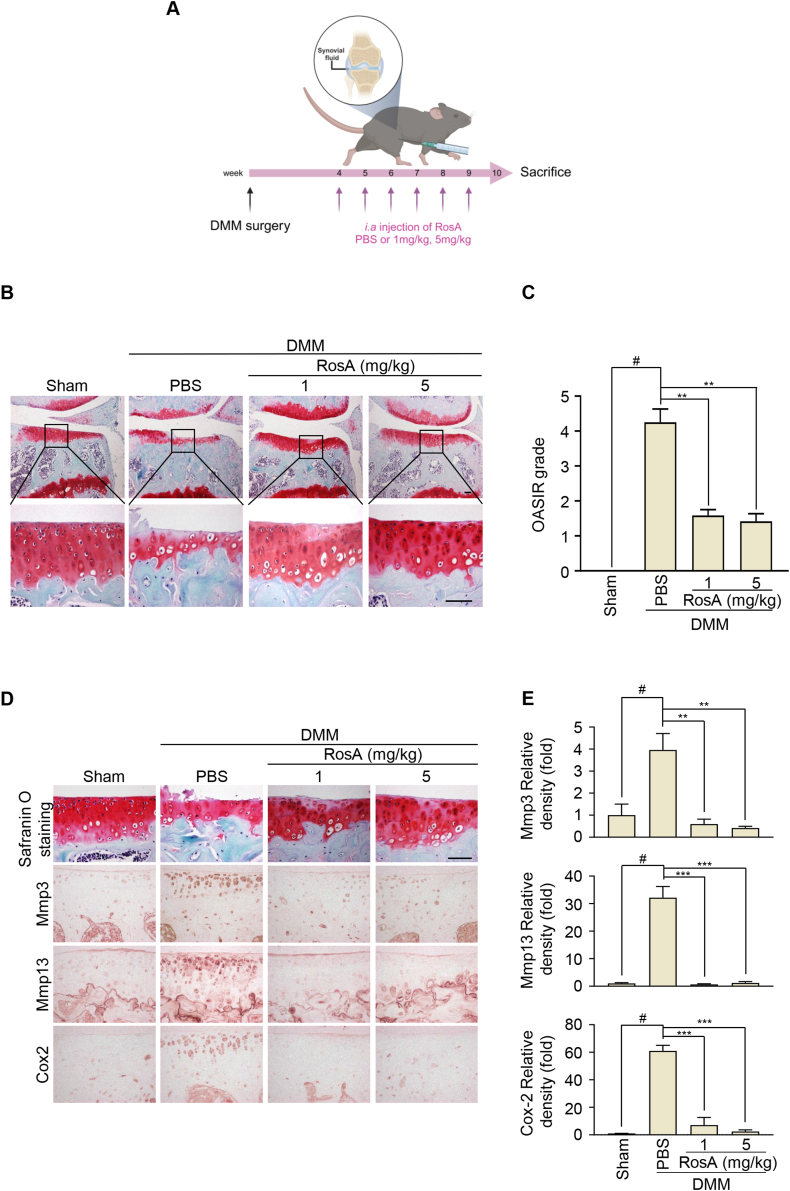
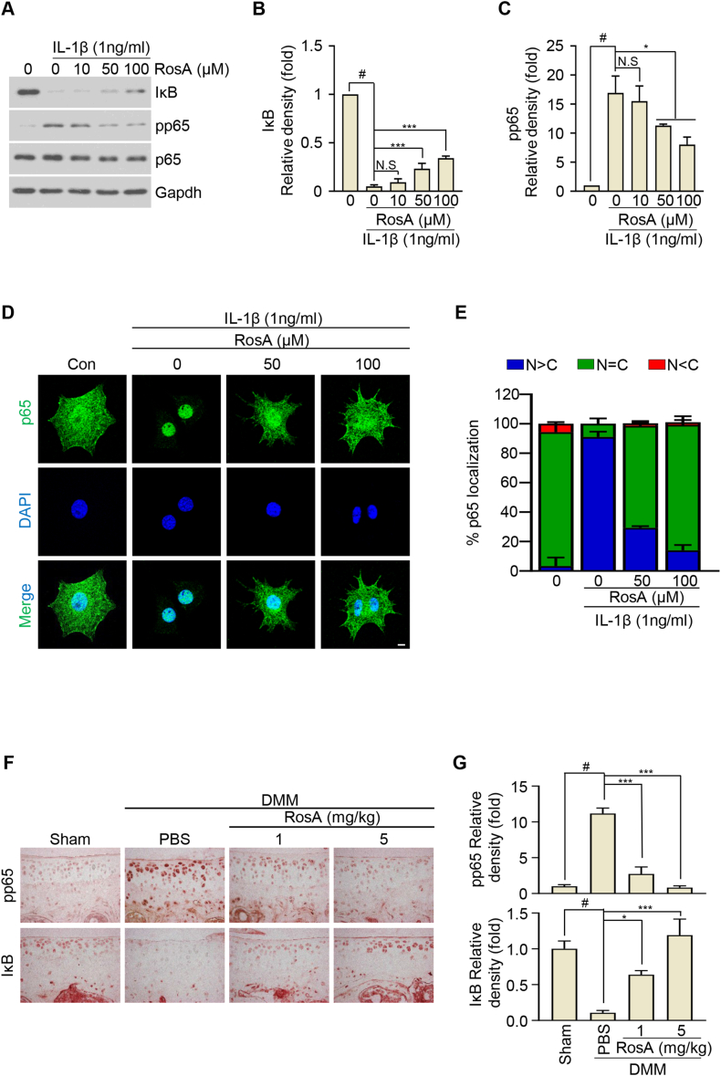
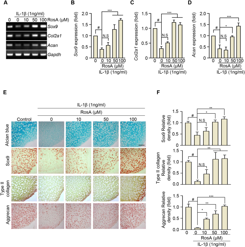
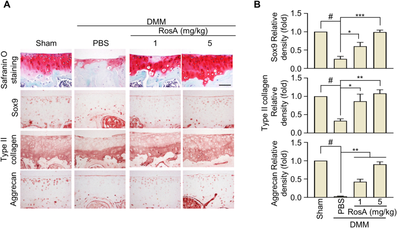
Methods: In vitro assessments were conducted using RT-PCR, qRT-PCR, Western blotting, and ELISA to measure the effects of RosA. The molecular mechanisms of RosA were determined by analyzing the translocation of p65 into the nucleus using immunocytochemistry (ICC). Histological analysis of cartilage explant was performed using alcian blue staining and immunohistochemistry (IHC). For in vivo analysis, the destabilization of the medial meniscus (DMM)-induced OA mouse model was utilized to evaluate cartilage destruction through Safranin-O staining. The expression of catabolic and anabolic factors in mice knee joints was quantified by immunohistochemistry.

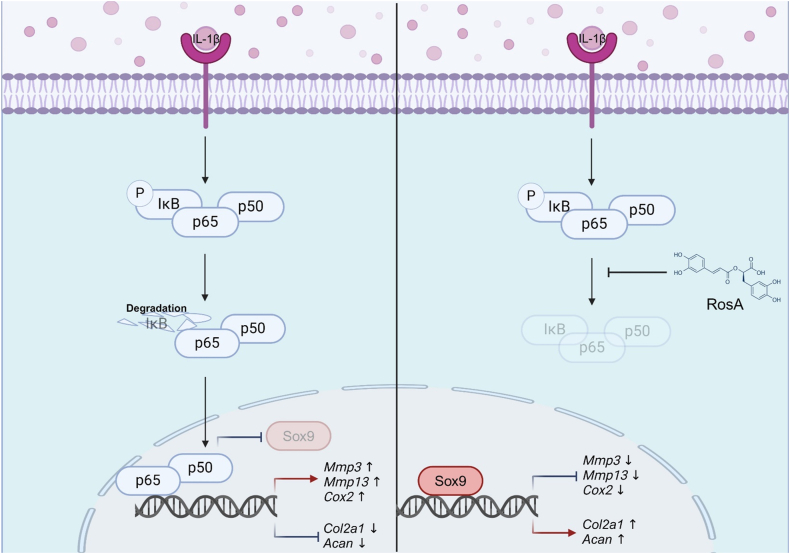
Results: The expression of catabolic factors in chondrocytes was significantly impeded by RosA. It also suppressed the NF-κB signaling pathway by decreasing phosphorylation of p65 and reducing degradation of IκB protein. In ex vivo experiments, RosA protected sulfated proteoglycan erosion triggered by IL-1β and suppressed the catabolic factors in cartilage explant. RosA treatment in animal models resulted in preventing cartilage destruction and reducing catabolic factors in the cartilage. RosA was also found to promote the expression of Sox9, Col2a1, and Acan in vitro, ex vivo, and in vivo analyses.

Conclusions: RosA attenuated the OA progression by suppressing the catabolic factors expression. These effects were facilitated through the suppression of the NF-κB signaling pathway. Additionally, it promotes cartilage regeneration by inducing anabolic factors. Therefore, RosA shows potential as an effective therapeutic agent for treating OA.

Keywords: Extracellular matrix; NF-κB; Osteoarthritis; Rosmarinic acid; Sox9.


Conflict of interest statement

The authors declare that they have no known competing financial interests or personal relationships that could have appeared to influence the work reported in this paper.

Figures

Fig. 1
Fig. 1
RosA inhibits Mmp3, Mmp13, and Cox2 expression induced by IL-1β in articular chondrocytes. (A, B, C, D) Chondrocytes were co-treated with IL-1β and RosA for 24 h Mmp3, Mmp13, and Cox2 expression were evaluated by RT-PCR (A), densitometry (B; Mmp3, C; Mmp13, and D; Cox2), western blot (E) and densitometry (F; Mmp3, G; Mmp13, H; Cox2) analysis. Collagenase activity (I) and PGE2 production (J) were detected in chondrocytes cultured medium. Values are presented as the mean ± standard deviation. One-way analysis of variance followed by Bonferroni's test was used to determine significant differences. None significant (N.S), ∗P < 0.05, ∗∗P < 0.01, ∗∗∗P < 0.001, #P < 0.05 compared to the control group. Original gel (Fig. S6) and blots (Fig. S7) can be seen in supplementary figures file.
Fig. 2
Fig. 2
RosA protects against extracellular matrix (ECM) degradation in cartilage explants. (A) Mouse knee cartilage explants were co-treated with IL-1β and RosA at the indicated concentration. The accumulation of sulfated-proteoglycans was evaluated by Alcian blue staining. (B) Expression of Mmp3, Mmp13, and Cox2 in cartilage explant after co-treated with IL-1β and RosA assessed by Immunohistochemical staining and (C) quantification (Mmp3; upper panel, Mmp13; middle panel, Cox2; lower panel). Scale bars, 100 μm. Values are presented as the mean ± standard deviation. One-way analysis of variance followed by Bonferroni's test was used to determine significant differences ∗∗P < 0.01, ∗∗∗P < 0.001, #P < 0.05 compared to the control group.
Fig. 3
Fig. 3
RosA suppresses cartilage degradation in the DMM-induced OA mice model. (A) Experimental framework outlining the establishment of the DMM-induced OA model. (B) Following DMM induction, mice received intra-articular (i.a) injections of RosA for 6 weeks. The degree of cartilage degradation was assessed using Safranin-O staining. (C) OA severity was quantified utilizing the OARSI scoring system at 10 weeks post-DMM induction. (D) Immunohistochemical staining was performed to assess the Mmp3, Mmp13, and Cox2 protein levels in experimental OA mice after i,a injection of RosA and (E) quantification. Scale bars, 100 μm. Values are presented as the mean ± standard deviation. One-way analysis of variance followed by Bonferroni's test was used to determine significant differences ∗∗P < 0.01, ∗∗∗P < 0.001, #P < 0.05 compared to the control group.
Fig. 4
Fig. 4
RosA suppresses the expression of catabolic factors via the NF-κB signaling pathway. (A, B, C) Chondrocytes were exposed to RosA at the indicated concentration for 24 h before being treated to IL-1β (1 ng/ml) for 15 min. Western blot analysis was performed to evaluate the Phosphorylation of p65 and the degradation of IκB (A) and densitometry analysis (B; IκB, C; pp65). (D) Chondrocytes were subjected to immunofluorescence staining for p65 (green) and 4′, 6-diamidino-2-phenylindole (DAPI) (blue) was used for nuclear staining. (E) The nuclear (N)-cytoplasmic (C) ratio of p65 was analyzed in three randomly chosen regions from three separate experiments (n = 100 cells per field). Scale bars, 10 μm. (F) The pp65 and IκB protein levels in OA mice cartilage after i,a-injection of RosA assessed by Immunohistochemical staining and (G) quantification. Scale bars, 100 μm. Values are presented as the mean ± standard deviation. One-way analysis of variance followed by Bonferroni's test was used to determine significant differences. None significant (N.S), ∗P < 0.05, ∗∗∗P < 0.001, #P < 0.05 compared to the control group. Original blots (Fig. S8) can be seen in supplementary figures file.
Fig. 5
Fig. 5
RosA restores the expression of Type II collagen, Aggrecan and Sox9 reduced by OA progression. (A, B, C, D) Chondrocytes were co-treated with IL-β and RosA for 24 h Sox9, Col2a1, and Acan expressions were evaluated by RT-PCR (A) and densitometry (B; Sox9, C; Col2a1, and D; Acan) analysis. (E) Sox9, Type II collagen and Aggrecan expressions in cartilage explant after being co-treated with IL-1β and RosA measured by Immunohistochemical staining and (F) quantification (Sox9; upper panel, Type II collagen; middle panel, Aggrecan; lower panel). Values are presented as the mean ± standard deviation. One-way analysis of variance followed by Bonferroni's test was used to determine significant differences. None significant (N.S), ∗P < 0.05, ∗∗P < 0.01, ∗∗∗P < 0.001, and #P < 0.05 compared to the control group. Original gel (Fig. S9) can be seen in supplementary figures file.
Fig. 6
Fig. 6
RosA induced the expression of Type II collagen, Aggrecan and Sox9 protein in DMM-induced OA mice model. (A) The Sox9, Type II collagen, and Aggrecan, protein levels were assessed through Immunohistochemical staining in the cartilage of OA mice following i,a - injection of RosA, and (B) quantification was performed. Scale bars, 100 μm. Values are presented as the mean ± standard deviation. One-way analysis of variance followed by Bonferroni's test was used to determine significant differences. None significant (N.S), ∗P < 0.05, ∗∗P < 0.01, ∗∗∗P < 0.001, #P < 0.05 compared to the Sham group.
Fig. 7
Fig. 7
A schematic abstract illustrating RosA's effects on both inhibiting catabolic factors and promoting ECM synthesis through the inhibition of the NF-κB pathway.

References

    1. Yao Q., Wu X., Tao C., Gong W., Chen M., Qu M., Zhong Y., He T., Chen S., Xiao G. Osteoarthritis: pathogenic signaling pathways and therapeutic targets. Signal Transduct. Targeted Ther. 2023;8(1):56. doi: 10.1038/s41392-023-01330-w. - DOI - PMC - PubMed
    1. Rapp A.E., Zaucke F. Cartilage extracellular matrix-derived matrikines in osteoarthritis. Am J Physiol Cell Physiol. 2023;324(2):C377–c394. doi: 10.1152/ajpcell.00464.2022. - DOI - PubMed
    1. Berenbaum F., Griffin T.M., Liu-Bryan R. Review: metabolic regulation of inflammation in osteoarthritis. Arthritis Rheumatol. 2017;69(1):9–21. doi: 10.1002/art.39842. - DOI - PMC - PubMed
    1. Fujii Y., Liu L., Yagasaki L., Inotsume M., Chiba T., Asahara H. Cartilage homeostasis and osteoarthritis. Int. J. Mol. Sci. 2022;23(11) doi: 10.3390/ijms23116316. - DOI - PMC - PubMed
    1. Mehana E.E., Khafaga A.F., El-Blehi S.S. The role of matrix metalloproteinases in osteoarthritis pathogenesis: an updated review. Life Sci. 2019;234 doi: 10.1016/j.lfs.2019.116786. - DOI - PubMed

LinkOut - more resources