. 2024 Oct 4;10(19):e38957.
doi: 10.1016/j.heliyon.2024.e38957. eCollection 2024 Oct 15.

Typical NF2 and LTZR1 mutations are retained in an immortalized human schwann cell model of schwannomatosis

Affiliations

Typical NF2 and LTZR1 mutations are retained in an immortalized human schwann cell model of schwannomatosis

Valentina Melfi et al. Heliyon. .

Abstract

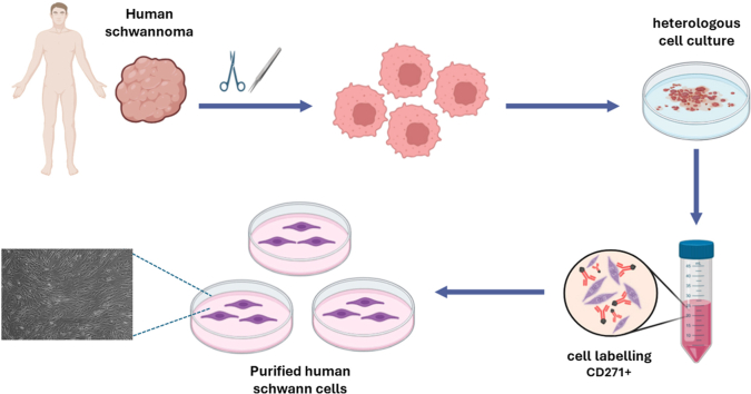
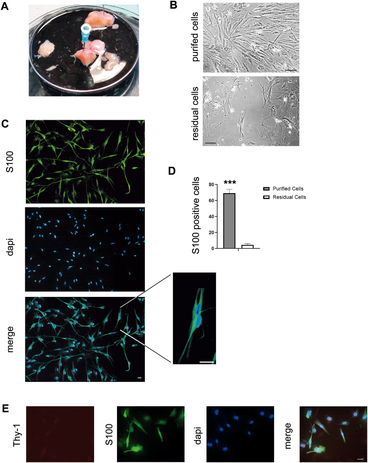
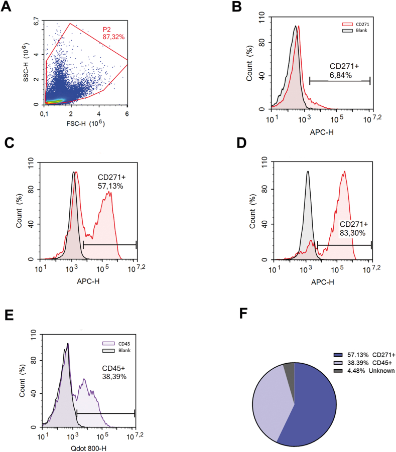
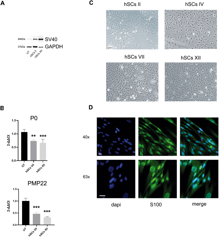
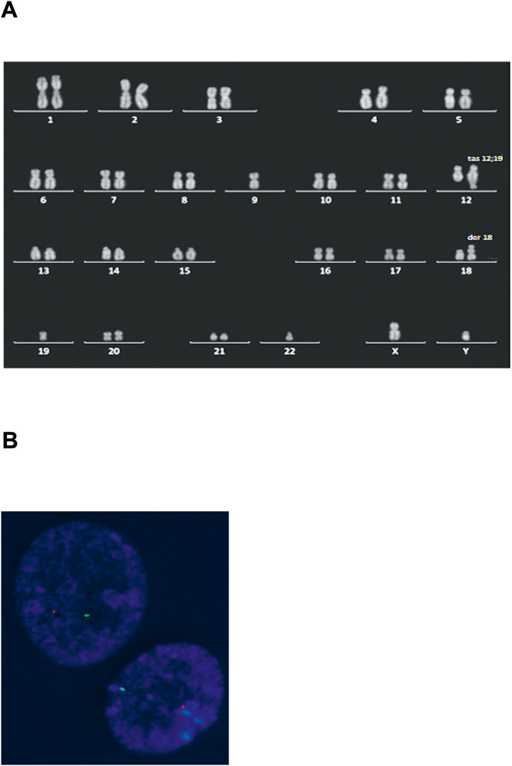
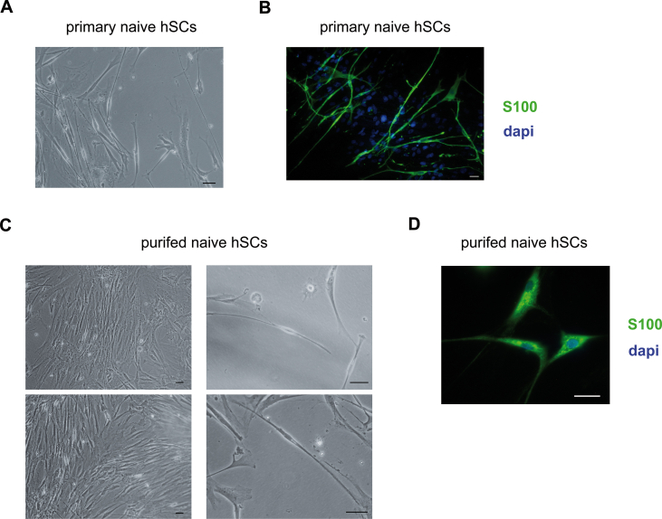
Human SCs play a primary role in SWN, a rare genetic disorder in which patients develop multiple schwannomas. So that, their isolation and immortalization could represent an irreplaceable tool to investigate the disease etiopathology. Although few clones of tumoural SCs have been obtained, unfortunately they present genetic, morphological and biological characteristics that do not fully represent the original cells. Herein we isolated, characterized and immortalized primary SCs from human schwannomas. Our immortalized human SCs present typical NF2 and LTZR1 genetic mutations of SWN and retain original phenotype characteristics, representing a valuable tool for further genetic, functional and biomolecular in vitro studies.


Conflict of interest statement

The authors declare the following financial interests/personal relationships which may be considered as potential competing interests: Valerio Magnaghi reports financial support was provided by the Ministry of University and Research (MUR). Other authors declare that they have no known competing financial interests or personal relationships that could have appeared to influence the work reported in this paper.

Figures

Image 1
Graphical abstract
Fig. 1
Fig. 1
Assessment of primary hSCs from schwannoma. A) Representative image of different encapsulated schwannomas, that underwent cell isolation procedures; B) Image of primary hSCs in culture following positive p75 receptor immunoselection. The fraction enriched in purified cells presented the typical spindle-shape morphology, that was not evidenced in the residual cells; scale bar 10 μm; C) IFL images showing the immunopositivity for the typical SCs marker S100 (green). Dapi stained the nuclei (blue); merge images were double-stained. See also a magnification of some cells, showing the fusiform morphology, typical of differentiated SCs. Scale bar 10 μm. D) Quantitative morphometric analysis of S100+ cells in purified vs. residual hSCs cultures. Around 70 % of S100+ cells (69.05 ± 4.93 %; n = 6; ∗∗∗p < 0.001 vs. residual) were found in purified SCs culture, while in the residual cell the percentage was low (4.46 ± 1.66 %; n = 6). E) Images of IFL analysis for the fibroblast marker Thy-1 (in red). SCs labelled for S100 (in green). Dapi stained the nuclei (blue); merge images were triple-stained. Scale bar 10 μm. (For interpretation of the references to colour in this figure legend, the reader is referred to the Web version of this article.)
Fig. 2
Fig. 2
Flow cytometry characterization of hSCs from schwannoma. A) Representative cytofluorimetric diagram dot-plot forward scatter (FSC-H) vs. side scatter (SSC-H; 106) of pre-purified cell population obtained from primary cell preparation. The P2 area (highlighted in red) represents the total cell population analysed (87.32 %), from which cell debris has been excluded; B) Representative cytofluorimetric dot-histogram of CD271+ cells (red peak), showing that just after post-tumour digestion only the 6.84 % of cells were immunopositive; C) The CD271+ cells (red peak) rose to 57.13 % one week after culturing; D) Then following purification the CD271+ cells (red peak) increased to 83.30 %; E) Before purification, the 38.39 % of the mixed culture was composed by CD45+ cells (violet peak). In all dot-histograms (B, C, D, E), results are expressed as fluorescent intensity vs. number of events; for each analysis 104 event were counted; APC-H, allophycocyanine fluorescent label; Qdot 800-H, fluorescent label (Thermo Fischer); F) Chart representing the percentage of cells of different origin in the mixed culture, before purification: 57.13 % CD271+ cells; 38.39 % CD45+ cells; 4.48 % unknown cells. (For interpretation of the references to colour in this figure legend, the reader is referred to the Web version of this article.)
Fig. 3
Fig. 3
Immortalization of hSCs from schwannoma. A) Representative immunoblot of the SV40 protein (94 kDa) in hSCs following immortalization, at different cell passages (II to XII). The housekeeping was GAPDH; control SCs (UT) were not transformed with SV40; B) Expression levels of the typical SCs markers P0 and PMP22 were significantly expressed [∗∗p < 0.01; ∗∗∗p < 0.001 vs. control untransfected SCs (UT)] at different cell passages in vitro (VI, VII, XII)); Values are means ± s.e.m. (n = 6). All statistical analysis were done with one-way ANOVA and Dunnett's post-hoc test; C) Images of immortalized hSCs at progressive culturing passages (II to XII passage); cells retained the typical spindle-shape morphology; scale bar 10 μm; D) IFL images showing the immunopositivity for the S100 (in green), until XII passage following immortalization. Nuclei were stained with dapi (in blue); merge images were double-stained. Scale bar 10 μm. (For interpretation of the references to colour in this figure legend, the reader is referred to the Web version of this article.)
Fig. 4
Fig. 4
Cytogenetic characterization of immortalized hSCs from schwannoma. A) Representative image of a Q-banded metaphase showing monosomy of chromosome 22 and two TAS: the first between the chromosome 12 long arm and the chromosome 19 short arm; the second between the chromosome 9 short arm and the chromosome 18 long arm; B) iFISH of two nuclei demonstrating the presence of only one green (ARSA control locus) and one orange signal (LSI TUPLE1). (For interpretation of the references to colour in this figure legend, the reader is referred to the Web version of this article.)
figs1
figs1

Similar articles

References

    1. Koontz N.A., Wiens A.L., Agarwal A., Hingtgen C.M., Emerson R.E., Mosier K.M. Schwannomatosis: the overlooked neurofibromatosis? AJR Am. J. Roentgenol. 2013;200(6):W646–W653. - PubMed
    1. Plotkin S.R., Messiaen L., Legius E., Pancza P., Avery R.A., Blakeley J.O., Babovic- Vuksanovic D., Ferner R., Fisher M.J., Friedman J.M., Giovannini M., Gutmann D.H., Hanemann C.O., Kalamarides M., Kehrer-Sawatzki H., Korf B.R., Mautner V.F., MacCollin M., Papi L., Rauen K.A., Riccardi V., Schorry E., Smith M.J., Stemmer-Rachamimov A., Stevenson D.A., Ullrich N.J., Viskochil D., Wimmer K., Yohay K., C. International Consensus Group on Neurofibromatosis Diagnostic, Huson S.M., Wolkenstein P., Evans D.G. Updated diagnostic criteria and nomenclature for neurofibromatosis type 2 and schwannomatosis: an international consensus recommendation. Genet. Med. 2022;24(9):1967–1977. - PubMed
    1. Gripp K.W., Baker L., Kandula V., Piatt J., Walter A., Chen Z., Messiaen L. Constitutional LZTR1 mutation presenting with a unilateral vestibular schwannoma in a teenager. Clin. Genet. 2017;92(5):540–543. - PubMed
    1. Smith M.J., Bowers N.L., Bulman M., Gokhale C., Wallace A.J., King A.T., Lloyd S.K., Rutherford S.A., Hammerbeck-Ward C.L., Freeman S.R., Evans D.G. Revisiting neurofibromatosis type 2 diagnostic criteria to exclude LZTR1-related schwannomatosis. Neurology. 2017;88(1):87–92. - PMC - PubMed
    1. Evans D.G., Bowers N.L., Tobi S., Hartley C., Wallace A.J., King A.T., Lloyd S.K.W., Rutherford S.A., Hammerbeck-Ward C., Pathmanaban O.N., Freeman S.R., Ealing J., Kellett M., Laitt R., Thomas O., Halliday D., Ferner R., Taylor A., Duff C., Harkness E.F., Smith M.J. Schwannomatosis: a genetic and epidemiological study. J. Neurol. Neurosurg. Psychiatry. 2018;89(11):1215–1219. - PubMed