Review
. 2024 Aug 22:44:101009.
doi: 10.1016/j.lanepe.2024.101009. eCollection 2024 Sep.

Fluid biomarkers in multiple sclerosis: from current to future applications

Affiliations
Review

Fluid biomarkers in multiple sclerosis: from current to future applications

Massimiliano Di Filippo et al. Lancet Reg Health Eur. .

Abstract

Multiple sclerosis (MS) is an immune-mediated inflammatory and degenerative disorder of the central nervous system (CNS) with heterogeneous clinical manifestations. In the last decade, the landscape of cerebrospinal fluid (CSF) and blood biomarkers as potential key tools for MS diagnosis, prognosis and treatment monitoring has evolved considerably, alongside magnetic resonance imaging (MRI). CSF analysis has the potential not only to provide information on the underlying immunopathology of the disease and exclude differential diagnoses, but also to predict the risk of future relapses and disability accrual, guide therapeutic decisions and thus improve patient outcomes. This Series article overviews the biological framework and current applicability of fluid biomarkers for MS, exploring their potential role in the molecular characterisation of the disease. We discuss recent advances in the field of neurochemistry that enabled the detection of brain-derived proteins in blood, opening the door to much more efficient longitudinal disease monitoring. Furthermore, we identify the current challenges in the application of fluid biomarkers for MS in a real-world setting, while offering recommendations for harnessing their full potential as key paraclinical tools to improve patient management and personalise treatment.

Keywords: Biomarkers; Blood; Cerebrospinal fluid; Multiple sclerosis.


Conflict of interest statement

Massimiliano Di Filippo participated on advisory boards and steering committees for and received speaker or writing honoraria, research support and funding for travelling from Alexion, BMS, Bayer, Biogen Idec, Genzyme, Horizon, Janssen, Merck, Mylan, Novartis, Roche, Siemens Healthineers, Teva and Viatris. Lorenzo Gaetani participated on advisory boards for, and received writing honoraria and travel grants from Almirall, Biogen, Euroimmun, Fujirebio, Lilly, Merck, Mylan, Novartis, Roche, Sanofi, Siemens Healthineers and Teva. Diego Centonze is an advisory board member of Almirall, Bayer Schering, Biogen, GW Pharmaceuticals, Merck Serono, Novartis, Roche, Sanofi-Genzyme, and Teva, and received honoraria for speaking or consultation fees from Almirall, Bayer Schering, Biogen, GW Pharmaceuticals, Merck Serono, Novartis, Roche, Sanofi-Genzyme, and Teva. He is also the principal investigator in clinical trials for Bayer Schering, Biogen, Merck Serono, Mitsubishi, Novartis, Roche, Sanofi-Genzyme, and Teva. His preclinical and clinical research was supported by grants from Bayer Schering, Biogen Idec, Celgene, Merck Serono, Novartis, Roche, Sanofi-Genzyme and Teva. Harald Hegen has participated in meetings sponsored by, received speaker honoraria or travel funding from Bayer, Biogen, Bristol Myers Squibb, Horizon, Janssen, Merck, Novartis, Sanofi-Genzyme, Siemens, Teva, and received honoraria for acting as consultant for Biogen, Bristol Myers Squibb, Novartis, Roche, Sanofi-Genzyme and Teva. He is associate editor of Frontiers in Neurology. Jens Kuhle received speaker fees, research support, travel support, and/or served on advisory boards by Swiss MS Society, Swiss National Research Foundation (320030_189140/1), University of Basel, Progressive MS Alliance, Alnylam, Bayer, Biogen, Bristol Myers Squibb, Celgene, Immunic, Merck, Neurogenesis, Novartis, Octave Bioscience, Quanterix, Roche, Sanofi, Stata DX. Charlotte E. Teunissen performed contract research for Acumen, ADx Neurosciences, AC-Immune, Alamar, Aribio, Axon Neurosciences, Beckman–Coulter, BioConnect, Bioorchestra, Brainstorm Therapeutics, Celgene, Cognition Therapeutics, EIP Pharma, Eisai, Eli Lilly, Fujirebio, Grifols, Instant Nano Biosensors, Merck, Novo Nordisk, Olink, PeopleBio, Quanterix, Roche, Toyama, Vivoryon. She is editor in chief of Alzheimer Research and Therapy, and serves on editorial boards of Medidact Neurologie/Springer, and Neurology: Neuroimmunology & Neuroinflammation. She had speaker contracts for Eli Lilly, Grifols, Novo Nordisk, Olink and Roche. Mar Tintoré has received compensation for consulting services, speaking honoraria and research support from Almirall, Bayer Schering Pharma, Biogen-Idec, Genzyme, Janssen, Merck-Serono, Novartis, Roche, Sanofi-Aventis, Viela Bio and Teva Pharmaceuticals. Data Safety Monitoring Board for Parexel and UCB Biopharma, Relapse Adjudication Committee for IMCYSE SA. Luisa Maria Villar participated on advisory boards and for and received speaker or writing honoraria, research support and funding for travelling from BMS, Bayer, Binding Site, Biogen, Horizon, Janssen, Merck, Novartis, Roche, and Sanofi. Eline Willemse declared no competing interests. Henrik Zetterberg has served at scientific advisory boards and/or as a consultant for Abbvie, Acumen, Alector, Alzinova, ALZPath, Annexon, Apellis, Artery Therapeutics, AZTherapies, Cognito Therapeutics, CogRx, Denali, Eisai, Merry Life, Nervgen, Novo Nordisk, Optoceutics, Passage Bio, Pinteon Therapeutics, Prothena, Red Abbey Labs, reMYND, Roche, Samumed, Siemens Healthineers, Triplet Therapeutics, and Wave, has given lectures in symposia sponsored by Alzecure, Biogen, Cellectricon, Fujirebio, Lilly, and Roche, and is a co-founder of Brain Biomarker Solutions in Gothenburg AB (BBS), which is a part of the GU Ventures Incubator Program (outside submitted work). Lucilla Parnetti declared no competing interests.

Figures

Fig. 1
Fig. 1
The evolution of diagnostic criteria for multiple sclerosis. ∗In the 1983 Poser criteria, the inclusion of CSF IgG OCB or IgG index was necessary to establish the definition of laboratory supported definite MS. This designation was considered a more conservative level of diagnosis when compared to clinically definite MS. For the latter, fulfilment of the criteria required either two distinct attacks with clinical evidence of two separate lesions or two attacks with clinical evidence of one lesion and paraclinical evidence of another separate lesion. In contrast, the former necessitated any of the following combinations: i) two attacks with either clinical or paraclinical evidence of one lesion along with CSF OCB/IgG index; ii) one attack with clinical evidence of two separate lesions and CSF OCB/IgG index; iii) one attack with clinical evidence of one lesion and paraclinical evidence of another separate lesion, along with CSF OCB/IgG index. In cases where two attacks and CSF OCB or IgG index were present, a diagnosis of laboratory-supported probable MS was permissible, even in the absence of MRI data. ∗∗ According to the 2001 and 2005 McDonald criteria, abnormalities in CSF analysis could offer supportive evidence of the immune and inflammatory nature of lesion(s). This became particularly valuable when imaging criteria proved insufficient, lacked specificity (as in the case of older patients), or when the clinical presentation was atypical. DIS had to be demonstrated by the presence of three of the following: i) One gadolinium-enhancing lesion or nine T2-hyperintense lesions if no gadolinium-enhancing lesion was present; ii) At least one infratentorial lesion; iii) At least one juxtacortical lesion; iv) At least three periventricular lesions. Alternatively, dissemination in space could also be established by two MRI lesions consistent with the suspicion of MS and CSF IgG OCB or IgG index. ∗∗∗ According to the 2001 McDonald criteria, the presence of abnormalities in CSF analysis was mandatory to diagnose MS in cases of insidious neurological progression suggestive of MS. § According to the 2005 revision of the McDonald criteria, in cases of insidious neurological progression suggestive of MS, MS could be diagnosed if there was one year of disease progression (retrospectively or prospectively determined) and two of the following: i) positive brain MRI (nine T2 lesions or four or more T2 lesions with positive VEP); ii) positive spinal cord MRI (two focal T2 lesions); iii) CSF IgG OCB or IgG index. CSF was therefore not necessary for the diagnosis of progressive MS. §§ According to the 2010 and 2017 revisions of the McDonald criteria, in cases of insidious neurological progression suggestive of MS, MS could be diagnosed it there was one year of disease progression (retrospectively or prospectively determined) and two of the following: i) evidence for DIS in the brain based on ≥ 1 T2 lesions in at least 1 area characteristic for MS (periventricular, juxtacortical, or infratentorial); ii) evidence for DIS in the spinal cord based on ≥ 1 T2 lesions in the cord; iii) positive CSF (isoelectric focusing evidence of IgG OCB and/or elevated IgG index). CSF was therefore not necessary for the diagnosis of progressive MS. # According to the 2017 revision of the McDonald criteria, the evidence of CSF IgG OCB and/or elevated IgG index could substitute for the evidence of DIT. Abbreviations. CSF: Cerebrospinal fluid. DIS: Dissemination in space. DIT: Dissemination in time. IgG: Immunoglobulin G. MRI: Magnetic resonance imaging. MS: Multiple sclerosis. OCB: Oligoclonal bands. VEP: Visual evoked potentials.
Fig. 2
Fig. 2
Pathophysiological basis of fluid biomarkers in multiple sclerosis. (1) In the early stages of the disease, immune cells infiltrate the central nervous system (CNS) through the blood–brain barrier. This includes macrophages, CD8+ T cells, CD4+ T cells, B cells, and plasma cells. T and B cells are primed in the periphery and attracted to the CNS by chemotactic factors, like chemokine (C-X-C motif) ligand 13 (CXCL13) for B cells. (2) Within the CNS, T cells and B cells interact closely, with B cells serving as antigen-presenting cells. (3) Activated B cells can mature into plasma cells, secreting IgG and IgM antibodies into the intrathecal space. This process also results in the release of free light chains (FLC) due to a mismatch between immunoglobulin light and heavy chains synthesis. (4–5) The inflammatory process leads to axonal damage, and the release of neuronal markers like neurofilament light chain (NfL) into the interstitial space, cerebrospinal fluid (CSF), and bloodstream. (6) Focal axonal injury induces axonal die-back or retrograde degeneration as well as Wallerian or anterograde degeneration contributing to neuronal loss. (7–8) CNS resident immune cells such as microglia and astrocytes become activated, impacting axon and synaptic integrity and function. Activated microglia and astrocytes release various mediators into the CSF, including soluble triggering receptor expressed on myeloid cells 2 (sTREM2), chitinase 1 (CHIT1) and chitinase-3-like protein 1 (CHI3L1). Additionally, astrocytic injury results in the release of structural proteins like glial fibrillary acidic protein (GFAP) into both the CSF and bloodstream. (9) Failure to resolve inflammation adequately over time leads to sustained immune response, resulting in persistent meningeal inflammation with formation of lymphoid structures. Abbreviations. CHI3L1: chitinase-3-like protein 1. CHIT1: chitinase 1. CNS: central nervous system. CSF: cerebrospinal fluid. CXCL13: chemokine (C-X-C motif) ligand 13. FLC: free light chain. GFAP: glial fibrillary acidic protein. NfL: neurofilament light chain. sTREM2: soluble triggering receptor expressed on myeloid cells 2. TREM2: cell surface triggering receptor expressed on myeloid cells 2.
Fig. 3
Fig. 3
Applications of fluid biomarkers in the clinical management of multiple sclerosis. Upper Panel: schematic representation of the multiple sclerosis (MS) clinical course and underlying events. The vertical axis depicts the level of clinical disability, while the horizontal axis represents time. The “sea level” corresponds to the threshold of clinical detection. Green lines represent episodes of acute focal inflammation in the central nervous system (CNS). Acute inflammation can manifest as asymptomatic (below the clinical threshold) or symptomatic (above the clinical threshold). During the preclinical phase of the disease, asymptomatic acute inflammations occur. The first episode of acute inflammation crossing the clinical threshold represents the clinical onset of the disease. Over time, clinical relapses may be followed by incomplete recovery. In parallel with recurring events of acute focal inflammation in the CNS, since the early phase of the disease a “smouldering” pathological process of persistent low-grade inflammation occur (red line), eventually leading to clinically detectable continuous worsening of disability. Lower Panel: MS biomarkers and their potential role in different disease phases. Biomarkers within bars with a solid black frame are those currently used on a large scale in clinical practice. Abbreviations. CHI3L1: chitinase-3-like protein 1. CHIT1: chitinase 1. CIS: clinically isolated syndrome. CNS: central nervous system. CSF: cerebrospinal fluid. CXCL13: chemokine (C-X-C motif) ligand 13. DMT: disease-modifying treatment. FLC: free light chain. GFAP: glial fibrillary acidic protein. NfL: neurofilament light chain. sTREM2: soluble triggering receptor expressed on myeloid cells 2.

References

    1. Jakimovski D., Bittner S., Zivadinov R., et al. Multiple sclerosis. Lancet (London, England) 2023 doi: 10.1016/S0140-6736(23)01473-3. - DOI - PubMed
    1. Portaccio E., Magyari M., Havrdova E.K., et al. Multiple sclerosis: emerging epidemiological trends and redefining the clinical course. Lancet Reg Health Eur. 2024;44:100977. doi: 10.1016/j.lanepe.2024.100977. - DOI
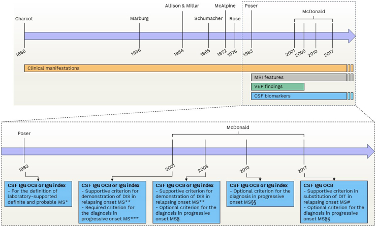
    1. Thompson A.J., Banwell B.L., Barkhof F., et al. Diagnosis of multiple sclerosis: 2017 revisions of the McDonald criteria. Lancet Neurol. 2018;17:162–173. - PubMed
    1. Rocca M.A., Preziosa P., Barkhof F., et al. MRI in the diagnosis and prognosis of multiple sclerosis. Lancet Reg Heal Eur. 2024;44:100978. doi: 10.1016/j.lanepe.2024.100978. - DOI
    1. Judge D., Roberts J., Khandker R.K., Ambegaonkar B., Black C.M. Physician practice patterns associated with diagnostic evaluation of patients with suspected mild cognitive impairment and Alzheimer's disease. Int J Alzheimer's Dis. 2019;2019 - PMC - PubMed

LinkOut - more resources