

Ultrasound assessment of insular development in adequate-for-gestational-age fetuses and fetuses with early-onset fetal growth restriction using 3D-ICRV technology
- PMID: 39444811
- PMCID: PMC11496279
- DOI: 10.3389/fmed.2024.1393115
Ultrasound assessment of insular development in adequate-for-gestational-age fetuses and fetuses with early-onset fetal growth restriction using 3D-ICRV technology
Abstract
Objective: This study aimed to evaluate the growth trajectory of the insula in adequate-for-gestational-age (AGA) and early-onset fetal growth restriction (FGR) fetuses and analyze the difference between the two groups using three-dimensional inversion crytal and realistic vue technique (3D-ICRV).
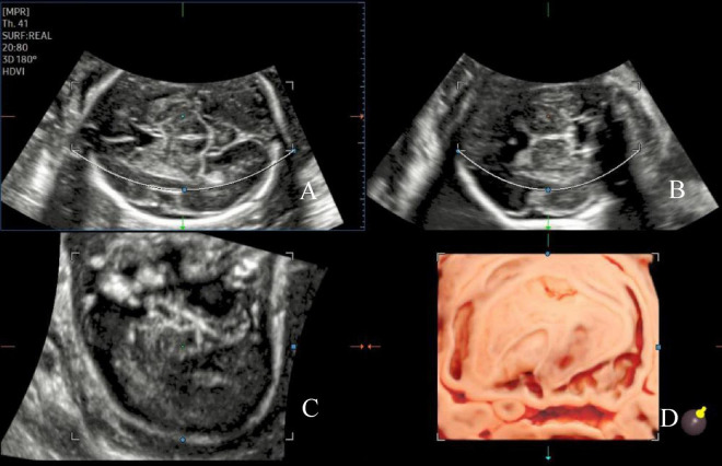
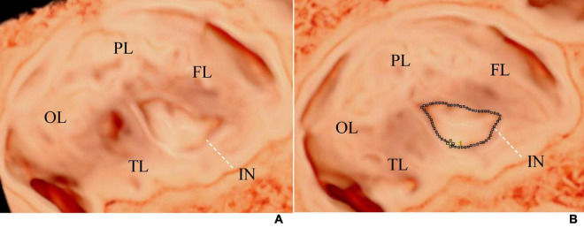
Methods: Singleton pregnant women, with a gestational age ranging from 20 to 32+6 weeks, who underwent routine examinations at Shandong Maternal and Child Care Hospital between March 2023 and December 2023 were included. The participants were divided into two groups: the FGR and AGA fetuses. Three-dimensional volumes were obtained using transabdominal ultrasound in the transverse section of the fetal hypothalamus based on different gestational ages. 3D-ICRV rendering technology was used for 3D imaging of the fetal insula. Volumes with a clear display of the insula were selected. We observed the morphology of the insula, and measured the area and circumference of the insula. By evaluating the growth trajectory of the insula in AGA and FGR fetuses, differences in insular development between the two groups were compared.
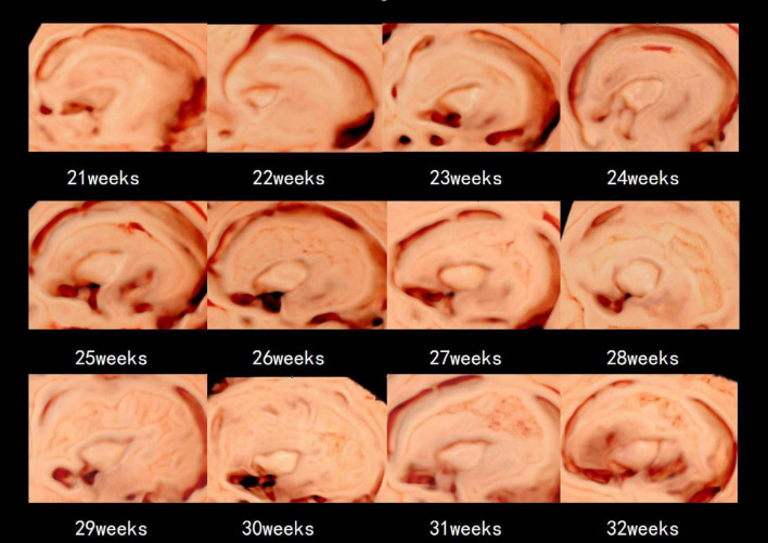
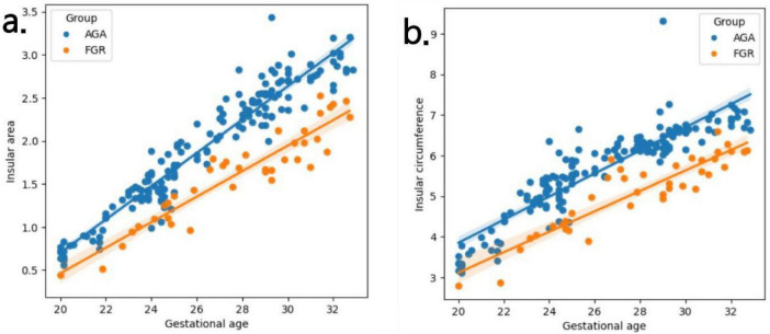
Results: Overall, 203 participants were included in this study, with 164 and 39 in the AGA and FGR groups, respectively. The 3D volumes were successfully acquired, and the area and circumference of the insula were measured using 3D-ICRV imaging technology. We found that as gestational age increased, the area and circumference of the insula gradually increased and showed positive correlations with the gestational age, with no significant changes in morphology. The growth rate of insular area and insular circumference in the FGR group is slower than that in the AGA group (insular area: 0.15 vs 0.19 cm2 / week, insular circumference: 0.25 vs 0.28 cm / week). The area and circumference of the insula in the FGR group were significantly different from those in the AGA group (insular area: p = 0.003, insular circumference: p = 0.004).
Conclusion: The measured values of the insula using 3D-ICRV identify the differences in insular development between the FGR and AGA fetuses. The findings of this study have important implications for the prenatal evaluation of cortical development and maturity in FGR fetuses and further clinical consultation and management.
Keywords: 3D-ICRV; cortical development and maturity; fetal growth restriction; fetus; insula.
Copyright © 2024 Xue, Xue, Ru, Zhang, Yin and Liu.
Conflict of interest statement
The authors declare that the research was conducted in the absence of any commercial or financial relationships that could be construed as a potential conflict of interest.
Figures
Similar articles
-
Fetal insular measurements in pregnancy with estimated fetal weight <10th centile and childhood neurodevelopmental outcomes.Am J Obstet Gynecol. 2024 Jan;230(1):85.e1-85.e15. doi: 10.1016/j.ajog.2023.07.009. Epub 2023 Jul 11. Am J Obstet Gynecol. 2024. PMID: 37442246
-
Sonographic demonstration of the sulci and gyri on the convex surface in normal fetuses using 3D-ICRV rendering technology.Ultraschall Med. 2023 Dec;44(6):e284-e295. doi: 10.1055/a-2122-6182. Epub 2023 Jul 4. Ultraschall Med. 2023. PMID: 37402405 English.
-
Fetal neurosonography detects differences in cortical development and corpus callosum in late-onset small fetuses.Ultrasound Obstet Gynecol. 2021 Jul;58(1):42-47. doi: 10.1002/uog.23592. Ultrasound Obstet Gynecol. 2021. PMID: 33438307
-
Two-dimensional Speckle tracking echocardiography in Fetal Growth Restriction: a systematic review.Eur J Obstet Gynecol Reprod Biol. 2020 Nov;254:87-94. doi: 10.1016/j.ejogrb.2020.08.052. Epub 2020 Sep 2. Eur J Obstet Gynecol Reprod Biol. 2020. PMID: 32950891
-
Fetal brain development in fetal growth restriction using MRI: a systematic review.BMC Pregnancy Childbirth. 2025 Feb 26;25(1):208. doi: 10.1186/s12884-024-07124-4. BMC Pregnancy Childbirth. 2025. PMID: 40012049 Free PMC article.
Cited by
-
Detecting microcephaly and macrocephaly from ultrasound images using artificial intelligence.BMC Med Imaging. 2025 May 26;25(1):183. doi: 10.1186/s12880-025-01709-x. BMC Med Imaging. 2025. PMID: 40419983 Free PMC article.
-
Risk of congenital anomalies associated with psychotropic medications: a review of neonatal reports in the FDA adverse event reporting System (FAERS).Arch Womens Ment Health. 2025 Aug;28(4):911-918. doi: 10.1007/s00737-024-01540-3. Epub 2024 Dec 23. Arch Womens Ment Health. 2025. PMID: 39710735
References
-
- Streeter GL. The development of the nervous system. In: Keibel F, Mall F. editors. Manual of human embryology. (1912) Philadelphia, PA: J. B. Lippincott Company.
LinkOut - more resources
Full Text Sources
Miscellaneous