. 2024 Jul 17;9(9):1088-1100.
doi: 10.1016/j.jacbts.2024.05.013. eCollection 2024 Sep.

Structure-Based Probe Reveals the Presence of Large Transthyretin Aggregates in Plasma of ATTR Amyloidosis Patients

Affiliations

Structure-Based Probe Reveals the Presence of Large Transthyretin Aggregates in Plasma of ATTR Amyloidosis Patients

Rose Pedretti et al. JACC Basic Transl Sci. .

Abstract

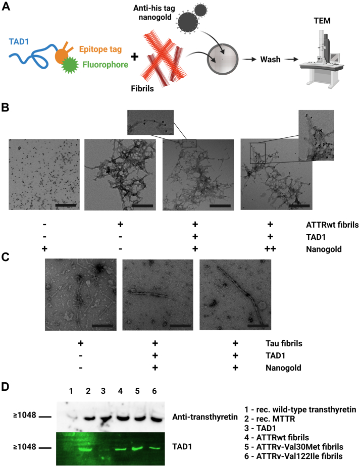
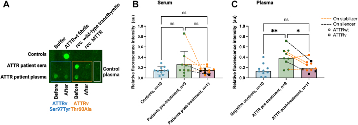
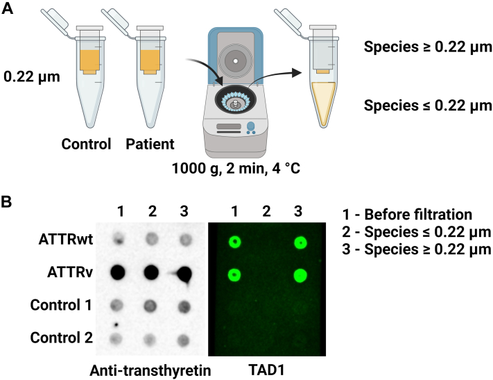
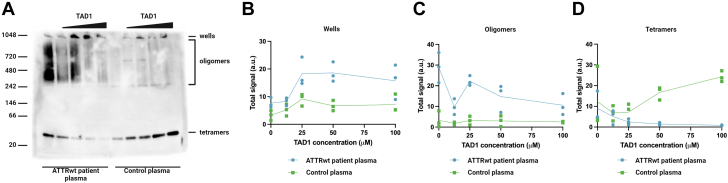
Amyloidogenic transthyretin (ATTR) amyloidosis is a relentlessly progressive disease caused by the misfolding and systemic accumulation of amyloidogenic transthyretin into amyloid fibrils. These fibrils cause diverse clinical phenotypes, mainly cardiomyopathy and/or polyneuropathy. Little is known about the aggregation of transthyretin during disease development and whether this has implications for diagnosis and treatment. Using the cryogenic electron microscopy structures of mature ATTR fibrils, we developed a peptide probe for fibril detection. With this probe, we have identified previously unknown aggregated transthyretin species in plasma of patients with ATTR amyloidosis. These species are large, non-native, and distinct from monomeric and tetrameric transthyretin. Observations from our study open many questions about the biology of ATTR amyloidosis and reveal a potential diagnostic and therapeutic target.

Keywords: amyloid; amyloidogenic transthyretin; biomarker; transthyretin.


Conflict of interest statement

This work was supported by the American Heart Association Career Development Award (847236) (to Dr Saelices), the National Institutes of Health (NIH) Director's New Innovator Award (DP2-HL163810-01) (to Dr Saelices), the Welch Foundation Research Award (I-2121-20220331) (to Dr Saelices), the NIH R01 (R01-HL160892) (to Dr Grodin), and the NIH grant 1S10OD021685-01A1 received by the Electron Microscopy Core of UTSW. R. Pedretti and Dr Saelices are inventors on a patent application (Provisional Patent Application 63/352,521) submitted by the University of Texas Southwestern Medical Center that covers the composition and structure-based diagnostic methods related to cardiac ATTR amyloidosis. Dr Grodin has received honoraria for scientific consulting from Alnylam, Eidos/BridgeBio, Intellia, Pfizer, Alexion, AstraZeneca, and Tenax Therapeutics; and has received research funding from Pfizer, Eidos/BridgeBio, the Texas Health Resources Clinical Scholars fund, and the National Heart, Lung, and Blood Institute. Dr Masri has received research funding from Pfizer, Ionis/Akcea, Attralus, and Cytokinetics; and has received consulting fees from Cytokinetics, Bristol Myers Squibb, Eidos, Pfizer, Ionis, Lexicon, Alnylam, Attralus, Haya, Intellia, BioMarin, and Tenaya. Dr Saelices has received honoraria for scientific consulting for Intellia and Attralus; and has served as a member of the Advisory Board of Alexion. All other authors have reported they have no relationships relevant to the contents of this paper to disclose.PerspectivesCOMPETENCY IN MEDICAL KNOWLEDGE: ATTR amyloidosis is an under-recognized cause of heart failure due to lack of tools that can effectively distinguish between native and amyloidogenic transthyretin. Using structures of mature ATTR fibrils derived from patient tissue, we have developed a novel detection probe that addresses this unmet need. This probe not only binds ATTR fibrils with high affinity, but also large ATTR aggregates in plasma of patients with ATTR amyloidosis. TRANSLATIONAL OUTLOOK: Combined with previous results, our data unveils a novel biomarker for cardiac ATTR amyloidosis that has the potential to be exploited as a novel diagnostic and/or therapeutic target. Our results may be leveraged to develop a screening tool using blood samples to identify disease onset earlier than the current diagnostic standard. This will enable timely treatment, improve patient prognosis, and reduce burden on the health care system.

Figures

None
Graphical abstract
Figure 1
Figure 1
Development and Screening of TAD Candidates (A) Rational design development of peptide probes based on previously published cryogenic electron microscopy structures of amyloidogenic transthyretin fibrils. Fibril structure is shown top-down as black lines, with the amino acid residues corresponding to β-strands F and H highlighted as the intended target for transthyretin aggregation detector (TAD) peptides. (B) Thioflavin T (ThT) screening of third-generation transthyretin aggregation binders (TABs) to assess candidate peptide binding to fibrils. Patient-derived seeds were incubated with 0.5 mg/mL monomeric transthyretin (MTTR) in the presence of 90 μmol/L peptide inhibitor. Fibril elongation was measured through ThT fluorescence. TAB3-12 was shown to be the most effective at halting fibril elongation. (C) Further characterization of probe candidate TAB3-12 compared to previous generation peptide inhibitors. Peptides were added to seeding reactions in a sub–stoichiometric ratio of 12 MTTR molecules to 1 peptide of interest. TAB3-12 was highly effective at inhibiting seeding compared to previously published inhibitors, even at sub–stoichiometric ratios. (D) Electron microscopy images of ThT reactions selected from C. Bar = 0.2 μm. (E) Assessment of TAD fibril binding activity through ThT assay. TAD1 was found to be the most effective at binding fibrils. (F) Dot blotting of recombinant transthyretin aggregation over time probing for total protein (top), aggregated species in total sample with TAD1 (middle), and insoluble components after centrifugation (bottom). TAD1 signal correlates with insoluble transthyretin accumulating in the reaction. FH2 = second generation TAB inhibitor that targets strands F and H of transthyretin.
Figure 2
Figure 2
Validation of TAD1 Binding to ATTR Fibrils (A) Scheme for labeling experiments. 5 μmol/L TAD1 was incubated with patient-derived wild-type amyloidogenic transthyretin (ATTRwt) fibrils or tau fibrils and nanogold beads containing an antibody directed against the polyhistidine tag of TAD1. Samples were visualized using electron microscopy. (B) Labeling of ATTRwt fibrils using TAD1 and either 50 μmol/L (+) or 250 μmol/L of nanogold (++). TAD1 binds the tips of ex vivo fibrils and along the fibril surface. Bar = 0.5 μm. (C) Labeling of tau fibrils extracted from brain using TAD1 and 50 μmol/L nanogold. There is no TAD1 binding to tau fibrils. Bar = 0.5 μm. (D) Native gel electrophoresis of TAD1, recombinant (rec) transthyretin and amyloid extracted from patients with ATTR amyloidosis, blotted and probed with an anti-transthyretin antibody (top) and TAD1 (bottom). TAD1 binds only high molecular weight transthyretin species, including those made from recombinant MTTR. Full unedited gels can be found in the Supplemental Appendix. ATTRv = variant amyloidogenic transthyretin; TEM = transmission electron microscopy; other abbreviations as in Figure 1.
Figure 3
Figure 3
TAD1 Detects Unique ATTR Species in Plasma But Not in Serum (A) Dot blotting of recombinant protein controls and extracted ATTRwt fibrils (top row), serum (middle row), and plasma (bottom row) from patients with ATTRv amyloidosis, before and after treatment with either protein stabilizers (Vyndamax 61 mg) or protein expression silencers (Patisiran 0.3 mg/kg, Vutrisiran 25 mg, or Eplontersen 45 mg), and control individuals. Samples were dotted onto nitrocellulose membrane and incubated with 5 μmol/L TAD1 overnight. Excess unbound peptide was washed off and TAD1 binding to ATTR species was measured through fluorescence intensity. TAD1 binds ATTR species present in serum and plasma. (B) Quantification of TAD1 fluorescence intensity of serum samples. Circles represent patient samples with ATTRwt amyloidosis, whereas squares represent patients with ATTRv amyloidosis. Dotted lines connect paired samples pre- and post-treatment either on tetramer stabilizers (orange) or protein expression silencers (black). Experiments were performed as in A. There is no statistically significant difference in TAD1 binding to serum samples. (C) Quantification of TAD1 fluorescence intensity of binding to plasma samples. Experimental protocol and symbols and are as in B. There is a significant increase in TAD1 signal when examining ATTR amyloidosis patients pre-treatment, and this signal is reduced after patients have been treated. Sample numbers are included in the labels. Each dot represents the average of 3 technical replicates from the same patient. Groups are shown as the mean ± SD and compared using analysis of variance with Tukey post hoc test. ∗P < 0.05; ∗∗P < 0.01. AU = absorbance units; other abbreviations as in Figures 1 and 2.
Figure 4
Figure 4
TAD1 Binds Large ATTR Species in ATTR Amyloidosis Patient Plasma (A) Scheme for filtration experiment. Patient and control plasma was passed through a 0.22-μm filter tube at 1,000g at 4 °C for 2 minutes. Unfiltered plasma (1) plasma that passed through the filter (2), and plasma that did not pass through the filter (3) were dotted onto nitrocellulose membrane and probed with 10 μmol/L TAD1. (B) Filtered plasma of patients with ATTR amyloidosis and control subjects probing with an anti-transthyretin antibody (left) and TAD1 (right). TAD1 only recognizes ATTR species too large to pass through the filter, indicating that these species are large. Abbreviations as in Figures 1 and 2.
Figure 5
Figure 5
TAD1 Binds High Molecular Weight Species in ATTR Amyloidosis Plasma (A) Protein band shift assay of ATTRwt patient plasma and control plasma with increasing concentrations of TAD1, probed with an anti-transthyretin antibody. Increasing TAD1 concentrations in patient plasma resulted in the accumulation of high molecular weight species (1,048 kDa) and the decrease in tetrameric and oligomeric transthyretin. (B) Quantification of transthyretin well signal from gel shift assay. Increasing TAD1 concentration resulted in more high molecular weight species in wells of patient plasma and little change in control plasma. (C) Quantification of transthyretin oligomer signal from gel shift assay. Increasing TAD1 concentration resulted in less oligomeric species in patient plasma and little change in control plasma. (D) Quantification of transthyretin tetramer signal from gel shift assay. Increasing TAD1 concentration resulted in less tetrameric transthyretin in patient plasma and more tetramer in control plasma. All replicates are included. Blue circles represent ATTRwt patient plasma, whereas green squares represent control plasma. Abbreviations as in Figures 1, 2 and 3.

References

    1. Benson M.D. Pathogenesis of transthyretin amyloidosis. Amyloid. 2012;19(suppl 1):14–15. - PubMed
    1. Colon W., Kelly J.W. Partial denaturation of transthyretin is sufficient for amyloid fibril formation in vitro. Biochemistry. 1992;31(36):8654–8660. - PubMed
    1. Schonhoft J.D., Monteiro C., Plate L., et al. Peptide probes detect misfolded transthyretin oligomers in plasma of hereditary amyloidosis patients. Sci Transl Med. 2017;9(407) - PMC - PubMed
    1. Jiang X., Labaudinière R., Buxbaum J.N., et al. A circulating, disease-specific, mechanism-linked biomarker for ATTR polyneuropathy diagnosis and response to therapy prediction. Proc Natl Acad Sci U S A. 2021;118(9) - PMC - PubMed
    1. Nguyen B.A., Singh V., Afrin S., et al. Structural polymorphism of amyloid fibrils in ATTR amyloidosis revealed by cryo-electron microscopy. Nat Commun. 2024;15(1):581. - PMC - PubMed

LinkOut - more resources