

Evaluation of the Conventional Acid-Etching System and the Self-Etching Primer in Bonding Metallic Orthodontic Brackets: An In-Vitro and In-Vivo Study
- PMID: 39445044
- PMCID: PMC11498908
- DOI: 10.7759/cureus.72226
Evaluation of the Conventional Acid-Etching System and the Self-Etching Primer in Bonding Metallic Orthodontic Brackets: An In-Vitro and In-Vivo Study
Abstract
Objective: This study aimed to compare the shear bond strength (SBS) of enamel-bonded orthodontic brackets with the conventional acid etching (CAE) system and the self-etching primer (SEP) in vitro and to compare the clinical performance of both systems when used in the treatment of malocclusion patients.
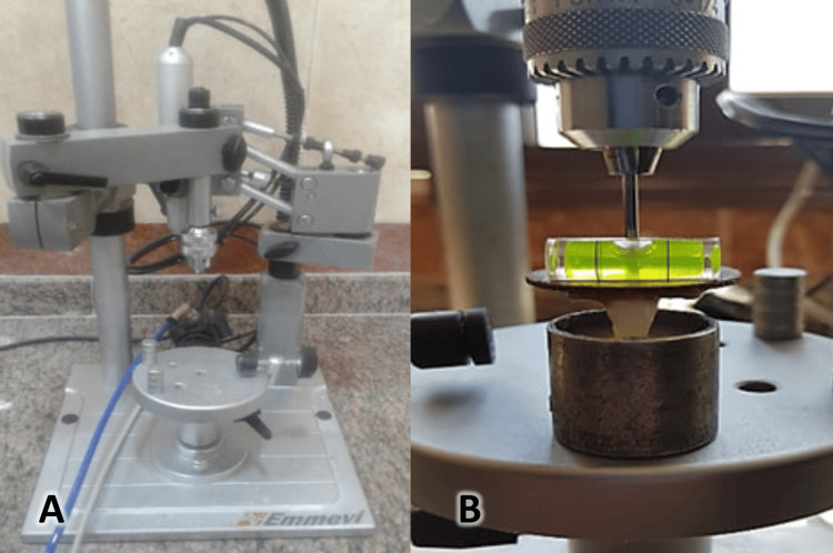
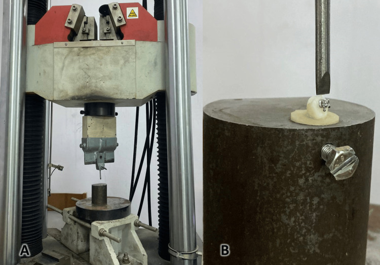
Materials and methods: In the first part of the study, 40 extracted human premolars were randomly divided into two groups containing 20 teeth. The first group (the conventional enamel etching group) employed 37% phosphoric acid before bonding the metallic brackets (0.022-inch slot, MBT prescription, American Orthodontics, Sheboygan, WI, USA). The etching system was Tetric 5th (Ivoclar Vivadent, Schaan, Liechtenstein). The second group used a SEP (Sep Tetric 7th, Ivoclar Vivadent, Schaan, Liechtenstein) to bond the same brackets. In the first part of the study, SBS was evaluated, followed by the adhesive remnant index (ARI) assessment. The second part of the study (i.e., the clinical part) assessed a cohort of 30 patients during a 6-month observation period. The upper 10 teeth (from the second premolar on the right side to the second on the right side) were bonded using the chosen method for each patient in the clinical assessment. That is, 150 teeth in each group were evaluated regarding the failure rate. The ARI was assessed for those teeth that lost their brackets.
Results: The mean SBS was greater in the SEP group compared to the CAE group (17.93 MPa and 16.60 MPa, respectively; P = 0.014). The difference was not statistically significant. Conversely, the failure rate was lower in the CAE group compared to the SEP group, with a failure rate of 6% and 14.7%, respectively. The difference was statistically significant (P = 0.014). However, the ARI showed no statistically significant difference in in-vivo and in-vitro analyses, as most bracket failures were at the adhesive level.
Conclusion: Laboratory results showed no statistical difference in the SBS mean values between the two groups. Clinically, the SEP group showed a greater failure rate than the CAE group, but both failure rates in the two groups were within the clinically acceptable range. The ARI did not show any difference between the two groups in terms of the failure site when the evaluation was conducted in vivo and in vitro, as most of the areas of failure were concentrated in the material itself.
Keywords: adhesive remnant index; bonding strength; composite; conventional acid etching technique; debonding; failure rate; metallic brackets; premolars; self-etching primer; shear bond strength.
Copyright © 2024, Alomar et al.
Conflict of interest statement
Human subjects: Consent was obtained or waived by all participants in this study. Local Research Ethics Committee at the National Dental Center, Ministry of Health issued approval Approval Number: 423. Ethical approval was obtained from the Local Research Ethics Committee at the National Dental Center (Reference number: 423 dated 27.5.2023). Animal subjects: All authors have confirmed that this study did not involve animal subjects or tissue. Conflicts of interest: In compliance with the ICMJE uniform disclosure form, all authors declare the following: Payment/services info: All authors have declared that no financial support was received from any organization for the submitted work. Financial relationships: All authors have declared that they have no financial relationships at present or within the previous three years with any organizations that might have an interest in the submitted work. Other relationships: All authors have declared that there are no other relationships or activities that could appear to have influenced the submitted work.
Figures
References
-
- Phosphoric acid incorporated with acidulated phosphate fluoride gel etchant effects on bracket bonding. Kim MJ, Lim BS, Chang WG, Lee YK, Rhee SH, Yang HC. https://meridian.allenpress.com/angle-orthodontist/article/75/4/678/5815.... Angle Orthod. 2005;75:678–684. - PubMed
-
- Effect of an acidic primer on shear bond strength of orthodontic brackets. Bishara SE, Gordan VV, VonWald L, Olson ME. Am J Orthod Dentofacial Orthop. 1998;114 - PubMed
LinkOut - more resources
Full Text Sources