. 2025 Feb;41 Suppl 1(Suppl 1):72-79.
doi: 10.1111/edt.13002. Epub 2024 Oct 24.

Transient Apical Breakdown: Incidence, Pathogenesis, and Healing

Affiliations

Transient Apical Breakdown: Incidence, Pathogenesis, and Healing

Mitsuhiro Tsukiboshi et al. Dent Traumatol. 2025 Feb.

Abstract

Background/aims: Transient apical breakdown (TAB) is a phenomenon that indicates temporary apical periodontal destruction and root resorption after tooth luxation injuries, followed by the healing process of the dental pulp. Andreasen in 1986, reported that TAB was seen in approximately 4.2% of all luxation injuries. However, there have been no reports thereafter on the types and frequency of the luxation traumatic injuries in which TAB occurs. Therefore, this retrospective study was aimed to assess the incidence and pathogenesis of dental trauma-induced TAB and to suggest a possible mechanism of subsequent healing based on a series of cases.

Methods: Data analysis included mature teeth (n = 56) of 49 patients aged 9-30 years who presented in a private dental office over a period of 10 years (2012-2022) to investigate the incidence and healing sequala of TAB.

Results: TAB was observed in 43.8% of subluxation, 62.5% of extrusive luxation, and 75% of lateral luxation injuries. The average age of patients who developed TAB was 14.5 years, ranging from 9 to 28 years old.

Conclusions: TAB can be expected in many cases of luxation injuries with minimal dislocation. Therefore, mild injuries (subluxation, extrusion, and lateral luxation), may exhibit spontaneous healing, recovery of dark discoloration of the crown, disappearance of a periapical radiolucent lesion and return to normal response to EPT as long as 12 months after the traumatic injury. Thus, a decision to perform endodontic treatment in these cases might be postponed until clear evidence for an infection exists.

Keywords: apical periodontitis; avulsion; concussion; dental trauma; intrusion; luxation; resorption.


Conflict of interest statement

The authors declare no conflicts of interest.

Figures
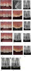
FIGURE 1
FIGURE 1
(a–q). TAB after lateral luxation of the left right maxillary central incisor in a 15‐year and 5‐month‐old male who suffered a bicycle accident. (a–c) First visit 1.5 h following the trauma. Clinical photograph is showing anterior maxillary teeth (a). There is a noncomplicated crown fractures on the right maxillary central and the left maxillary lateral incisors. There was a positive response EPT (+) on both teeth. First visit PA radiograph showing no obvious displacement of the teeth (c). CBCT sagittal view of the left maxillary central incisor, showing lateral luxation (b), and it had no response on EPT (−). The anterior teeth had an existing palatal metal wire splint following a past orthodontic treatment before the trauma. (d–f) One‐month follow‐up. Clinical photographs showing apparent brown discoloration of the left maxillary central incisor on the buccal (d) and palatal (e) views. PA radiograph showing no obvious change around the apex (f). There was still no response on EPT (−). (g–i) Four‐month follow‐up. Clinical photographs of the buccal (g) and palatal (h) views showing a slight reduction in the crown discoloration of the left maxillary central incisor. PA radiograph, showing slight apical root resorption of the left maxillary central incisor (i). There was still no response on EPT (−). (j, k) Eleven‐month follow‐up clinical photograph of the buccal view (j) still showing discoloration of the left maxillary central incisor. PA radiograph showing evidence for an enlarged apical foramen on the left maxillary central incisor (k). There was still no response on EPT (−). (l) One‐year and 4‐month follow‐up. PA radiograph showing beginning of PCO in the left maxillary central incisor. At this time sensibility test turned into positive, with EPT (+). (m–o) Three‐year follow‐up. Clinical photographs of the buccal (m) and palatal (n) views of the left maxillary central incisor showing obvious reduction in coronal discoloration. PA radiograph showing progression of PCO in the left maxillary central incisor (o). (p, q) PA radiographs comparing the root length, immediately after trauma, before TAB (p) and 3 years after trauma (q). Resorption and shortening of the root on the left maxillary central incisor are evident (q).
FIGURE 2
FIGURE 2
(a–t). TAB after extrusive luxation of the right maxillary central incisor in a 15‐year and 10‐month‐old male. The trauma was caused by a baseball direct hit on the mouth and the visited the clinic 5.5 h following the trauma. (a–) The day of trauma, buccal view photograph showing an extrusive luxation of the right maxillary central incisor (a). CBCT sagittal view of the right maxillary central incisor, showing extrusive luxation of the right maxillary central incisor. The root is fully developed with a closed apex (b). PA radiograph showing an extrusive luxation of the right maxillary central incisor (c). There was no response on EPT (−) on the right maxillary central incisor. (d, e) Three weeks after trauma follow‐up. Buccal view photograph showing slight dark (brown) discoloration of the crown of right maxillary central incisor (d). Palatal view photograph showing slight dark discoloration of the crown of right maxillary central incisor (e). The discoloration is more apparent on the palatal view (e) compared to the buccal view (d). There was no response on EPT (−) on the right maxillary central incisor. (f–h) Six‐month after trauma follow‐up. The right maxillary central incisor was repositioned and splinted for 3 weeks. The discoloration that was evident at 3 weeks after the trauma appears to be reduced as shown on the buccal (f) and palatal (g) views. At this time, the tooth responded to EPT (+). The PA radiograph shows apical root resorption with a widened apical foramen on the right maxillary central incisor (h). (i–k) One‐year after trauma follow‐up. The discoloration is reduced as shown in the clinical photograph of the buccal view of the right maxillary central incisor (i). CBCT (j) and PA (k) images show the apex of the right maxillary central incisor is shortened with a widened apical foramen, and a lateral canal. (l–n) Two years after trauma follow‐up. The crown discoloration is reduced as shown in the clinical photograph of the buccal view of the right maxillary central incisor (l). CBCT (m) and PA (n) radiographs PCO on the right maxillary central incisor. (o–q) Six years and 2 months after trauma. Yellow crown discoloration in the crown is noticeable as shown in the clinical photograph of the buccal (o) and palatal (p) views of the right maxillary central incisor. Further progression of PCO is noticeable on the PA radiograph (q). There are no pathological findings in the periarticular tissues. (r–t) Ten years and 5 months after trauma. Yellow discoloration of the right maxillary central incisor has progressed and is noticeable (r). The pulp canal of the right maxillary central incisor shows progression of PCO (s, t). The pulp canal of the right maxillary central incisor still has a positive response to EPT (+).
FIGURE 3
FIGURE 3
(a–n). TAB, after slight lateral luxation of the right maxillary central incisor in a 20‐year‐old male. The trauma was caused by a friend who bumped their head into the patient's mouth during gym class. (a, b) Clinical photograph (a) and PA (b) radiograph taken 6 days after trauma. Both, the right and left maxillary central incisors had a no response on EPT (−). (c–f) CBCT taken 6 days after the trauma showing both maxillary central incisors are involved in slight lateral luxation, as evident on the sagittal view (d, e) and axial (f) views, with fracture of the alveolar bone as evident on the axial view (f). (g–i) Six weeks after trauma follow‐up. Pink discoloration of the right maxillary central incisor is evident on the clinical photograph of the buccal aspect (g). Thickening of the periodontal ligament space is evident on the PA radiograph of both teeth (h). Both, the right and left maxillary central incisors, still had a no response on EPT (−). Cervical pulpotomy was performed on the right maxillary central incisor. The canal was rinsed with 2% sodium hypochlorite and saline followed by medication with calcium hydroxide (Vitapex, Neo Company, Tokyo, Japan) (i). (j) Ten months after trauma, PCO of the right maxillary central incisor has progressed as evident on the PA radiograph. The calcium hydroxide was replaced with MTA (Bio MTA, imported by Morita, Tokyo, Japan). Root resorption at the apex of left maxillary central incisor is observed. Both maxillary central incisors had a response on EPT (+) at this time. (k, l) One year and 5 months after trauma. PCO of the right maxillary central incisor has progressed as evident on the PA radiograph (k). Both maxillary central incisors still had a positive response on EPT (+). Yellow discoloration of the right maxillary central incisor has been treated by walking bleach performed 6 months earlier (l). (m, n) Comparison of root length between before and after TAB has occurred in the right maxillary central incisor. Six days after trauma (m). One year 5 months after trauma (n). The root of the left maxillary central incisor has been resorbed and shortened, with no changes on the right maxillary central incisor.
FIGURE 4
FIGURE 4
(a–d). TAB after subluxation of the left maxillary central incisor in a 12‐year‐old male due to a bicycle accident. Sagittal views of CBCT scans are presented below. (a) One day after subluxation. The left maxillary central incisor has closed apex and thickening of the apical periodontal space is evident. There was no response on EPT (−). (b) Three months after trauma follow‐up. The left maxillary central incisor shows a small periapical radiolucency, surface root resorption at the apex evident, and there is enlargement of the apical foramen. There was still no response on EPT (−). (c) Six months after trauma follow‐up. The apical foramen is kept wide, with no apical radiolucent lesion evident anymore. There was still no response on EPT (−). (d) Two years after trauma follow‐up. PCO in the left maxillary central incisor is evident. The left maxillary central incisor showed a positive response on EPT (+) since after 1 year and 2 months after trauma.
FIGURE 5
FIGURE 5
Illustration of a suggested mechanism of TAB in a mature maxillary incisor: (a) Immediately after trauma (subluxation). The pulp is ischemic but not infected. Blood vessels around the apex are raptured and bleeding occurs in a small area. (b) Bleeding induces inflammatory reaction as defense mechanism of the body, which causes osteoclast to disseminate from blood vessels. Osteoclasts cause bone and root resorption, which results in shortening of the root, enlargement of the apical foramen, and a radiolucent periapical lesion or thickening of the apical periodontal ligament. (c) Once the apical foramen becomes wider than 1 mm, revascularization may take place, and the ischemic tissues may be replaced with vital tissues. (d) If cells derived from pulp tissues repopulate the canal, then PCO takes place.

References

    1. Andreasen F. M., “Transient Apical Breakdown and Its Relation to Color and Sensibility Changes After Luxation Injuries to Teeth,” Endodontics & Dental Traumatology 2 (1986): 9–19. - PubMed
    1. Cohenca N., Karni S., and Rotstein I., “Transient Apical Breakdown Following Tooth Luxation,” Dental Traumatology 19 (2003): 289–291. - PubMed
    1. Boyd K. S., “Transient Apical Breakdown Following Subluxation Injury: A Case Report,” Endodontics & Dental Traumatology 11 (1995): 37–40. - PubMed
    1. Zhu Z., “Transient Apical Breakdown of a Discoloured Maxillary Central Incisor During Orthodontic Treatment: A Case Report,” Australian Endodontic Journal 49, no. S1 (2023): 476–480. - PubMed
    1. González O. L., Vera J., Orozco M. S., Mancera J. T., González K. V., and Malagón G. V., “Transient Apical Breakdown and Its Relationship With Orthodontic Forces: A Case Report,” Journal of Endodontia 40, no. 8 (2014): 1265–1267. - PubMed

LinkOut - more resources