

Comparison of B Cell Variable Region Gene Segment Characteristics in Neuro-autoantibodies
- PMID: 39446034
- PMCID: PMC11532373
- DOI: 10.4049/immunohorizons.2400037
Comparison of B Cell Variable Region Gene Segment Characteristics in Neuro-autoantibodies
Abstract
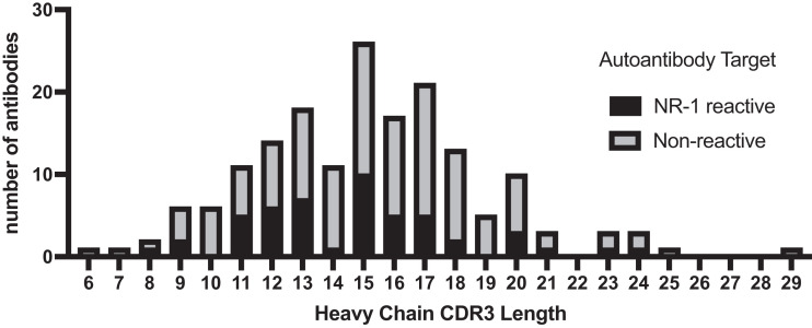
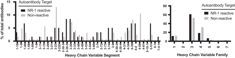
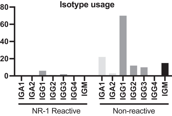
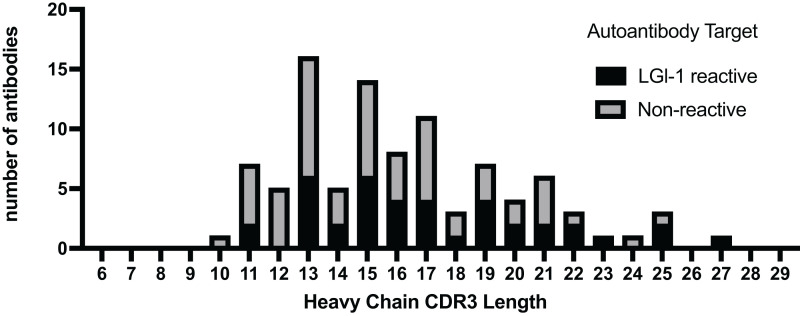
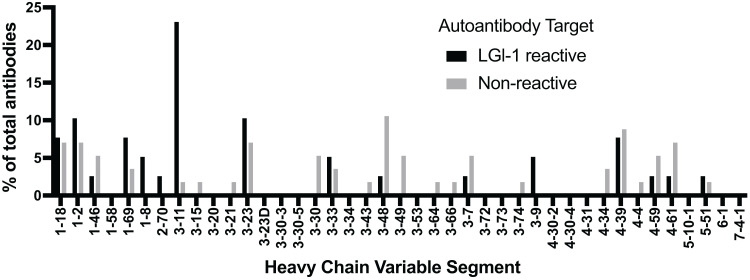
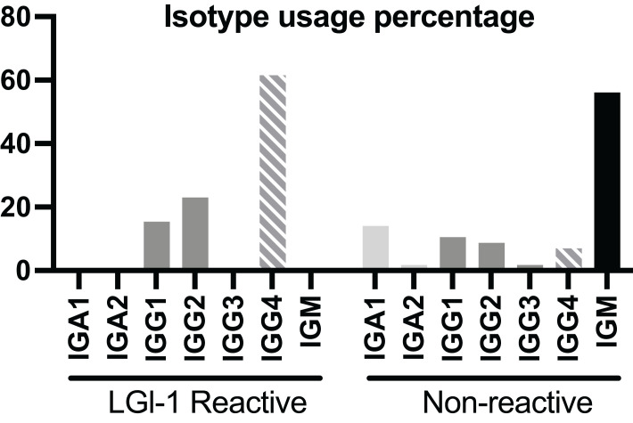
Autoimmune pediatric neurologic diseases have variable phenotypes and presentations, making diagnosis challenging. The pathologic mechanisms are also distinct, including cell-mediated and Ab-mediated autoimmunity, paraneoplastic syndromes, and postinfectious processes. In recent years a number of studies have described the characteristics of the autoantibodies involved in a number of these diseases. Some of the described Abs use a restricted set of variable gene segments. We sought to compare the Ab characteristics of autoantibodies related to some of the more common disorders to discover whether specific Ab signatures are universally associated with neuroautoimmune diseases. We initially performed a literature review to summarize the Ab characteristics of autoantibodies related to some of the more common disorders, including N-methyl-d-aspartate receptor (NMDAR) and leucine-rich, glioma-inactivated 1 (LGI-1). Next, we performed data analysis from selected studies that sequenced Ig genes to further characterize NMDAR and LGI-1 autoantibodies including CDR3 length distribution, variable gene sequence usage, and isotype use. We found that CDR3 length of NMDAR autoantibodies was normally distributed whereas the CDR3 length distribution of LGI-1 autoantibodies was skewed, suggesting that there is no global structural restriction on types of autoantibodies that can cause encephalitis. We also found that IgG1-IgG3 were the main NMDAR autoantibody isotypes detected, while IgG4 was the major isotype used in autoantibodies from LGI-1 encephalitis. These findings are useful for our understanding of autoimmune encephalitis and will help facilitate better diagnosis and treatment of these conditions in the future.
Copyright © 2024 The Authors.
Conflict of interest statement
The authors have no financial conflicts of interest.
Figures
References
-
- Gole, S., Anand A.. 2023. Autoimmune Encephalitis. In: StatPearls [Internet]. Treasure Island (FL): StatPearls Publishing. Available at: https://www.ncbi.nlm.nih.gov/books/NBK578203/. - PubMed
-
- Vora, N. M., Holman R. C., Mehal J. M., Steiner C. A., Blanton J., Sejvar J.. 2014. Burden of encephalitis-associated hospitalizations in the United States, 1998–2010. Neurology 82: 443–451. - PubMed
-
- Kitley, J., Waters P., Woodhall M., Leite M. I., Murchison A., George J., Küker W., Chandratre S., Vincent A., Palace J.. 2014. Neuromyelitis optica spectrum disorders with aquaporin-4 and myelin-oligodendrocyte glycoprotein antibodies: a comparative study. JAMA Neurol. 71: 276–283. - PubMed
-
- Glaser, C. A., Gilliam S., Schnurr D., Forghani B., Honarmand S., Khetsuriani N., Fischer M., Cossen C. K., Anderson L. J., California Encephalitis Project, 1998-2000 . 2003. In search of encephalitis etiologies: diagnostic challenges in the California Encephalitis Project, 1998–2000. Clin. Infect. Dis. 36: 731–742. - PubMed
Publication types
MeSH terms
Substances
Supplementary concepts
LinkOut - more resources
Full Text Sources
