Review
. 2025 Jan 6;222(1):e20232298.
doi: 10.1084/jem.20232298. Epub 2024 Oct 24.

MAIT cells: Conserved watchers on the wall

Affiliations
Review

MAIT cells: Conserved watchers on the wall

Lilou Germain et al. J Exp Med. .

Abstract

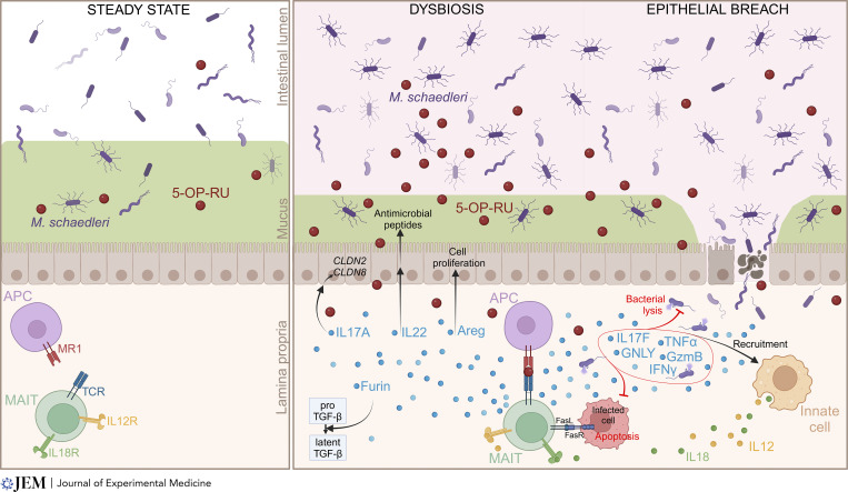
MAIT cells are innate-like T cells residing in barrier tissues such as the lung, skin, and intestine. Both the semi-invariant T cell receptor of MAIT cells and the restricting element MR1 are deeply conserved across mammals, indicating non-redundant functions linked to antigenic specificity. MAIT cells across species concomitantly express cytotoxicity and tissue-repair genes, suggesting versatile functions. Accordingly, MAIT cells contribute to antibacterial responses as well as to the repair of damaged barrier tissues. MAIT cells recognize riboflavin biosynthetic pathway-derived metabolites, which rapidly cross epithelial barriers to be presented by antigen-presenting cells. Changes in gut ecology during intestinal inflammation drive the expansion of strong riboflavin and MAIT ligand producers. Thus, MAIT cells may enable real-time surveillance of microbiota dysbiosis across intact epithelia and provide rapid and context-dependent responses. Here, we discuss recent findings regarding the origin and regulation of MAIT ligands and the role of MAIT cells in barrier tissues. We speculate on the potential reasons for MAIT cell conservation during evolution.


Conflict of interest statement

Disclosures: O. Lantz reported grants and personal fees from Biomunex during the conduct of the study and grants and personal fees from Biomunex outside the submitted work; in addition, O. Lantz had a patent to Car-T MAIT pending. No other disclosures were reported.

Figures

Figure 1.
Figure 1.
MAIT cell development in mice. 5-A-RU-derived MAIT ligands, mostly produced in the colon mucosa, travel to the thymus and contribute to the positive selection and/or intrathymic expansion of MAIT cells, in particular the MAIT17 subset. Upon positive selection by CD4+CD8+ DP thymocytes, MAIT cell precursors express PLZF and differentiate into either MAIT1 or MAIT17 cells. Expression of distinct homing proteins in each subset targets MAIT1 cells preferentially to the spleen and liver, while MAIT17 cells are home to barrier tissues such as lung, skin, and gut lamina propria. In addition to 5-A-RU-derived ligands, a bile acid metabolite primarily produced by bacterial bile salt hydrolases (BSH) can also be found in the thymus and modulate MAIT cell development. Created with Biorender.
Figure 2.
Figure 2.
Barrier-promoting and antimicrobial functions of MAIT cells at the intestinal barrier. Intestinal dysbiosis leads to an expansion of ribD-expressing bacteria, particularly M. schaedleri, resulting in the synthesis of MAIT ligands and the activation of MAIT cells in the lamina propria. During colitis, MAIT cells produce barrier-promoting factors such as IL17A, a cytokine that induces the expression of tight junction proteins (claudin 8 and 2) in epithelial cells; amphiregulin, which stimulates epithelial cell proliferation through the epidermal growth factor receptor; and Furin, involved in TGFβ maturation. MAIT cells also provide antimicrobial mediators, such as IL22, which stimulates epithelial production of antimicrobial peptides, and the inflammatory cytokines IL17F, TNFα, granulysine, granzyme B, and IFNγ, which promote the recruitment of innate immune cells and contribute to bacterial lysis. MAIT cells can also express FasL, leading to FasR-mediated apoptosis of infected cells. Created with Biorender.

Similar articles

Cited by

References

    1. Abbas, C.A., and Sibirny A.A.. 2011. Genetic control of biosynthesis and transport of riboflavin and flavin nucleotides and construction of robust biotechnological producers. Microbiol. Mol. Biol. Rev. 75:321–360. 10.1128/MMBR.00030-10 - DOI - PMC - PubMed
    1. Álvarez-Carretero, S., Tamuri A.U., Battini M., Nascimento F.F., Carlisle E., Asher R.J., Yang Z., Donoghue P.C.J., and Dos Reis M.. 2022. A species-level timeline of mammal evolution integrating phylogenomic data. Nature. 602:263–267. 10.1038/s41586-021-04341-1 - DOI - PubMed
    1. Ashoori, M., and Saedisomeolia A.. 2014. Riboflavin (vitamin B2) and oxidative stress: A review. Br. J. Nutr. 111:1985–1991. 10.1017/S0007114514000178 - DOI - PubMed
    1. Becker, J.M., Kauffman S.J., Hauser M., Huang L., Lin M., Sillaots S., Jiang B., Xu D., and Roemer T.. 2010. Pathway analysis of Candida albicans survival and virulence determinants in a murine infection model. Proc. Natl. Acad. Sci. USA. 107:22044–22049. 10.1073/pnas.1009845107 - DOI - PMC - PubMed
    1. Ben Youssef, G., Tourret M., Salou M., Ghazarian L., Houdouin V., Mondot S., Mburu Y., Lambert M., Azarnoush S., Diana J.-S., et al. . 2018. Ontogeny of human mucosal-associated invariant T cells and related T cell subsets. J. Exp. Med. 215:459–479. 10.1084/jem.20171739 - DOI - PMC - PubMed

MeSH terms

Substances

LinkOut - more resources