

Changes in tissues and organs through PMCTA carrier substances
- PMID: 39446159
- PMCID: PMC11732870
- DOI: 10.1007/s00414-024-03350-9
Changes in tissues and organs through PMCTA carrier substances
Abstract
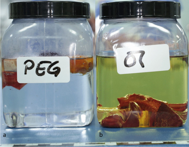
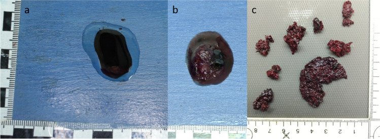
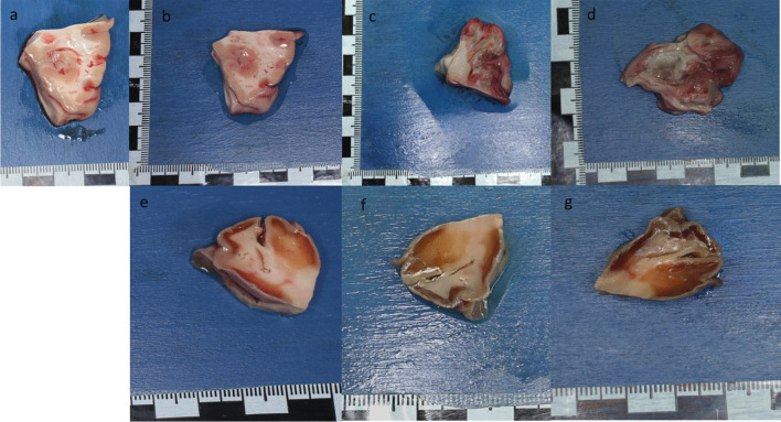
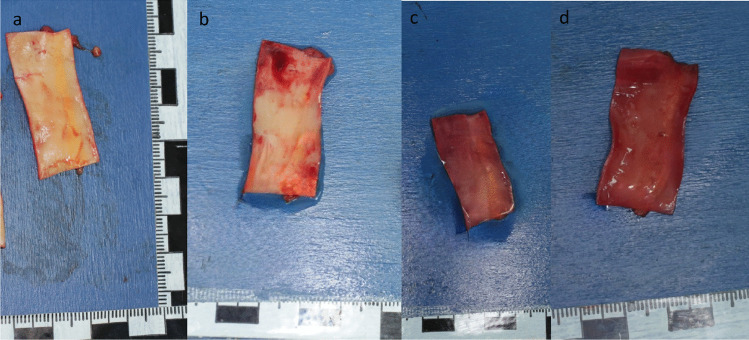
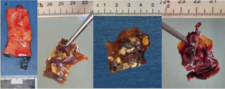
To date, lipophilic contrast agents mixed with oil, usually paraffin oil, are the most commonly used contrast agents in post-mortem computed tomography angiography (PMCTA). Iodine-based hydrophilic contrast media in combination with a water-soluble carrier, e.g. polyethylene glycol (PEG), are also common. However, their influence on different tissues and organs is poorly understood. In order to analyse the changes in the cadavers caused by the different carrier substances, we evaluated the effects of PEG 200 and oil on the different tissues and organs. Therefore, during a forensic autopsy, liquid femoral vein blood and samples of different organs and vessels were taken and preserved at room temperature in the two liquids mentioned. The condition of the samples was documented during the autopsy and 24, 48 and 72 h after preservation. Microscopic examination took place after 72 h. After 24 h, the samples placed in PEG 200 already showed a clear solidification of almost all structures. Crumbly blood agglomerates had formed in the previously liquid blood. In contrast, the samples stored in oil showed signs of classic cadaveric decomposition after 24 h, which increased with time. The microscopic and immunohistochemical evaluation of the samples stored in PEG showed a good diagnostic quality. The analysis of tissues stored in oil was much more difficult due to putrefaction. PEG and oil show significantly different effects on human tissues, mainly conservation and dehydration are affected. It is crucial to be aware of these differences in order to choose the most appropriate PMCTA method for each forensic case.
Keywords: Carrier substance; PEG; PMCTA; Paraffin oil; Polyethylenglycole.
© 2024. The Author(s).
Conflict of interest statement
Declarations. Ethical approval: Principal consent of the local ethics committee was obtained, as the committee stated, that “examinations of body materials and evidence from cadavers taken and examined on behalf of public prosecutors or investigating authorities [for scientific purpose] there is no obligation to seek advice of the Ethics Committee in case of scientific publication of anonymized results” (No. 22–0572-KB). Informed consent: Not applicable Disclosure of potential conflicts of interest: The authors declare no conflict of interest. Research involving human participants and/or animals: Not applicable (see Ethical Approval).
Figures


References
-
- Bornik A, Heinze S, Campana L, Rost T, Wittig H, Labudde D et al (2019) Theoretische Grundlagen der forensischen Bildgebung. Rechtsmedizin 29(1):1–12 - DOI
MeSH terms
Substances
LinkOut - more resources
Full Text Sources
