. 2024 Oct 24;20(10):e1011434.
doi: 10.1371/journal.pgen.1011434. eCollection 2024 Oct.

Exposure to benzyl butyl phthalate (BBP) leads to increased double-strand break formation and germline dysfunction in Caenorhabditis elegans

Affiliations

Exposure to benzyl butyl phthalate (BBP) leads to increased double-strand break formation and germline dysfunction in Caenorhabditis elegans

Ayana L Henderson et al. PLoS Genet. .

Abstract

Benzyl butyl phthalate (BBP), a plasticizer found in a wide range of consumer products including vinyl flooring, carpet backing, food packaging, personal care products, and children's toys, is an endocrine-disrupting chemical linked to impaired reproduction and development in humans. Despite evidence that BBP exposure perturbs the integrity of male and female gametes, its direct effect on early meiotic events is understudied. Here, using the nematode Caenorhabditis elegans, we show that BBP exposure elicits a non-monotonic dose response on the rate of X-chromosome nondisjunction measured using a high-throughput screening platform. From among the range of doses tested (1, 10, 100 and 500 μM BBP), we found that 10 μM BBP elicited the strongest effect on the germline, resulting in increased germ cell apoptosis and chromosome organization defects. Mass spectrometry analysis shows that C. elegans efficiently metabolizes BBP into its primary metabolites, monobutyl phthalate (MBP) and monobenzyl phthalate (MBzP), and that the levels of BBP, MBP, and MBzP detected in the worm are within the range detected in human biological samples. Exposure to 10 μM BBP leads to germlines with enlarged mitotic nuclei, altered meiotic progression, activation of a p53/CEP-1-dependent DNA damage checkpoint, increased double-strand break levels throughout the germline, chromosome morphology defects in oocytes at diakinesis, and increased oxidative stress. RNA sequencing analysis indicates that BBP exposure results in the altered expression of genes involved in xenobiotic metabolic processes, extracellular matrix organization, oocyte morphogenesis, meiotic cell cycle, and oxidoreduction. Taken together, we propose that C. elegans exposure to BBP leads to increased oxidative stress and double-strand break formation, thereby compromising germline genomic integrity and chromosome segregation.


Conflict of interest statement

The authors have declared that no competing interests exist.

Figures

Fig 1
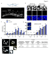
Fig 1. BBP dose-response curve reveals non-monotonic effects of BBP on rate of X-chromosome nondisjunction, apoptosis, and chromosome organization defects in the germline.
(A) The experimental workflow for high-throughput screening of X-chromosome nondisjunction. Pxol-1::gfp;col-121 worms were synchronized by hypochlorite treatment, grown to the L4 larval stage on NGM plates, and exposed on multi-well plates for 24 hours to 1, 10, 100, and 500 μM of BBP or DMSO (0.1%) vehicle control. Approximately 4,000 worms were screened in two biological repeats for each condition. Gated adults were sorted by GFP expression in embryos in utero. Frequency of X-chromosome nondisjunction in BBP-exposed worms was quantified as fold increase over DMSO. Gray bar indicates exposure to 100 μM DEET, an internal positive control. Diagram created with BioRender.com. (B) Schematic of C. elegans highlighting late pachytene as the stage where germ cell apoptosis was assessed. Representative images of acridine orange-stained germ cell corpses in late pachytene of DMSO or BBP-exposed worms in DIC (left) and TRITC (right). Arrowheads indicate germ cell corpses. Quantification of the mean number of germ cell corpses observed following exposure to each chemical condition. Error bars represent SEM. ***P<0.001, ****P<0.0001 by the two-tailed Mann-Whitney test, 95% C.I. N = 30 per condition. Four biological repeats. Diagram created with BioRender.com. (C) Carnoy’s fixed and DAPI-stained images of gonads at the pachytene stage following exposure to DMSO or BBP. Images represent examples of gonads with normal germline configuration (first panel) or various chromosome organization defects in the germline including laggers (second panel), aggregates (third panel), and gaps (fourth panel). Yellow arrowheads indicate the respective defect in each panel. Quantification of chromosome organization defects below. *P = 0.0106 for 10 μM BBP, *P = 0.0212 for 500μM BBP, Fisher’s exact test, 95% C.I. N = 27–31 gonads. Three biological repeats. Scale bar, 5 μm.
Fig 2
Fig 2. Mass spectrometry reveals physiologically relevant, internal levels of BBP and its metabolites within the worm.
(A) BBP is metabolized by lipases and esterases into its two primary metabolites: MBP and MBzP. Gas chromatography mass spectrometry was used to measure BBP, while liquid chromatography with tandem mass spectrometry was used to measure MBP and MBzP in the worm following chemical exposure. Chromatograms represent the detected concentrations of BBP (left), MBP (middle), and MBzP (right) following exposure to (B) 0.1% DMSO vehicle control, (C) 10 μM of BBP, and (D) 100 μM of BBP. X-axis represents retention time; y-axis represents abundance or intensity (cps).
Fig 3
Fig 3. BBP alters meiotic progression and leads to activation of a DNA damage checkpoint.
(A) High-resolution images of whole mounted dissected gonads stained with DAPI (blue) and immunostained against SUN-1 (pS8) (green) following DMSO or BBP exposure. Asterisks indicate the orientation of the gonad, starting from the premeiotic tip to late pachytene. Dashed lines mark the end of the rows with all nuclei showing SUN-1 (pS8) signal (end of the leptotene/zygotene stage). Arrowheads indicate “laggers”, which are pS8+ nuclei persisting into pachytene. Scale bar, 5 μm. (B) Quantification of the frequency of laggers detected per gonad with pSUN-1 staining in worms of the indicated chemical treatment. Error bars represent SEM. **P<0.01 by the two-tailed Mann-Whitney test, 95% C.I. N = 26 gonads per condition. Five biological repeats. (C) Quantification of the mean number of rows of nuclei in leptotene/zygotene following DMSO or BBP exposure. Error bars represent SEM. N = 30 gonads per condition. Three biological repeats. (D) Quantification of the mean number of rows of nuclei in the premeiotic tip following DMSO or BBP exposure. Error bars represent SEM. ****P<0.0001 by the two-tailed Mann-Whitney test, 95% C.I. N = 30 gonads per condition. Three biological repeats. (E) Left, high magnification representative images of nuclei in the premeiotic tip from hermaphrodites exposed to DMSO or BBP. Scale bar, 5 μm. Right, quantification of the mean nuclear diameter (μm) in worms following DMSO or BBP exposure. ****P<0.0001 by the two-tailed Mann-Whitney test, 95% C.I. N ≥ 211 nuclei from 6 gonads per condition. Two biological repeats. (F) Quantification of the mean number of germ cell corpses in cep-1 (-/-) or (+/-) hermaphrodites either unexposed, exposed to DMSO vehicle control, or exposed to 10 μM of BBP. Error bars represent SEM. ****P<0.0001 by the two-tailed Mann-Whitney test, 95% C.I. N = 30–31 gonads per condition. Four biological repeats.
Fig 4
Fig 4. Increased DSB formation throughout the germline and chromosome morphology defects at diakinesis in BBP-exposed worms.
(A) High-resolution image of a whole-mounted and DAPI-stained C. elegans hermaphrodite gonad. Zones 1 and 2 correspond to the premeiotic tip; zone 3, leptotene/zygotene; zone 4, early pachytene; zones 5 and 6, mid-pachytene; and zone 7, late pachytene. Scale bar, 5 μm. Diagram created with BioRender.com. (B) Representative images of premeiotic tip and mid-pachytene nuclei from hermaphrodite gonads exposed to either DMSO or BBP co-stained with DAPI (blue) and RAD-51 (green). Scale bar, 5 μm. (C) Quantification of the mean number of RAD-51 foci per nucleus in each zone of the germlines from DMSO- or BBP-exposed col-121 worms. Error bars represent SEM. *P<0.05, **P<0.01, ***P<0.001, ****P<0.0001 by the two-tailed Mann-Whitney test, 95% C.I. N = 5 gonads per condition. Two biological repeats. PMT, premeiotic tip; TZ, transition zone (leptotene/zygotene); EP, early pachytene; MP, mid-pachytene; LP, late pachytene. (D) Quantification of the mean number of RAD-51 foci per nucleus in each zone of the germlines from DMSO- or BBP-exposed rad-54.L;col-121 worms. Error bars represent SEM. *P<0.05, ****P<0.0001 by the two-tailed Mann-Whitney test, 95% C.I. N = 6 gonads per condition. Two biological repeats. PMT, premeiotic tip; TZ, transition zone (leptotene/zygotene); EP, early pachytene; MP, mid-pachytene; LP, late pachytene. Insets are representative high magnification images of nuclei in mid-pachytene from rad-54.L;col-121 hermaphrodites exposed to DMSO or BBP co-stained with DAPI (blue) and RAD-51 (green). Scale bar, 3 μm. (E) High-resolution images of -1 oocytes at diakinesis from DMSO- or BBP-exposed hermaphrodites. Insets shown to facilitate visualization of a chromosome fragment (blue arrowhead) and a frayed chromosome (yellow arrowhead) defect. Quantification of the chromosome morphology defects observed in -1 oocytes at diakinesis including chromosome fragments, chromatin bridges, and frayed chromosomes per chemical condition. For total number of defects scored, P = 0.27 by the Fisher’s exact test, 95% C.I. N = 26–29 gonads per condition. Three biological repeats. Scale bar, 5 μm.
Fig 5
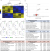
Fig 5. BBP exposure leads to differential gene expression.
(A) Heatmap of 344 differentially expressed genes identified by RNA-seq from whole worm lysates from DMSO- or BBP-exposed worms (four independent biological repeats). (B) Volcano plot highlighting the top 8 differentially expressed genes by effect size in BBP exposed worms compared to DMSO control. (C) Top 8 differentially expressed genes by effect size. Blue represents DMSO-exposed worms. Green represents BBP-exposed worms. Symbols indicate the batch or biological repeat. (D) Gene ontology enrichment analysis for biological processes in the upregulated gene population (top) or downregulated gene population (bottom). †External encapsulating structure organization; ‡negative regulation of muscle cell differentiation; §deadenylation-independent decapping of nuclear-transcribed mRNA; ~nuclear-transcribed mRNA catabolic process; ~~positive regulation of meiotic cell cycle process. (E) GO enrichment analysis for molecular functions in the upregulated gene population (left) or downregulated gene population (right). *Extracellular matrix structural constituent conferring tensile strength; **oxidoreduction-driven active transmembrane transporter activity; ***oxidoreductase activity, acting on paired donors, with incorporation or reduction of molecular oxygen, reduced flavin or flavoprotein as one donor, and incorporation of one atom of oxygen; #cyclin-dependent protein serine/threonine kinase regulator activity. FE, fold enrichment; FDR, false discovery rate.
Fig 6
Fig 6. BBP induces oxidative stress within the germline.
(A) Dissected gonads of gst-4p::gfp;col-121 worms exposed to DMSO, BBP, M9, or Paraquat. Dashed line used to outline the dissected gonads. (B) Quantification of the mean GFP intensity (a.f.u., arbitrary fluorescence units) measured in the gonads of gst-4p::gfp;col-121 exposed to DMSO, BBP, M9, or Paraquat. Error bars represent SEM. *P = 0.0392, ***P = 0.0003 by the two-tailed Mann-Whitney test, 95% C.I. N = 20–26 gonads per condition. Four biological repeats.

Similar articles

References

    1. Ford HB, Schust DJ. Recurrent pregnancy loss: etiology, diagnosis, and therapy. Rev Obstet Gynecol. 2009. Spring;2(2):76–83. - PMC - PubMed
    1. Hassold T, Abruzzo M, Adkins K, Griffin D, Merrill M, Millie E, et al.. Human aneuploidy: incidence, origin, and etiology. Environ Mol Mutagen. 1996;28(3):167–75. doi: 10.1002/(SICI)1098-2280(1996)28:3&lt;167::AID-EM2&gt;3.0.CO;2-B - DOI - PubMed
    1. Fujimoto VY, Kim D, vom Saal FS, Lamb JD, Taylor JA, Bloom MS. Serum unconjugated bisphenol A concentrations in women may adversely influence oocyte quality during in vitro fertilization. Fertil Steril. 2011. Apr;95(5):1816–9. doi: 10.1016/j.fertnstert.2010.11.008 - DOI - PubMed
    1. Zhou C, Gao L, Flaws JA. Exposure to an environmentally relevant phthalate mixture causes transgenerational effects on female reproduction in mice. Endocrinology. 2017. 158(6):1739–54. doi: 10.1210/en.2017-00100 - DOI - PMC - PubMed
    1. Grindler NM, Vanderlinden L, Karthikraj R, Kannan K, Teal S, Polotsky AJ, et al.. Exposure to phthalate, an endocrine disrupting chemical, alters the first trimester placental methylome and transcriptome in women. Sci Rep. 2018. Apr 17;8(1):6086. doi: 10.1038/s41598-018-24505-w - DOI - PMC - PubMed

MeSH terms