. 2024 Oct 24;19(10):e0312343.
doi: 10.1371/journal.pone.0312343. eCollection 2024.

Design of multi-modal antenna arrays for microwave hyperthermia and 1H/1⁹F MRI monitoring of drug release

Affiliations

Design of multi-modal antenna arrays for microwave hyperthermia and 1H/1⁹F MRI monitoring of drug release

Daniel Hernandez et al. PLoS One. .

Abstract

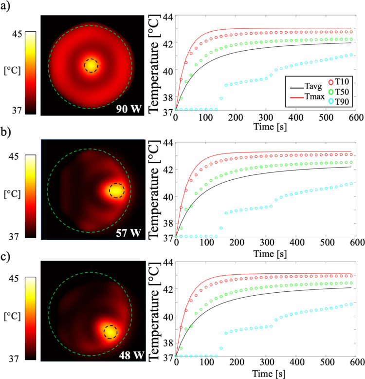
This simulation-based study presented a novel hybrid RF antenna array designed for neck cancer treatment within a 7T MRI system. The proposed design aimed to provide microwave hyperthermia to release 19F-labeled anticancer drugs from thermosensitive liposomes, facilitating drug concentration monitoring through 19F imaging and enabling 1H anatomical imaging and MR thermometry for temperature control. The design featured a bidirectional microstrip for generating the magnetic |B1|-fields required for 1H and 19F MR imaging, along with a patch antenna for localized RF heating. The bidirectional microstrip was operated at 300 MHz and 280 MHz through the placement of excitation ports at the ends of the antenna and an asymmetric structure along the antenna. Additionally, a patch antenna was positioned at the center. Based on this setup, an array of six antennas was designed. Simulation results using a tissue-mimicking simulation model confirmed the intensity and uniformity of |B1|-fields for both 19F and 1H nuclei, demonstrating the suitability of the design for clinical imaging. RF heating from the patch antennas was effectively localized at the center of the cancer model. In simulations with a human model, average |B1|-fields were 0.21 μT for 19F and 0.12 μT for 1H, with normalized-absolute-average-deviation values of 81.75% and 87.74%, respectively. Hyperthermia treatment was applied at 120 W for 600 s, achieving an average temperature of 40.22°C in the cancer model with a perfusion rate of 1 ml/min/kg. This study demonstrated the potential of a hybrid antenna array for integrating 1H MR, 19F drug monitoring, and hyperthermia.


Conflict of interest statement

The authors have declared that no competing interests exist.

Figures

Fig 1
Fig 1. A planar microstrip was used for demonstration purposes.
This bidirectional microstrip features dual-port excitations and a wing line (yellow) with variable positions along the microstrip. To create an asymmetric conductor line, the wing line is shifted to different positions with a step size of ΔZ = 20 mm.
Fig 2
Fig 2
The geometry of a single hybrid antenna with the bidirectional microstrip surface coil and patch antenna in a) the top view showing the excitation ports and the asymmetric wing lines, and b) the lateral view showing the dimensions of the dielectric materials.
Fig 3
Fig 3. The hybrid antenna array consists of six elements, along with the water pad, muscle, and cancer simulation models.
Different locations of the muscle and cancer simulation models were tested: a) in the central position, b) with the muscle simulation model shifted 60 mm to the left, and c) shifted 42.5 mm to the left and upward.
Fig 4
Fig 4
a) The human head and neck model with the cancer tissue shown as a pink sphere. b) The positioning of the array applicator around the neck. c) The X-Y view showing the position of the cancer inside the neck model.
Fig 5
Fig 5
The plots illustrate the following relationships: a) The change in impedance versus frequency as a function of the wing position. b) The resonance frequency as a function of the wing distance, viewed from ports 1 and 2. c) The frequency difference between ports 1 and 2 at each wing position. d) The |B1|-field produced by the microstrip when the wing is positioned 40 mm from port 1, showing the field at 344 MHz. e) The |B1|-field produced by the microstrip when the wing is positioned 40 mm from port 1, showing the field at 325 MHz at port 2.
Fig 6
Fig 6
a) Analysis of the relationship between the length of W and the resonance frequency. b) Computed S11 parameter after tuning and matching for the single module, combining the bidirectional microstrip operating at 280 MHz and 300 MHz, and the patch antenna operating at 550 MHz.
Fig 7
Fig 7
The plots show the scattering parameters S1j describing the coupling between each element, for the ports operating at the following frequencies: a) 280 MHz, b) 300 MHz, and c) 550 MHz.
Fig 8
Fig 8
The magnetic |B1|-field was computed with the array and simulation model for a) 19F at 280 MHz, and b) 1H at 300 MHz. c) The |E|-field at 550 MHz was calculated.
Fig 9
Fig 9
Temperature maps in the simulation model when placed at different positions: a) at the center, b) shifted to the left, and c) shifted to the upper left. Plots show the Tmax, T10, T50, and T90 values within the cancer model.
Fig 10
Fig 10
The computed |B1|-field for a) 19F at 280 MHz, b) 1H at 300 MHz, and c) electric field at 550 MHz on the Z-Y and X-Y planes within a human model.
Fig 11
Fig 11
Temperature maps for perfusion rates of 1 ml/min/kg at 120 W (a) and 3 ml/min/kg at 225 W (c). The corresponding temperature profiles within the cancer model are shown in (b) and (d). The dashed black circles indicate the cancer model.
Fig 12
Fig 12
SAR maps on the head and neck with the proposed antenna for each frequency: a) 280 MHz, b) 300 MHz, and c) 550 MHz.

Similar articles

Cited by

References

    1. Barsouk A., Aluru J.S., Rawla P., Saginala K., Barsouk A. “Epidemiology, risk factors, and prevention of head and neck squamous cell carcinoma.” Medical Sciences. 2023. Jun 13;11(2):42. - PMC - PubMed
    1. Magin R.L., Niesman M.R., “Temperature-Dependent Drug Release from Large Unilamellar Liposomes,” Cancer Drug Delivery. vol. 1, issue 2, pp.109–117, 1984. doi: 10.1089/cdd.1984.1.109 - DOI - PubMed
    1. Needham D., Dewhirst M.W. “The development and testing of a new temperature-sensitive drug delivery system for the treatment of solid tumors,” Advanced Drug Delivery Reviews. vol. 53, issue 3, pp.285–305, 2001. doi: 10.1016/s0169-409x(01)00233-2 - DOI - PubMed
    1. Ponce A.M. et al.., “Magnetic Resonance Imaging of Temperature-Sensitive Liposome Release: Drug Dose Painting and Antitumor Effects,” Journal of the National Cancer Institute. vol. 99, issue 1, pp.53–63, 2007. doi: 10.1093/jnci/djk005 - DOI - PubMed
    1. Dromi S. et al.., “Pulsed-High Intensity Focused Ultrasound and Low Temperature–Sensitive Liposomes for Enhanced Targeted Drug Delivery and Antitumor Effect,” Clinical Cancer Research, vol. 13, issue 9, pp. 2722–2727, 2007. doi: 10.1158/1078-0432.CCR-06-2443 - DOI - PMC - PubMed