. 2024 Oct 24;20(10):e1012006.
doi: 10.1371/journal.ppat.1012006. eCollection 2024 Oct.

Caspase-1 in Cx3cr1-expressing cells drives an IL-18-dependent T cell response that promotes parasite control during acute Toxoplasma gondii infection

Affiliations

Caspase-1 in Cx3cr1-expressing cells drives an IL-18-dependent T cell response that promotes parasite control during acute Toxoplasma gondii infection

Isaac W Babcock et al. PLoS Pathog. .

Abstract

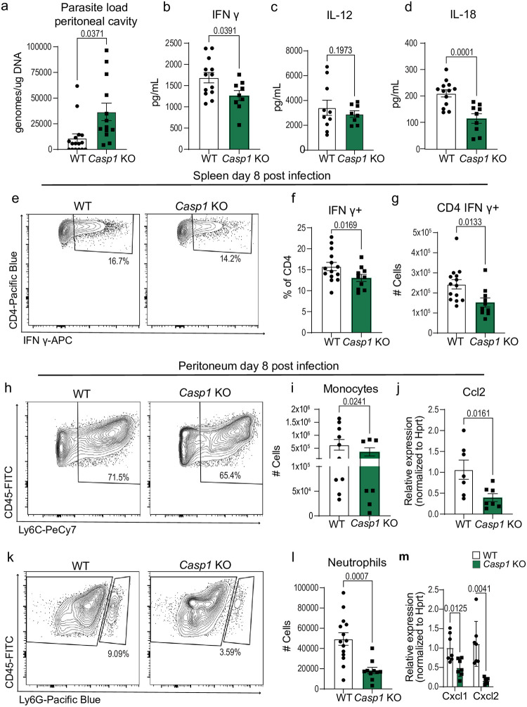
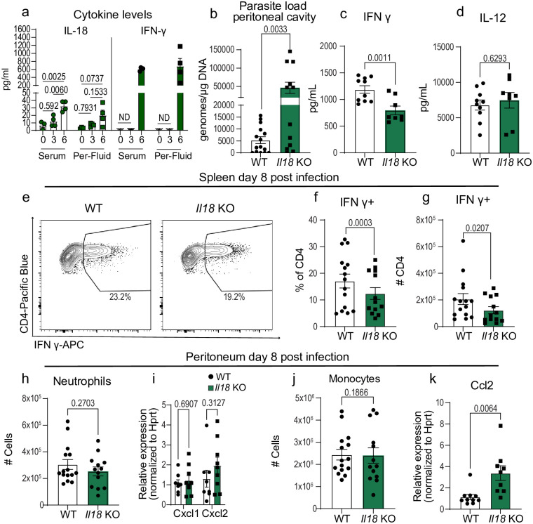
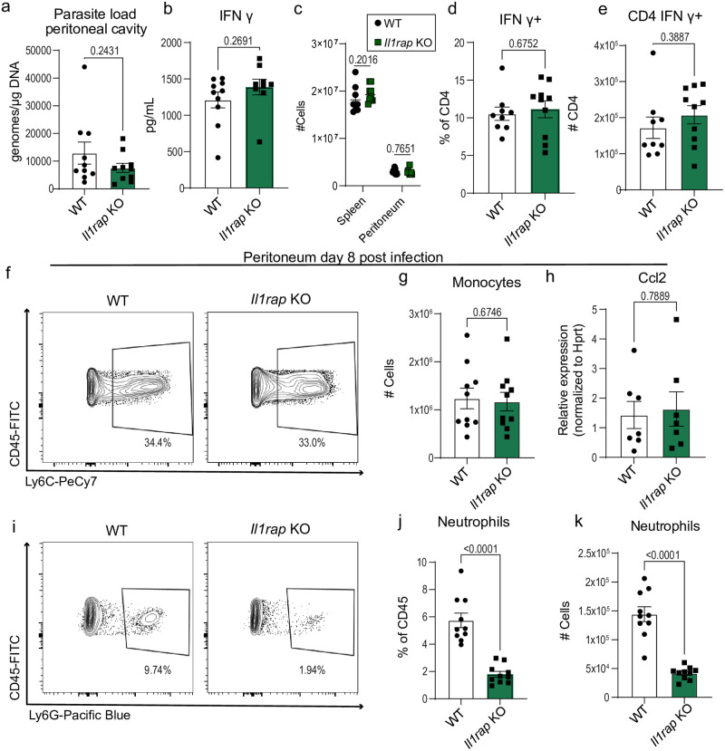
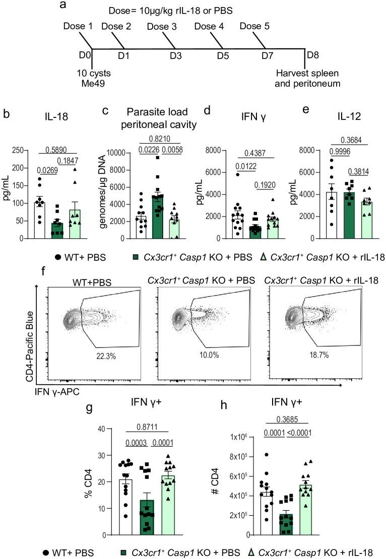
Inflammasome activation is a robust innate immune mechanism that promotes inflammatory responses through the release of alarmins and leaderless cytokines, including IL-1α, IL-1β, and IL-18. Various stimuli, including infectious agents and cellular stress, cause inflammasomes to assemble and activate caspase-1. Then, caspase-1 cleaves targets that lead to pore formation and leaderless cytokine activation and release. Toxoplasma gondii has been shown to promote inflammasome formation, but the cell types utilizing caspase-1 and the downstream effects on immunological outcomes during acute in vivo infection have not been explored. Here, using knockout mice, we examine the role of caspase-1 responses during acute T. gondii infection globally and in Cx3cr1-positive populations. We provide in vivo evidence that caspase-1 expression is critical for, IL-18 release, optimal interferon-γ (IFN-γ) production, monocyte and neutrophil recruitment to the site of infection, and parasite control. Specifically, we find that caspase-1 expression in Cx3cr1-positive cells drives IL-18 release, which potentiates CD4+ T cell IFN-γ production and parasite control. Notably, our Cx3cr1-Casp1 knockouts exhibited a selective T cell defect, mirroring the phenotype observed in Il18 knockouts. In further support of this finding, treatment of Cx3cr1-Casp1 knockout mice with recombinant IL-18 restored CD4+ T cell IFN-γ responses and parasite control. Additionally, we show that neutrophil recruitment is dependent on IL-1 receptor accessory protein (IL-1RAP) signaling but is dispensable for parasite control. Overall, these experiments highlight the multifaceted role of caspase-1 in multiple cell populations contributing to specific pathways that collectively contribute to caspase-1 dependent immunity to T. gondii.


Conflict of interest statement

The authors have declared that no competing interests exist.

Figures

Fig 1
Fig 1. Caspase-1 mediates innate and adaptive immune responses to T. gondii.
(a) qPCR analysis of T. gondii parasite load 8 days post-infection (8 dpi) in the peritoneum of wildtype WT C57BL/6 (n = 16) and Casp1 Knockout (Casp1 KO) (n = 13) mice, four experiments. (b-d) Serum cytokine levels 8 dpi, three experiments for (b) IFN-γ: WT (n = 13), Casp1 KO (n = 9); (c) IL-12: WT (n = 10), Casp1 KO (n = 8); and (d) IL-18: WT (n = 13), Casp1 KO (n = 9). (e-g) Flow cytometry of CD3+ CD4+ IFN-γ+ T cells in spleen at 8 dpi, three experiments, WT (n = 14), Casp1 KO (n = 10). (e) Representative flow cytometry plot. (f) Frequency of CD4+ T cells IFN-γ+. (g) Number of CD4+ T cells producing IFN-γ. (h) Representative flow cytometry plots of CD45+ CD11b+ Ly6C+ monocytes in the peritoneum 8dpi. (i) Number of CD45+ CD11b+ Ly6C+ monocytes in WT (n = 10), Casp1 KO (n = 7), two experiments. (j) RT-qPCR analysis of peritoneal Ccl2 expression in WT (n = 7) and Casp1 KO (n = 7), two experiments. (k) Representative flow cytometry plot of CD45+ CD11b+ Ly6G+ neutrophils in the peritoneum at 8 dpi. (l) Number of CD45+ CD11b+ Ly6G+ neutrophils in WT (n = 15), Casp1 KO (n = 9), three experiments. (m) RT-qPCR analysis of peritoneal Cxcl1 and Cxcl2 expression in WT (n = 7) and Casp1 KO (n = 7), two experiments. Data are mean ± s.e.m., p values by randomized-block ANOVA and post-hoc Tukey test (a-d, f-g, i-j, and l-m).
Fig 2
Fig 2. IL-18 promotes CD4+ T cell IFN-γ production but is dispensable for myeloid cell recruitment to the initial site of infection.
(a) IL-18 and IFN-γ levels in serum and peritoneal fluid (Per-Fluid) at 0 dpi (n = 3), 3 dpi (n = 4), 6 dpi (n = 4). (b) qPCR analysis of T. gondii parasite load 8 days post-infection (8 dpi) in the peritoneum of wildtype (WT) C57BL/6 (n = 15) and Il18 KO (n = 12) mice, three experiments. (c,d) Serum cytokine levels 8 dpi, two experiments for (c) IFN-γ: WT (n = 10), Il18 KO (n = 8); (d) IL-12: WT (n = 10), Il18 KO (n = 8). (e-g) Flow cytometry of CD3+ CD4+ IFN-γ+ T cells in spleen at 8 dpi, three experiments, WT (n = 15), Il18 KO (n = 13). (e) Representative flow cytometry plot. (f) Frequency of CD4+ T cells IFN-γ+. (g) Number of CD4+ T cells producing IFN-γ. (h) Number of CD45+ CD11b+ Ly6G+ neutrophils in WT (n = 15), Il18 KO (n = 13), three experiments. (i) RT-qPCR analysis of peritoneal Cxcl1 and Cxcl2 expression in WT (n = 7) and Il-18 KO (n = 9), two experiments. (j) Number of CD45+ CD11b+ Ly6C+ monocytes in WT (n = 15), Il18 KO (n = 13), three experiments. (k) RT-qPCR analysis of peritoneal Ccl2 expression in WT (n = 10) and Il18 KO (n = 9), two experiments. Data are mean ± s.e.m., p values by one-way ANOVA with post-hoc Tukey test (a) or randomized-block ANOVA and post-hoc Tukey test (b-d and f-k).
Fig 3
Fig 3. IL-1RAP signaling mediates neutrophil recruitment to the sight of infection but is dispensable for parasite control during acute T. gondii infection.
(a) qPCR analysis of T. gondii parasite load 8 days post-infection (8 dpi) in the peritoneum of Il1rap KO and wildtype (WT) mice, C57BL/6 (n = 10) and Il1rap KO (n = 10) mice, two experiments. (b) Serum cytokine levels 8 dpi, two experiments for IFN-γ: WT (n = 10), Il1rap KO (n = 10). (c) Cell counts from spleen and peritoneum: WT (n = 9), Il1rap KO (n = 10). (d-e) Flow cytometry of CD3+ CD4+ IFN-γ+ T cells in spleen at 8 dpi, two experiments, WT (n = 9), Il1rap KO (n = 10). (d) Frequency of CD4+ T cells IFN-γ+. (e) Number of CD4+ T cells producing IFN-γ. (f) Representative flow cytometry plots of CD45+ CD11b+ Ly6C+ monocytes in the peritoneum at 8dpi. (g) Number of CD45+ CD11b+ Ly6C+ monocytes in WT (n = 10), Il1rap KO (n = 10), two experiments. (h) RT-qPCR analysis of peritoneal Ccl2 expression in WT (n = 7) and Il1rap KO (n = 7), two experiments. (i) Representative flow cytometry plot of CD45+ CD11b+ Ly6G+ neutrophils in the peritoneum at 8 dpi. (j) Frequency and (k) Number of CD45+ CD11b+ Ly6G+ neutrophils in WT (n = 10) and Il1rap KO (n = 10), two experiments. Data are mean ± s.e.m., p values by randomized-block ANOVA and post-hoc Tukey test (a-e, g-h, and j-k).
Fig 4
Fig 4. Caspase-1 in Cx3cr1-expressing cells is necessary for parasite control and optimal CD4+T cell production of IFN-γ.
(a) qPCR analysis of T. gondii parasite load 8 days post-infection (8 dpi) in the peritoneum of Cx3cr1+/+ Caspase 1fl/fl (WT) (n = 13) and Cx3cr1Cre/+ Caspase 1fl/fl (Cx3cr1+ Casp1 KO) (n = 10) mice, three experiments. (b-d) Serum cytokine levels 8 dpi, three experiments for (b) IFN-γ: WT (n = 13), Cx3cr1+ Casp1 KO (n = 8); (c) IL-12: WT (n = 12), Cx3cr1+ Casp1 KO (n = 8); and (d) IL-18: WT (n = 10), Cx3cr1+ Casp1 KO (n = 9). (e-g) Flow cytometry of CD3+ CD4+ IFN-γ+ T cells in spleen at 8 dpi, two experiments, WT (n = 8), Cx3cr1+ Casp1 KO (n = 9). (e) Representative flow cytometry plot. (f) Frequency of IFN-γ-producing CD4+ T cells (g) Number of CD4+ T cells producing IFN-γ. (h) Representative flow cytometry plots of CD45+ CD11b+ Ly6C+ monocytes in the peritoneum 8 dpi. (i) Number of CD45+ CD11b+ Ly6C+ monocytes in WT (n = 8) and Cx3cr1+ Casp1 KO (n = 8), two experiments. (j) RT-qPCR analysis of peritoneal Ccl2 expression in WT (n = 7) and Cx3cr1+ Casp1 KO (n = 10), two experiments. (k) Representative flow cytometry plot of CD45+ CD11b+ Ly6G+ neutrophils in the peritoneum at 8 dpi. (l) Number of CD45+ CD11b+ Ly6G+ neutrophils in WT (n = 8), Cx3cr1+ Casp1 KO (n = 9), two experiments. (m) RT-qPCR analysis of peritoneal Cxcl1 and Cxcl2 expression in WT (n = 6) and Cx3cr1+ Casp1 KO (n = 10), two experiments. Data are mean ± s.e.m. p values by randomized-block ANOVA and post-hoc Tukey test (a-d, f-g, i-j, and l-m).
Fig 5
Fig 5. Recombinant IL-18 administration rescues CD4+ T cell IFN-γ production and parasite control in Cx3cr1+ caspase-1 deficient mice.
(a) Schematic of experimental design. (b) Serum cytokine levels of IL-18, two experiments, WT + PBS (n = 8), Cx3cr1+ Casp1 KO + PBS (n = 8), and Cx3cr1+Casp1 KO + rIL-18 (n = 8). (c) qPCR analysis of parasite load in peritoneal cavity, three experiments, WT + PBS (n = 11), Cx3cr1+ Casp1 KO + PBS (n = 12), and Cx3cr1+ Casp1 KO + rIL-18 (n = 10). (d) Serum cytokine levels of IFN-γ, three experiments, WT + PBS (n = 13), Cx3cr1+ Casp1 KO + PBS (n = 12), and Cx3cr1+ Casp1 KO + rIL-18 (n = 12). (e) Serum cytokine levels of IL-12, two experiments, WT + PBS (n = 8), Cx3cr1+Casp1 KO + PBS (n = 8), and Cx3cr1+ Casp1 KO + rIL-18 (n = 8). (f-h) Flow cytometry of CD3+ CD4+ IFN-γ+ T cells in spleen at 8 dpi, three experiments, WT + PBS (n = 13), Cx3cr1+ Casp1 KO + PBS (n = 12), and Cx3cr1+ Casp1 KO + rIL-18 (n = 12). (f) Representative flow cytometry plot. (g) Frequency of CD4+ T cells IFN-γ+. (h) Number of CD4+ T cells producing IFN-γ. Data are presented as mean ± s.e.m., p values by randomized-block ANOVA and post-hoc Tukey test (b-e, g-h).

References

    1. Takeuchi O, Akira S. Pattern Recognition Receptors and Inflammation. Cell. 2010;140(6):805–20. doi: 10.1016/j.cell.2010.01.022 - DOI - PubMed
    1. Kanneganti TD, Lamkanfi M, Núñez G. Intracellular NOD-like receptors in host defense and disease. Immunity. 2007;27(4):549–59. Epub 2007/10/31. doi: 10.1016/j.immuni.2007.10.002 . - DOI - PubMed
    1. Kesavardhana S, Malireddi RKS, Kanneganti TD. Caspases in Cell Death, Inflammation, and Pyroptosis. Annu Rev Immunol. 2020;38:567–95. Epub 2020/02/06. doi: 10.1146/annurev-immunol-073119-095439 - DOI - PMC - PubMed
    1. Suzuki Y, Orellana MA, Schreiber RD, Remington JS. Interferon-gamma: the major mediator of resistance against Toxoplasma gondii. Science. 1988;240(4851):516–8. Epub 1988/04/22. doi: 10.1126/science.3128869 . - DOI - PubMed
    1. Scharton-Kersten T, Contursi C, Masumi A, Sher A, Ozato K. Interferon consensus sequence binding protein-deficient mice display impaired resistance to intracellular infection due to a primary defect in interleukin 12 p40 induction. J Exp Med. 1997;186(9):1523–34. Epub 1997/11/14. doi: 10.1084/jem.186.9.1523 - DOI - PMC - PubMed

LinkOut - more resources