

Prevalence and Clinical Implications of Hemosiderin Deposits in Recent Small Subcortical Infarcts
- PMID: 39447100
- PMCID: PMC11510007
- DOI: 10.1212/WNL.0000000000209973
Prevalence and Clinical Implications of Hemosiderin Deposits in Recent Small Subcortical Infarcts
Abstract
Background and objectives: A quarter of ischemic strokes are of lacunar clinical subtype and have an underlying recent small subcortical infarct (RSSI), but their long-term outcomes remain poorly characterized. Hemosiderin deposits (HDs) have been noted in RSSIs at chronic stages and might mimic primary hemorrhage. We characterized HDs' morphology, frequency, and clinical relevance.
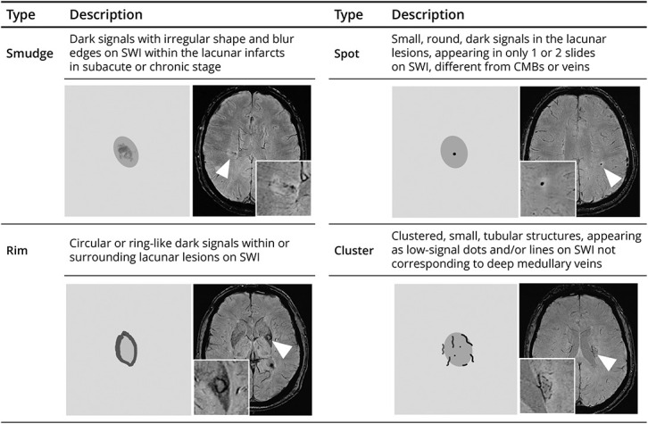
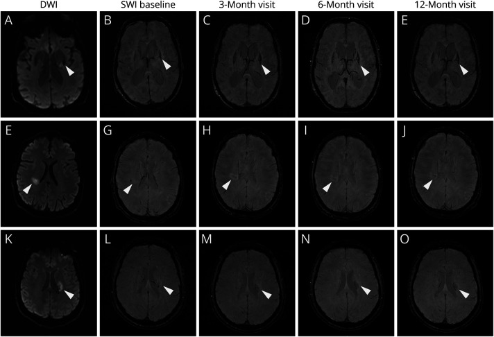
Methods: Participants with RSSIs were identified from a prospective longitudinal study and evaluated on 3T MRI including susceptibility-weighted imaging (SWI) from stroke diagnosis to 12 months. We categorized HDs in RSSIs on SWI at all available time points into 4 types (spots, smudge, rim, cluster) and assessed their associations with demographic factors, stroke-related factors, and image markers with adjusted logistic regression.
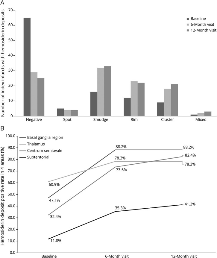
Results: HDs were observed in 43 (55.0%) of 108 participants within 3 months and 83 (76.9%) of 108 within 12 months after stroke onset. The mean time to first detection of HDs was 87 (interquartile range 53-164) days. A "rim" pattern (similar to late appearance of primary hemorrhage) occurred in at least 26.5% of RSSIs at all follow-up time points, mainly those located in the lentiform/internal capsule (50.0%) or thalamus (36.4%). Infarct volume (odds ratio [OR] 1.003, 95% CI 1.001-1.006; p = 0.004) and the total small vessel disease (SVD) score at baseline (OR 2.50, 95% CI 1.28-4.86, p = 0.007) independently predicted HDs at 12 months. HDs were positively associated with more lacunes (OR 1.60, 95% CI 1.13-2.26, p < 0.01), but not the Fazekas score, number of microbleeds, basal ganglia mineral deposit score, or clinical outcomes.
Discussion: HDs occur commonly in RSSIs and may be associated with infarct volume and SVD score. Hemosiderin "rim" is common in RSSIs, urging caution to avoid mistaking ischemic RSSI for primary hemorrhage in subacute and chronic stages.
Conflict of interest statement
The authors report no relevant disclosures. Go to
Figures
References
MeSH terms
Substances
LinkOut - more resources
Full Text Sources
Medical