. 2025 Jan 1;328(1):F1-F14.
doi: 10.1152/ajprenal.00177.2024. Epub 2024 Oct 24.

Sex differences in the adrenal circadian clock: a role for BMAL1 in the regulation of urinary aldosterone excretion and renal electrolyte balance in mice

Affiliations

Sex differences in the adrenal circadian clock: a role for BMAL1 in the regulation of urinary aldosterone excretion and renal electrolyte balance in mice

Hannah M Costello et al. Am J Physiol Renal Physiol. .

Abstract

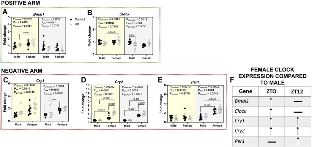
Brain and muscle ARNT-Like 1 (BMAL1) is a circadian clock transcription factor that regulates physiological functions. Male adrenal-specific Bmal1 (ASCre/+::Bmal1) KO mice displayed blunted serum corticosterone rhythms, altered blood pressure rhythm, and altered timing of eating, but there is a lack of knowledge in females. This study investigates the role of adrenal BMAL1 in renal electrolyte handling and urinary aldosterone levels in response to low salt in male and female mice. Mice were placed in metabolic cages to measure 12-h urinary aldosterone after a standard diet and 7 days low-salt diet, as well as daily body weight, 12-h food and water intake, and renal sodium and potassium balance. Adrenal glands and kidneys were collected at ZT0 or ZT12 to measure the expression of aldosterone synthesis genes and clock genes. Compared with littermate controls, ASCre/+::Bmal1 KO male and female mice displayed increased urinary aldosterone in response to a low-salt diet, although mRNA expression of aldosterone synthesis genes was decreased. Timing of food intake was altered in ASCre/+::Bmal1 KO male and female mice, with a blunted night/day ratio. ASCre/+::Bmal1 KO female mice displayed decreases in renal sodium excretion in response to low salt, but both male and female KO mice had changes in sodium balance that were time-of-day-dependent. In addition, sex differences were found in adrenal and kidney clock gene expression. Notably, this study highlights sex differences in clock gene expression that could contribute to sex differences in physiological functions.NEW & NOTEWORTHY Our findings highlight the importance of sex as well as time-of-day in understanding the role of the circadian clock in the regulation of homeostasis. Time-of-day is a key biological variable that is often ignored in research, particularly in preclinical rodent studies. Our findings demonstrate important differences in several measures at 6 AM compared with 6 PM. Consideration of time-of-day is critical for the translation of findings in nocturnal rodent physiology to diurnal human physiology.

Keywords: adrenal gland; brain and muscle ARNT-like 1; circadian rhythms; kidney; sex differences.


Figures

Figure 1.
Figure 1.. Increased urinary aldosterone in ASCre/+::Bmal1 KO male and female mice in response to a low salt diet.
Twelve-hour urinary aldosterone between (A) ZT0 and ZT12 (inactive period, sun) and between (B) ZT12 and ZT0 (active period, moon) in male and female control (closed black circles, n=5–9) and ASCre/+::Bmal1 KO (open grey circles, n=7–9) mice following standard diet. Urinary aldosterone during the inactive (C) and active (D) periods following the acute response to a low salt diet (1 day) in male and female control and KO mice. Urinary aldosterone during the inactive (E) and active (F) periods following a more chronic response to a low salt diet (7 days) in male and female control and KO mice. Data presented as mean±SD. Genotype, sex, and interaction effects were determined by 2-way ANOVA with significant Šidák post hoc comparisons shown.
Figure 2.
Figure 2.. Male and female ASCre/+::Bmal1 KO mice exhibit altered timing of eating behavior.
Averaged 12-hour food intake between (A) ZT0 and ZT12 (inactive period, sun) and between (B) ZT12 and ZT0 (active period, moon), (C) night/day food intake ratio, and (D) 24-hour food intake in male and female control (closed black circles, n=5–9) and ASCre/+::Bmal1 KO (open grey circles, n=7–9) mice following a standard diet. Averaged 12-hour food intake during inactive (E) and active (F) period, (G) night/day food intake ratio, and (H) 24-hour food intake in male and female control and KO mice following a low salt diet. Data presented as mean±SD. Genotype, sex and interaction effects were determined by 2-way ANOVA with significant Šidák post hoc comparisons shown.
Figure 3.
Figure 3.. Sex and genotype differences in renal sodium handling in response to a low salt diet.
Averaged 12-hour Na excretion, Na excretion normalized to body weight (bw) and Na balance between ZT0 and ZT12 (inactive period, sun) and between ZT12 and ZT0 (active period, moon) in male and female control (closed black circles, n=5–9) and ASCre/+::Bmal1 KO (open grey circles, n=7–9) mice following a standard diet (Na excretion A,B; Na excretion normalized to bw C,D; Na balance E,F) or 7 days low salt diet (Na excretion G,H; Na excretion normalized to bw I,J; Na balance K,L). Data presented as mean±SD. Genotype, sex and interaction effects were determined by 2-way ANOVA with significant Šidák post hoc comparisons shown.
Figure 4.
Figure 4.. Differences in renal sodium handling in the response to a low salt diet during steady state in male and female ASCre/+::Bmal1 KO mice.
Averaged 12-hour sodium (Na) excretion, Na excretion normalized to body weight (bw) and estimated Na balance between ZT0 and ZT12 (inactive period, sun) and between ZT12 and ZT0 (active period, moon) over days 4–7 (steady state) of a low salt diet in male and female control (closed black circles, n=5–9) and ASCre/+::Bmal1 KO (open grey circles, n=7–9) mice (Na excretion A,B; Na excretion normalized to bw C,D; estimated Na balance E,F). Data presented as mean±SD. Genotype, sex and interaction effects were determined by 2-way ANOVA with significant Šidák post hoc comparisons shown.
Figure 5.
Figure 5.. Sex differences in potassium balance in response to a low salt diet.
Averaged 12-hour K excretion, K excretion normalized to body weight (bw) and estimated K balance between ZT0 and ZT12 (inactive period, sun) and between ZT12 and ZT0 (active period, moon) in male and female control (closed black circles, n=5–9) and ASCre/+::Bmal1 KO (open grey circles, n=7–9) mice following a standard diet (K excretion A,B; K excretion normalized to bw C,D; estimated K balance E,F) or 7 days low salt diet (K excretion G,H; K excretion normalized to bw I,J; estimated K balance K,L). Data presented as mean±SD. Genotype, sex and interaction effects were determined by 2-way ANOVA with significant Šidák post hoc comparisons shown.
Figure 6.
Figure 6.. Decreased adrenal expression of aldosterone synthesis genes in male and female ASCre/+::Bmal1 KO mice.
Adrenal glands were collected from ASCre/+::Bmal1 KO male and female mice with littermate controls at ZT0 (inactive period; day; yellow shading) or ZT12 (active period; night; grey shading) following either a standard or low salt diet. RNA was isolated and converted to cDNA from one adrenal gland and relative mRNA expression of steroidogenic acute regulatory protein (StAR) and aldosterone synthase (CYP11B2), which are involved in aldosterone synthesis (A), were measured using a TaqMan assay with Actb as the reference gene. Data expressed as fold change relative to control male mice. Relative gene expression of Star (B) and Cyp11b2 (C) at ZT0 and ZT12 in male and female control (closed black circles, n=8) and KO (open grey circles, n=7–8) mice following a standard diet. Relative gene expression of Star (D) and Cyp11b2 (E) at ZT0 and ZT12 in male and female control and KO mice following a low salt diet. Data presented as mean±SD. Genotype, sex and interaction effects for each time point were determined by 2-way ANOVA with significant Šidák post hoc comparisons shown.
Figure 7.
Figure 7.. Time-of-day-dependent sex differences in adrenal clock gene expression.
Adrenal glands were collected from ASCre/+::Bmal1 KO male and female mice with littermate controls at ZT0 (inactive period; day; yellow shading) or ZT12 (active period; night; grey shading) following a standard diet. RNA was isolated and converted to cDNA from one adrenal gland and relative mRNA expression was measured using qPCR for Clock (A; part of positive arm of the clock), Cry1, Cry2, Per1 (B-D; part of the negative arm of the clock), Dbp, Rev-erba and Rora (E-G; part of the regulatory loops of the clock) with Actb as the reference gene in male and female control (closed black circles, n=7–8) and KO (open grey circles, n=7–8) mice. Data expressed as fold change relative to control male mice. Data presented as mean±SD. Genotype, sex and interaction effects were determined by 2-way ANOVA with significant Šidák post hoc comparisons shown. Panel H illustrates table of summarized findings of female clock expression to male clock expression.
Figure 8.
Figure 8.. Evidence of adrenal-kidney clock crosstalk.
Kidneys were collected from ASCre/+::Bmal1 KO male and female mice with littermate controls at ZT0 (inactive period; day; yellow shading) or ZT12 (active period; night; grey shading) following a standard diet. Kidney medulla was dissected and RNA was isolated and converted to cDNA and relative mRNA expression was measured using qPCR for Bmal1, Clock (A and B; part of positive arm of the clock), Cry1, Cry2, and Per1 (C-E; part of the negative arm of the clock) with Actb as the reference gene in male and female control (closed black circles, n=7–8) and KO (open grey circles, n=7–8) mice. Data expressed as fold change relative to control male mice. Data presented as mean±SD. Genotype, sex and interaction effects were determined by 2-way ANOVA with significant Šidák post hoc comparisons shown. Panel F illustrates table of summarized findings of female clock expression to male clock expression.

References

    1. Riede SJ, van der Vinne V, Hut RA. The flexible clock: predictive and reactive homeostasis, energy balance and the circadian regulation of sleep–wake timing. Journal of Experimental Biology 220: 738–749, 2017. doi: 10.1242/jeb.130757. - DOI - PubMed
    1. Cox KH, Takahashi JS. Circadian clock genes and the transcriptional architecture of the clock mechanism. J Mol Endocrinol 63: R93–R102, 2019. doi: 10.1530/JME-19-0153. - DOI - PMC - PubMed
    1. Zhang R, Lahens NF, Ballance HI, Hughes ME, Hogenesch JB. A circadian gene expression atlas in mammals: implications for biology and medicine. Prot Natl Acad Sci USA 111: 16219–16224, 2014. doi: 10.1073/PNAS.1408886111. - DOI - PMC - PubMed
    1. Partch CL, Green CB, Takahashi JS. Molecular architecture of the mammalian circadian clock. Trends Cell Biol 24: 90–99, 2014. - PMC - PubMed
    1. Costello HM, Sharma RK, McKee AR, Gumz ML. Circadian Disruption and the Molecular Clock in Atherosclerosis and Hypertension. . - PMC - PubMed

MeSH terms

LinkOut - more resources