. 2024 Nov 27;187(24):6966-6980.e23.
doi: 10.1016/j.cell.2024.09.043. Epub 2024 Oct 23.

Adaptive multi-epitope targeting and avidity-enhanced nanobody platform for ultrapotent, durable antiviral therapy

Affiliations

Adaptive multi-epitope targeting and avidity-enhanced nanobody platform for ultrapotent, durable antiviral therapy

Yufei Xiang et al. Cell. .

Abstract

Pathogens constantly evolve and can develop mutations that evade host immunity and treatment. Addressing these escape mechanisms requires targeting evolutionarily conserved vulnerabilities, as mutations in these regions often impose fitness costs. We introduce adaptive multi-epitope targeting with enhanced avidity (AMETA), a modular and multivalent nanobody platform that conjugates potent bispecific nanobodies to a human immunoglobulin M (IgM) scaffold. AMETA can display 20+ nanobodies, enabling superior avidity binding to multiple conserved and neutralizing epitopes. By leveraging multi-epitope SARS-CoV-2 nanobodies and structure-guided design, AMETA constructs exponentially enhance antiviral potency, surpassing monomeric nanobodies by over a million-fold. These constructs demonstrate ultrapotent, broad, and durable efficacy against pathogenic sarbecoviruses, including Omicron sublineages, with robust preclinical results. Structural analysis through cryoelectron microscopy and modeling has uncovered multiple antiviral mechanisms within a single construct. At picomolar to nanomolar concentrations, AMETA efficiently induces inter-spike and inter-virus cross-linking, promoting spike post-fusion and striking viral disarmament. AMETA's modularity enables rapid, cost-effective production and adaptation to evolving pathogens.

Keywords: COVID; IgM antibody; SARS-CoV-2; antibody engineering; antiviral therapy; broadly neutralizing antibodies; cryotomography; multi-specific antibodies; nanobody; virus cross-linking.


Conflict of interest statement

Declaration of interests Y.X. and Y.S. are co-inventors on a provisional patent of AMETA technology filed by Icahn School of Medicine at Mount Sinai. The A.G.-S. laboratory has received research support from GSK, Pfizer, Senhwa Biosciences, Kenall Manufacturing, Blade Therapeutics, Avimex, Johnson & Johnson, Dynavax, 7Hills Pharma, Pharmamar, ImmunityBio, Accurius, Nanocomposix, Hexamer, N-fold LLC, Model Medicines, Atea Pharma, Applied Biological Laboratories, and Merck, outside of the reported work. A.G.-S. has consulting agreements for the following companies involving cash and/or stock: Castlevax, Amovir, Vivaldi Biosciences, Contrafect, 7Hills Pharma, Avimex, Pagoda, Accurius, Esperovax, Applied Biological Laboratories, Pharmamar, CureLab Oncology, CureLab Veterinary, Synairgen, Paratus, Pfizer, and Prosetta, outside of the reported work. A.G.-S. has been an invited speaker in meeting events organized by Seqirus, Janssen, Abbott, and AstraZeneca. A.G.-S. is an inventor of patents and patent applications on the use of antivirals and vaccines for the treatment and prevention of virus infections and cancer, owned by the Icahn School of Medicine at Mount Sinai, New York, outside of the reported work. Y.X. and Y.S. are co-inventors on a patent of Nbs filed by Univ. of Pittsburgh. Y.S. is a co-founder of Antenna Biotech Inc.

Figures
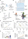
Figure 1.
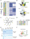
Figure 1.. A modular system to produce multivalent nanobodies with enhanced avidity.
(A) Schematics of conjugation and purification of Nb-IgM-Fc constructs. Nanobodies and recombinant IgM-Fc were incubated with a 30:1 molar ratio at 4°C overnight. Excessive nanobodies were removed through centrifugation using a 100 kDa molecular weight cutoff filter. Nb-IgM-Fc conjugates were efficiently recovered from the filter. (B) Denatured SDS and native protein gel analyses showing the complete conjugation and high purity of the conjugates. (C) Mass photometry analysis of a conjugated Nb-IgM-Fc. The total measurement mass is 731±143 kDa, including the predicted molecular weight of 708 kDa from amino acids and glycosylations on the IgM-Fc. (D) Summary of five major epitope classes targeted by RBD nanobodies, based on high-resolution cryo-EM structures. (E) Correlation analysis of neutralization potency (EC50s) of Nb-IgM-Fc and the corresponding Nb monomers against the pseudotyped SARS-CoV-2 Wuhan-Hu-1 (D614G) strain. Each dot represents an Nb. (F) Plots summary of EC50s of Nb monomers and their corresponding Nb-IgM-Fc against pseudotyped SARS-CoV-2 variants and SARS-CoV. Nanobodies targeting different epitope classes were plotted separately. (G) Summary of neutralization potencies (EC50s) of Nb-IgM-Fc conjugates against pseudotyped SARS-CoV-2, variants and SARS-CoV. Welch’s t-tests were used for comparison of EC50s of Nb-IgM-Fc targeting different epitopes. *: p < 0.05, ***: p < 0.001, ****: p < 0.0001. (H) A plot of average solvent-accessible surface areas (SASA) of four major epitopes of RBD Nbs during the transition of an RBD from the “close” to fully open state. SASA of individual Nb can be found in Figure S4. The transition was modeled by Molecular Dynamics (MD) simulation (Methods). The exposed area was presented as a function of the distance between the centroids of Subdomain 1 (SD1, amino acids 531–592) and the RBD (amino acids 336–518) across different protomers of the Spike protein. All calculations of accessible surface area were conducted using a 7-Å probe.
Figure 2.
Figure 2.. Engineering and production of AMETA constructs against sarbecoviruses.
(A) Structure-guided design of Nb dimers to improve cooperative binding and neutralization activities. Sequence conservation is based on 19 sarbecovirus RBD sequences (Methods). The fold improvement of a dimer over the corresponding monomers was based on their average EC50s against the SARS-CoV-2 Omicron variants and SARS-CoV (Table S4). (B) Models of the dimeric nanobodies on the SARS-CoV-2 spike glycoprotein. Upper panel: cooperative binding within a single RBD. Lower panel: cooperative binding between two RBDs. (C) Schematic presentation of a representative AMETA construct. (D) Design of four AMETA constructs and their potential coverage on the conserved RBD surface residues. (E) Denatured SDS and native protein gel analyses showing the complete conjugation and high purity of the AMETA constructs. (F) Mass photometry analysis of AMETA constructs.
Figure 3.
Figure 3.. In vitro neutralization activities of the AMETA constructs.
(A) Summary of neutralization potencies of AMETA against a panel of SARS-CoV-2 variants and SARS-CoV. (B)-(E) Radar plots comparing the neutralization potencies (EC50s) of four AMETA constructs and their corresponding single-epitope Nb-IgM-Fc. The highest tested concentration was 100 nM.
Figure 4.
Figure 4.. Biodistribution and in vivo efficacy of AMETA4 in mouse models.
(A) Intranasal administration of 89Zr-labeled AMETA4 (275.5 ± 43.5 mCi) in C57BL/6 mice (N=5) was followed by a 20-minute PET scan to monitor AMETA4’s distribution. Representative 3D reconstruction images are shown. (B) Mice were euthanized 72 hours post-injection, and tissues were analyzed for radioactivity. Gamma-counting determined the residual tracer, corrected for decay, and expressed as a percentage of the injected dose per gram of tissue (%ID/g). (C) Experimental design summary: mouse-adapted SARS-CoV-2 (1 × 10⁴ PFU) was intranasally administered to three groups of S/129 mice (N=8). AMETA4 (2.2 nmole/kg or 2 mg/kg) was delivered intranasally either 6 hours before (green squares) or 6 hours after (purple triangles) infection. A control group received isotype IgM (gray circles, 2.2 nmole/kg), and a group of uninfected animals served as additional controls. Daily monitoring of animal weight changes was conducted, and animals were euthanized for lung tissue viral titer analysis on day 3. (D) Body weight changes over time, expressed as a percentage. Significant differences are marked with *** (P < 0.001). (E) Viral titers in lung tissues at 3 days post-infection (d.p.i.), showing significant reductions in treated groups compared to controls. Significance levels are indicated as **** (P < 0.0001) and * (P < 0.05). (F) Lung pathology scores, comparing treated and control groups.
Figure 5.
Figure 5.. The architecture of an AMETA construct.
(A) Structure model based on single-particle cryo-EM illustrating the full architecture of an AMETA construct (AMETA3). Bi-epitope Nb dimers S36–182 (targeting epitope III and I, respectively), as well as 132–118 (targeting epitope IV and II, respectively), are distinguished by varying colors. Additional components such as the Cμ domains, J chain, IgΔNSpyCatcher003 domain, and peptide linkers are annotated. (B) Structural model of a single clamp of AMETA3 derived from a 50 ns full-atom Molecular Dynamics (MD) simulation, encompassing 100 frames. Models from all the frames were aligned to the initial conformations of Cμ3 and Cμ4 domains (amino acids 404–728 and 1159–1484) to generate the figure. The Nb sequences (S36, 182, 118, and 132) are colored in pink, green, yellow, and blue, respectively. (C) MD simulation displaying the root-mean-square fluctuation (RMSF) of AMETA3 at the amino acid level, highlighting the notable flexibility of the Nb dimers, IgΔNSpyCatcher003 and the linkers.
Figure 6.
Figure 6.. In situ cryo-electron tomography (cryo-ET) analysis of AMETA binding to authentic SARS-CoV-2.
Various concentrations of AMETA4 (10 pM - 1nM, A to C) or an isotype control of recombinant IgM-Fc protein (D) were incubated with the egressed SARS-CoV-2 (Victoria) in tissue culture before fixation and vitrification for subsequent cryo-ET analysis. The tomographic slices presented are 3.6 nm in thickness. Blue arrows indicate pre-fusion spikes, red arrows post-fusion spikes, purple arrows the AMETA4 densities crosslinking the spikes, and yellow arrows dense AMETA surrounding the viruses. Scale bar: 50 nm. (E) Segmentation of the slice (Figure A, lower panel), showing AMETA4 at a concentration of 10 pM. Pre-fusion spikes are in blue, and partially resolved AMETA densities are shown in purple. (F) Segmentation of slice (Figure B, lower panel) corresponding to AMETA4 at 100 pM concentration. The virus ribonucleoproteins (RNPs) are in beige. Unassigned densities in cyan imply either dissociated spike or AMETA4. (G) Quantification of the average number of identifiable spikes per viral particle under each condition (see details in Table S6). (H) Quantification of the percentage of spike confirmation (pre-/post-fusion) per identifiable spike under each condition (see details in Table S6).

References

    1. Blandino R, and Baumgarth N (2019). Secreted IgM: New tricks for an old molecule. J Leukoc Biol 106, 1021–1034. 10.1002/JLB.3RI0519-161R. - DOI - PMC - PubMed
    1. Boni MF, Lemey P, Jiang X, Lam TT, Perry BW, Castoe TA, Rambaut A, and Robertson DL (2020). Evolutionary origins of the SARS-CoV-2 sarbecovirus lineage responsible for the COVID-19 pandemic. Nat Microbiol 5, 1408–1417. 10.1038/s41564-020-0771-4. - DOI - PubMed
    1. Cao Y, Yisimayi A, Jian F, Song W, Xiao T, Wang L, Du S, Wang J, Li Q, Chen X, et al. (2022). BA.2.12.1, BA.4 and BA.5 escape antibodies elicited by Omicron infection. Nature 608, 593–602. 10.1038/s41586-022-04980-y. - DOI - PMC - PubMed
    1. Carabelli AM, Peacock TP, Thorne LG, Harvey WT, Hughes J, Consortium C-GU, Peacock SJ, Barclay WS, de Silva TI, Towers GJ, and Robertson DL (2023). SARS-CoV-2 variant biology: immune escape, transmission and fitness. Nat Rev Microbiol 21, 162–177. 10.1038/s41579-022-00841-7. - DOI - PMC - PubMed
    1. Case DA, Cheatham TE 3rd, Darden T, Gohlke H, Luo R, Merz KM Jr., Onufriev A, Simmerling C, Wang B, and Woods RJ (2005). The Amber biomolecular simulation programs. J Comput Chem 26, 1668–1688. 10.1002/jcc.20290. - DOI - PMC - PubMed

MeSH terms

LinkOut - more resources