. 2024 Nov:534:113768.
doi: 10.1016/j.jim.2024.113768. Epub 2024 Oct 22.

Human monoclonal antibody cloning and expression with overlap extension PCR and short DNA fragments

Affiliations

Human monoclonal antibody cloning and expression with overlap extension PCR and short DNA fragments

Zachary Ende et al. J Immunol Methods. 2024 Nov.

Abstract

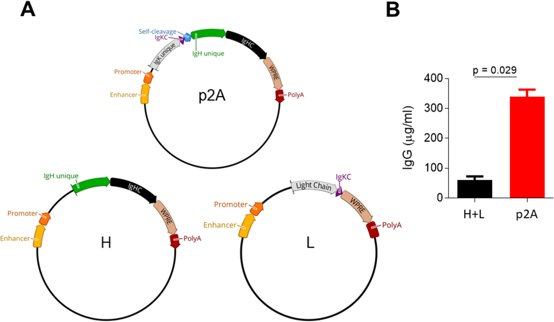
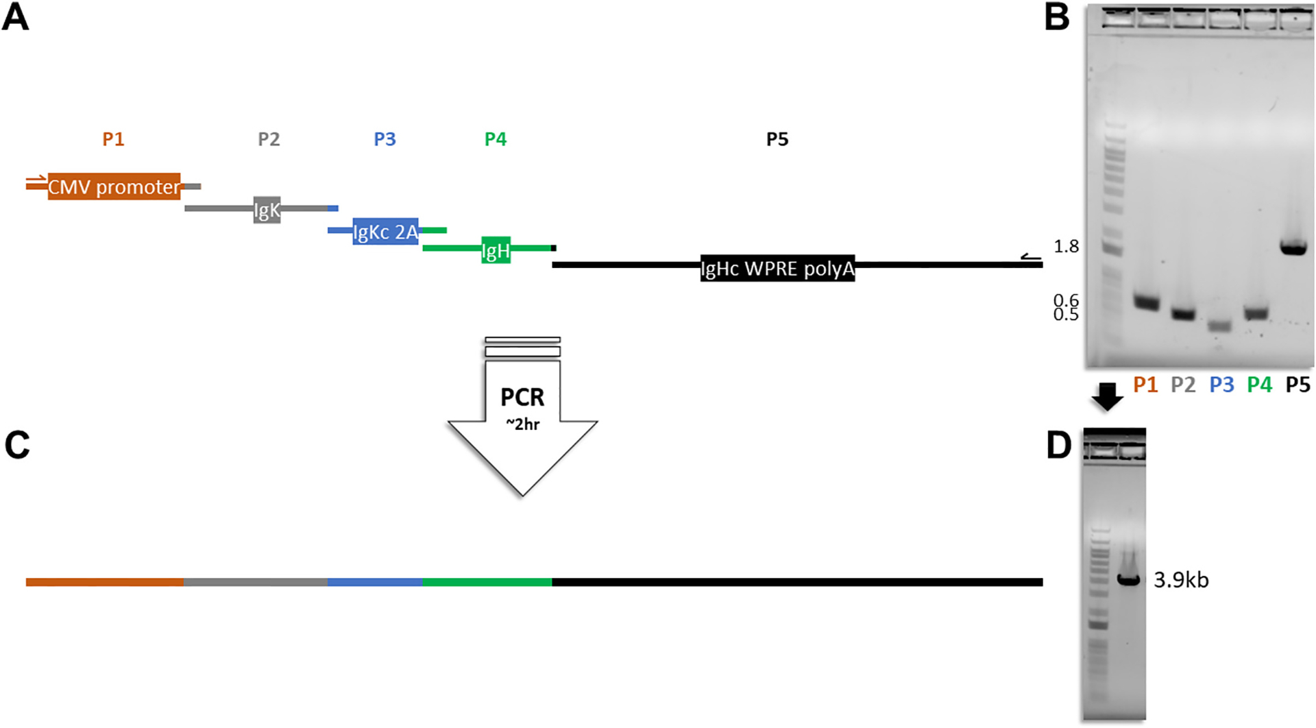
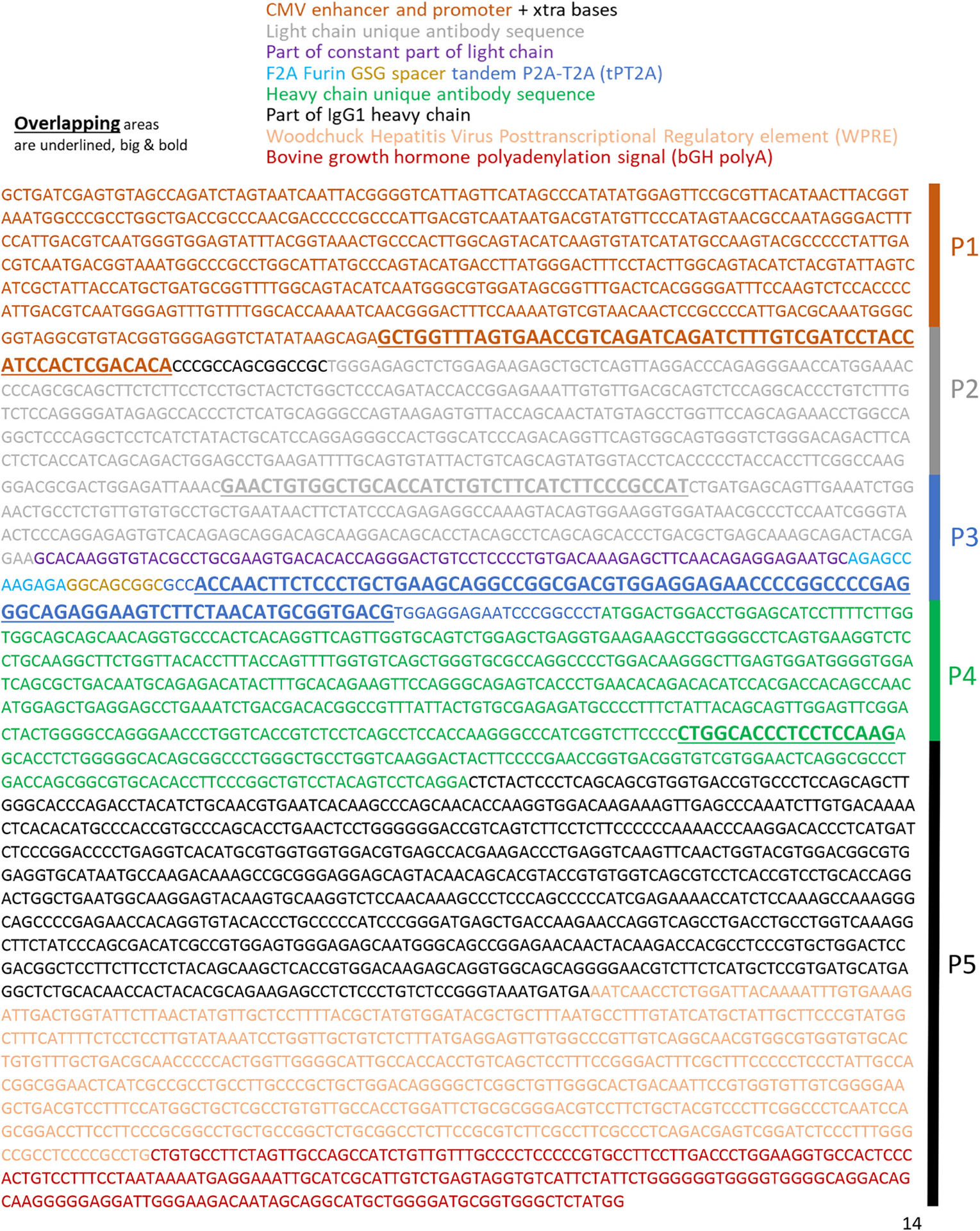
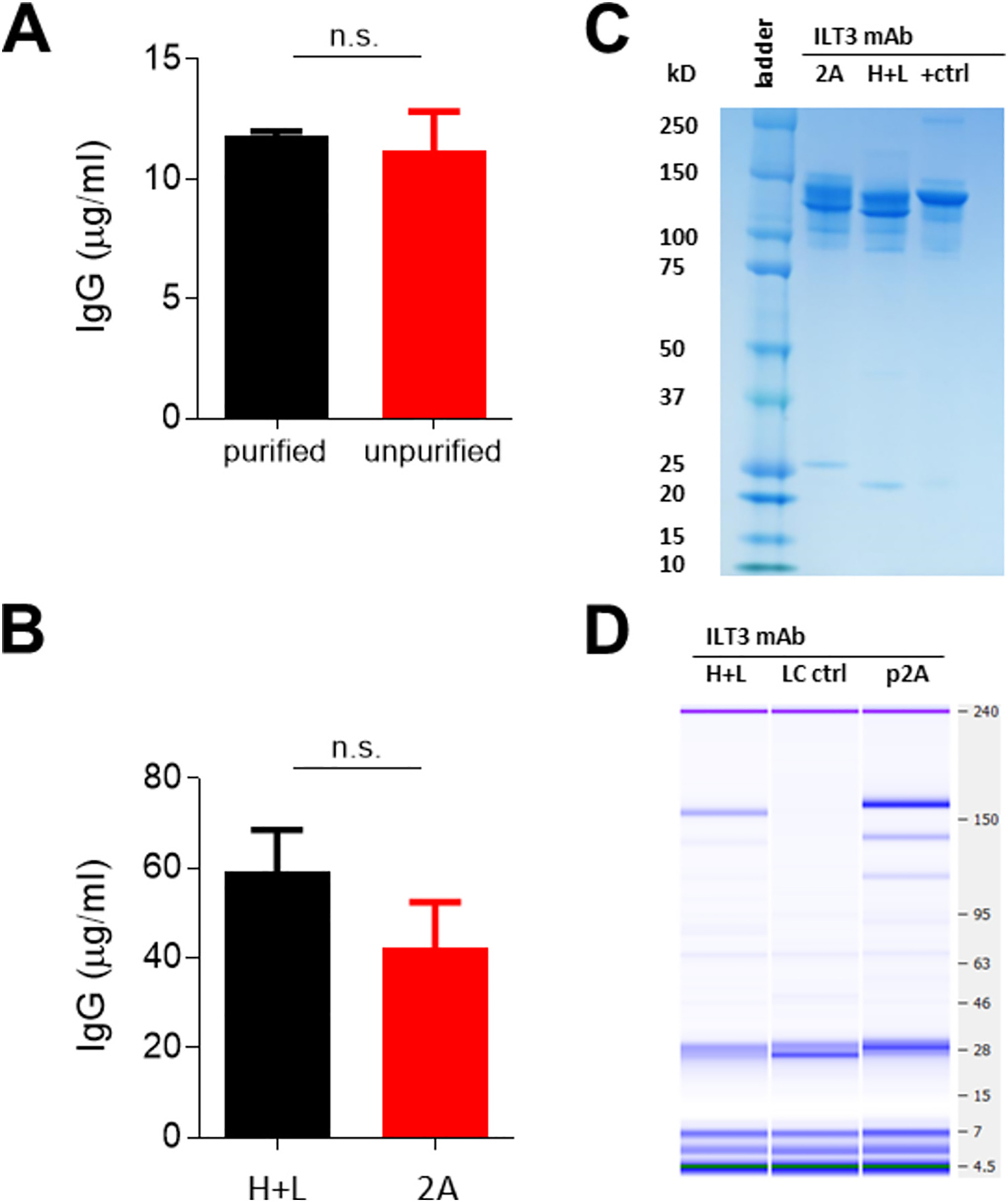
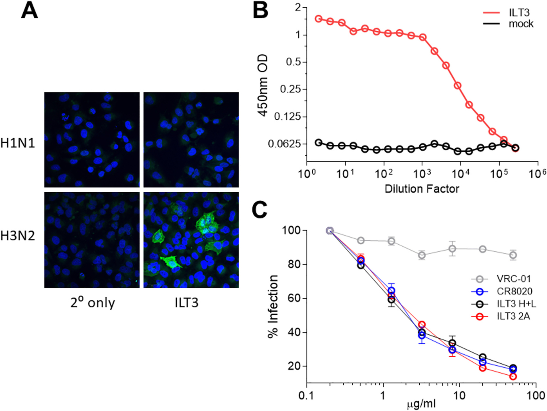
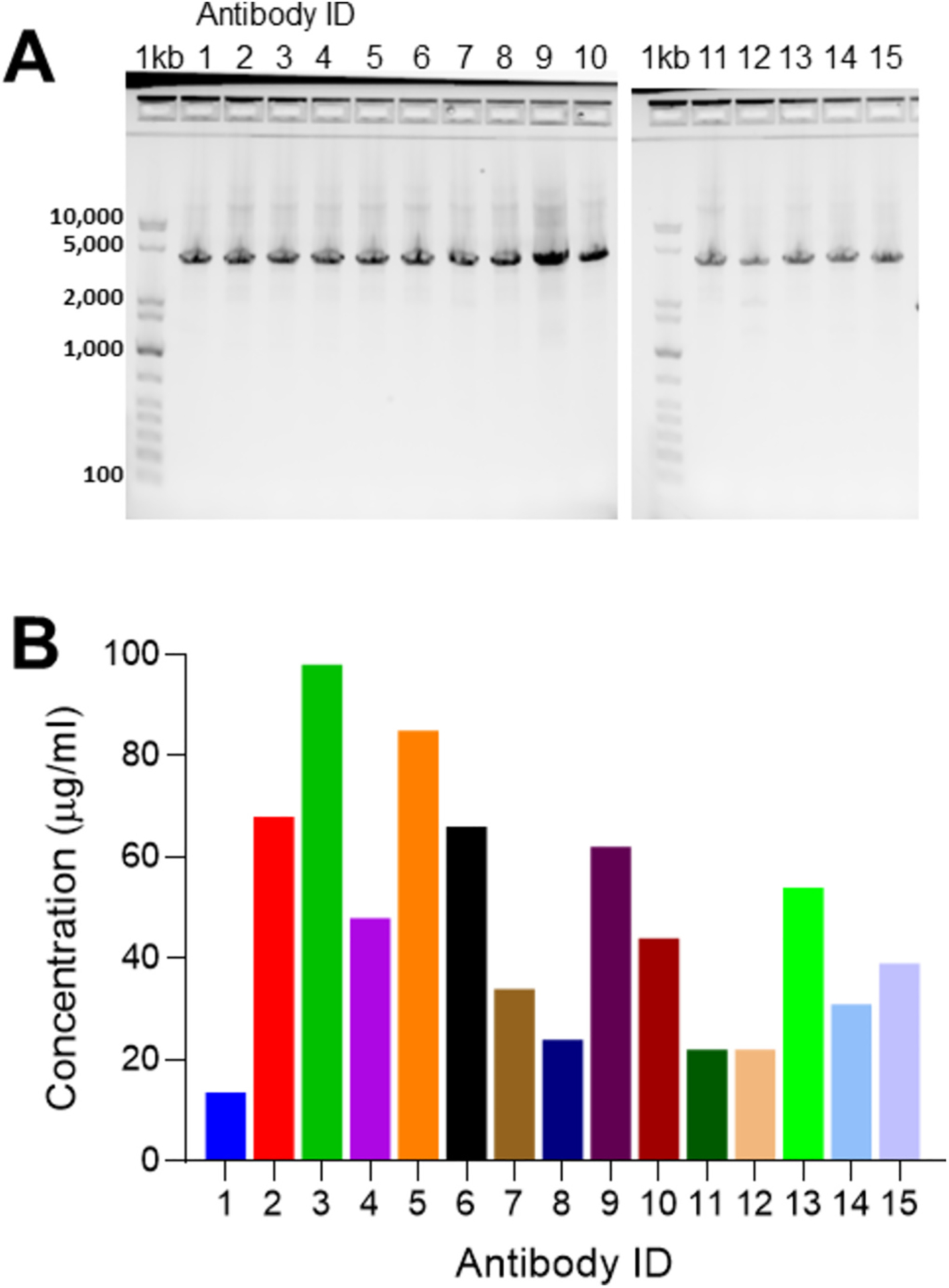
Monoclonal antibodies are powerful therapeutic, diagnostic, and research tools. Methods utilized to generate monoclonal antibodies are evolving rapidly. We created a transfectable linear antibody expression cassette from a 2-h high-fidelity overlapping PCR reaction from synthesized DNA fragments. We coupled heavy and light chains into a single linear sequence with a promoter, self-cleaving peptide, and poly(A) signal to increase the flexibility of swapping variable regions from any sequence available in silico. Transfection of the linear cassette tended to generate similar levels to the two-plasmid system and generated an average of 47 μg (14-98 μg) after 5 days in 2 ml cultures with 15 unique antibody sequences. The levels of antibodies produced were sufficient for most downstream applications in less than a week. The method presented here reduces the time, cost, and complexity of cloning steps.

Keywords: B cells; Biolayer interferometry; Confocal microscopy; Influenza; Monoclonal antibodies.


Conflict of interest statement

Declaration of competing interest The authors declare to have no financial and non-financial competing interests.

Figures

Fig. 1.
Fig. 1.. Plasmid maps and expression levels.
a, Single plasmid (p2A; top) antibody expression cassette along with heavy (H; bottom left) and light (L; bottom right) chain plasmids from the two-plasmid system. b, IgG expressedfrom plasmids as either co-transfected heavy and light chains (black; H + L) or a single expression cassette (red; p2 A) with 1 μg/ml transfections. Plasmid elements include the following: Enhancer and Promoter = CMV; IgK unique = immunoglobulin kappa (light) chain sequence different for each antibody; IgKC = kappa (light) constant; Self-cleavage = Furin site, GSG spacer, and tandem P2 A T2 A sequence; IgHC = heavy chain constant; WPRE = woodchuck posttranscriptional regulatory element; PolyA = poly(A) signal; IgG = immunoglobulin G isotype.
Fig. 2.
Fig. 2.. Generation of a linear antibody expression cassette from an overlapping PCR.
a, Diagrammatic illustration showing the five pieces in correct proportional length to base pair sizes that go into making the linear expression cassette: (P1) CMV promoter and enhancer; (P2) immunoglobulin kappa chain variable portion (IgK); (P3) immunoglobulin kappa chain constant (IgKc) portion, Furin 2 A recognition site, GSG spacer, tandem P2A-T2A (2A) self-cleaving peptide; (P4) immunoglobulin heavy chain variable (IgH) portion; (P5) immunoglobulin heavy chain constant (IgHc) region, woodchuck hepatitis virus posttranscriptional regulatory element (WPRE), and bovine growth hormone polyadenylation signal (polyA). b, P1-P5 DNA segments run on an agarose gel with a 1 kb plus DNA ladder prior to overlapping PCR; Nearest band size numbers are shown in kilobases. c, Full linear antibody expression cassette containing P1-P5 and d, run on an agarose gel with a 1 kb plus DNA ladder.
Fig. 3.
Fig. 3.. Detailed example of an antibody expression cassette DNA sequence.
An example antibody expression cassette sequence that is colour-coded to the sections listed above the sequence. Underlined in large bold text are the overlapping portions for the 5-piece overlapping PCR along with the scheme from Fig. 1 on the right. Naturally occurring signal peptides derived from each antibody sequence were included for both heavy and light chains at the coding region starts (ATG). Translation is shown in Fig. S1.
Fig. 4.
Fig. 4.. Antibody expression from different transfection conditions.
a, Concentration of IgG from purified (black) and unpurified (red) overlapping PCR products. b, IgG for mAb ILT3 from co-transfected heavy and light chain plasmids (black; H + L) and linear expression cassette (red; 2 A) from 0.5 μg/ml transfections showing standard error of the mean for error bars. c, Coomassie stained protein gel (+ctrl = VRC01) of antibodies and d, bioanalyzer protein gel under non-reducing conditions (LC ctrl = light chain only supernatant) with transfection supernatants. Bands at the size of the paired antibody (~150 kD) gave 124 μg/ml (H + L) and 296 μg/ml (p2 A). mAb = monoclonal antibody. P-values are from the Mann-Whitney test.
Fig. 5.
Fig. 5.. Anti-influenza antibody expression and binding to influenza hemagglutinin.
a, Immunofluorescence assay showing ILT3 binding to H1N1 or H3N2 infected MDCK cells in vitro (DAPI, blue; HA, green). b, Binding to H3N2 HA by ILT3 anti-influenza mAb containing supernatant (red) from cells transfected with the linear antibody expression cassette put together by overlapping PCR and mock (black). c, Microneutralization assay using an H3N2 virus and the following mAbs: positive control CR8020 (+ ctrl; blue), negative control VRC-01 (− ctrl; grey), ILT3 from heavy and light chains (H + L; black) or expression cassette (2 A; red). Significance determined by Mann-Whitney test.
Fig. 6.
Fig. 6.. Antibody generation and expression of 15 unique sequences.
a, GelPilot® 1 kb Plus DNA ladder (1 kb) and 15 amplifications of 15 unique mAb sequences all at 3.9 kb all the way across, and b, their concentrations measured by biolayer interferometry 5 days post transfection in μg/ml from 4 ml total. mAb = monoclonal antibody.

Similar articles

References

    1. Almagro JC, Mellado-Sanchez G, Pedraza-Escalona M, Perez-Tapia SM, 2022. Evolution of anti-SARS-CoV-2 therapeutic antibodies. Int. J. Mol. Sci 23. 10.3390/ijms23179763. - DOI - PMC - PubMed
    1. Borrebaeck CA, 2000. Antibodies in diagnostics - from immunoassays to protein chips. Immunol. Today 21, 379–382. 10.1016/s0167-5699(00)01683-2. - DOI - PubMed
    1. Briney B, Inderbitzin A, Joyce C, Burton DR, 2019. Commonality despite exceptional diversity in the baseline human antibody repertoire. Nature 566, 393–397. 10.1038/s41586-019-0879-y. - DOI - PMC - PubMed
    1. Chng J, et al., 2015. Cleavage efficient 2A peptides for high level monoclonal antibody expression in CHO cells. MAbs 7, 403–412. 10.1080/19420862.2015.1008351. - DOI - PMC - PubMed
    1. Corti D, Purcell LA, Snell G, Veesler D, 2021. Tackling COVID-19 with neutralizing monoclonal antibodies. Cell 184, 3086–3108. 10.1016/j.cell.2021.05.005. - DOI - PMC - PubMed

Publication types

MeSH terms

LinkOut - more resources