Multicenter Study
. 2025 Apr 10;96(5):453-461.
doi: 10.1136/jnnp-2024-334263.

MYH7-related myopathies: clinical, myopathological and genotypic spectrum in a multicentre French cohort

Affiliations
Multicenter Study

MYH7-related myopathies: clinical, myopathological and genotypic spectrum in a multicentre French cohort

Marie Bahout et al. J Neurol Neurosurg Psychiatry. .

Abstract

Background: Myosin heavy chain 7 (MYH7)-related myopathies (MYH7-RMs) are a group of muscle disorders linked to pathogenic variants in the MYH7 gene, encoding the slow/beta-cardiac myosin heavy chain, which is highly expressed in skeletal muscle and heart. The phenotype is heterogeneous including distal, predominantly axial or scapuloperoneal myopathies with variable cardiac involvement.

Methods: We retrospectively analysed the clinical, muscle MRI, genetic and myopathological features of 57 MYH7 patients. Patients received a thorough neurological (n=57, 100%), cardiac (n=51, 89%) and respiratory (n=45, 79%) assessment. Muscle imaging findings and muscle biopsies were reappraised in 19 (33%) and 27 (47%) patients, respectively.

Results: We identified three phenotypes with varying degrees of overlap: distal myopathy (70%), scapuloperoneal (23%) and axial with peculiar cervical spine rigidity called the 'sphinx' phenotype (7%). 14% of patients had either dilated cardiomyopathy, hypertrophic cardiomyopathy or left ventricular non-compaction cardiomyopathy. 31% of patients had prominent respiratory involvement, including all patients with the 'sphinx' phenotype. Muscle MRI showed involvement of tibialis anterior, followed by quadriceps, and erector spinae in patients with axial phenotype. Cores represented the most common myopathological lesion. We report 26 pathogenic variants of MYH7 gene, 9 of which are novel.

Conclusions: MYH7-RMs have a large phenotypic spectrum, including distal, scapuloperoneal or axial weakness, and variable cardiac and respiratory involvement. Tibialis anterior is constantly and precociously affected both clinically and on muscle imaging. Cores represent the most common myopathological lesion. Our detailed description of MYH7-RMs should improve their recognition and management.

Keywords: GENETICS; MRI; MYOPATHY; NEUROPATHOLOGY.


Conflict of interest statement

Competing interests: None declared.

Figures
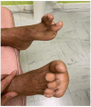
Figure 1
Figure 1. Distal involvement. Bilateral hanging big toe sign as peculiar feature in a patient with Laing distal phenotype.
Figure 2
Figure 2. Sphinx phenotype: main clinical features. Bilateral ptosis (A). Weakness of orbicularis oculi (B). Masseter retraction (C). Forward displacement of the trunk due to extensor spinae global weakness (D–F). Severe scoliosis treated with arthrodesis (blue arrow) leading to an amelioration of the dorsal bending (E).
Figure 3
Figure 3. MRI pattern of muscle involvement. Mercuri score modified by Fisher: 0: normal appearance; 1: mild involvement, fatty infiltration <30%; 2: moderate involvement, fatty infiltration between 30% and 60%; 3: severe involvement, fatty infiltration >60%; 4: end-stage appearance, completely infiltrated.
Figure 4
Figure 4. Muscle MRI patterns T1-weighed axial MRI images. Arms and trunk muscles (A); pelvic girdle (B); thighs (C); legs (D). (1) Laing distal phenotype. (C, D): fatty replacement of tibialis anterior, vastus lateralis, medialis and intermedius and gastrocnemius medialis (Mercuri grade 4, arrows). Mild fatty replacement of adductors magnus (Mercuri grade 2). (A, B): Pelvic girdle muscles, trunk and arms are well preserved. (2) Sphinx phenotype. (A, B): note the severe paravertebral fatty involvement (Mercuri grade 4) leading to major scoliosis and vertebrae rotation. (C, D): Fatty involvement of quadriceps, adductors magnus (Mercuri grade 3) whereas gracilis and hamstrings are better preserved. In legs, the fatty involvement is more pronounced in the tibialis anterior (Mercuri grade 3) than in the posterior compartment (Mercuri grade 2). (3) Atypical MRI phenotype (clinically Laing distal phenotype) (C, D) Atypical pattern of asymmetric fatty replacement of vastus intermedius and medialis, more pronounced on the left side (Mercuri grades 4 and 3, respectively). Note the fatty involvement of the left gastrocnemius medialis (Mercuri grade 3). (A, B): Pelvic girdle muscles, trunk and arms demonstrate normal appearance.
Figure 5
Figure 5. Myopathological findings. (A)SDH technique showing eccentric cores. (B) SDH, fibres showing either single or multiple cores. (C) NADH, type 1 fibres are smaller than type 2 fibres, corresponding to a significant type 1 fibre atrophy or CFTD (congenital fibre-type disproportion). The small lesion dark blue lesions in NADH (D) and reddish in mGT (E) correspond to tubular aggregates red (arrows). (F) ATPase 9.40 showing congenital type 1 fibre disproportion.
SDH: succinate dehydrogenase mGT: modified Gömöri thricrome NADH: nicotinamide adenine dinucleotide dehydrogenase
Figure 6
Figure 6. MYH7 pathogenic variants. Schematic representation of the distribution and nature of the pathogenic MYH7-variants identified in the reported cohort on the MYH7 corresponding protein (bMHC). The three main domains of the protein are indicated: the N-terminal globular myosin head is shown in grey, the neck domain in blue and the C-terminal myosin tail in orange: *indicates never reported variants, and the blue heart indicates variants associated with cardiomyopathy in the patients. Missense variants are indicated in black, in frame codon deletions in pink and splicing variants in blue.

References

    1. Sellers JR. Myosins: a diverse superfamily. Biochim Biophys Acta. 2000;1496:3–22. doi: 10.1016/S0167-4889(00)00005-7. - DOI - PubMed
    1. Tajsharghi H, Oldfors A. Myosinopathies: pathology and mechanisms. Acta Neuropathol. 2013;125:3–18. doi: 10.1007/s00401-012-1024-2. - DOI - PMC - PubMed
    1. Geisterfer-Lowrance AA, Kass S, Tanigawa G, et al. A molecular basis for familial hypertrophic cardiomyopathy: a beta cardiac myosin heavy chain gene missense mutation. Cell. 1990;62:999–1006. doi: 10.1016/0092-8674(90)90274-i. - DOI - PubMed
    1. Hershkovitz T, Kurolap A, Ruhrman-Shahar N, et al. Clinical diversity of MYH7-related cardiomyopathies: Insights into genotype-phenotype correlations. Am J Med Genet A. 2019;179:365–72. doi: 10.1002/ajmg.a.61017. - DOI - PubMed
    1. Tajsharghi H, Thornell L-E, Lindberg C, et al. Myosin storage myopathy associated with a heterozygous missense mutation in MYH7. Ann Neurol. 2003;54:494–500. doi: 10.1002/ana.10693. - DOI - PubMed

Publication types

MeSH terms