. 2024 Oct 24;148(1):56.
doi: 10.1007/s00401-024-02813-y.

Cryo-EM structure of a natural prion: chronic wasting disease fibrils from deer

Affiliations

Cryo-EM structure of a natural prion: chronic wasting disease fibrils from deer

Parvez Alam et al. Acta Neuropathol. .

Abstract

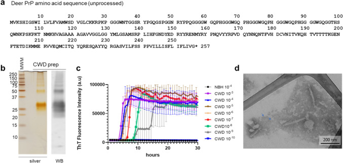
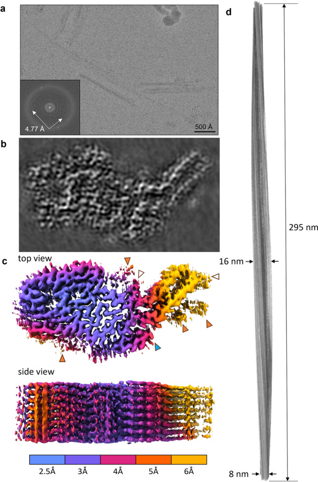
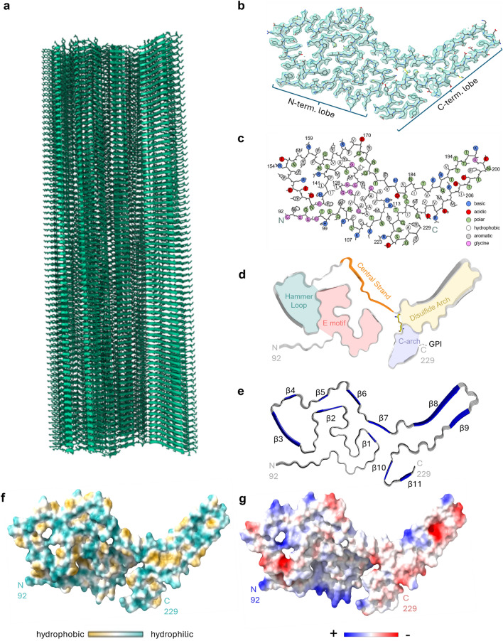
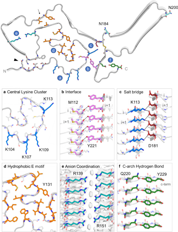
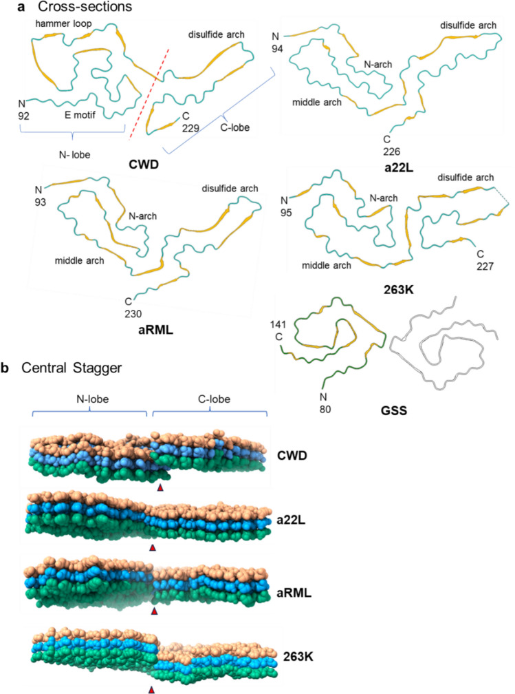
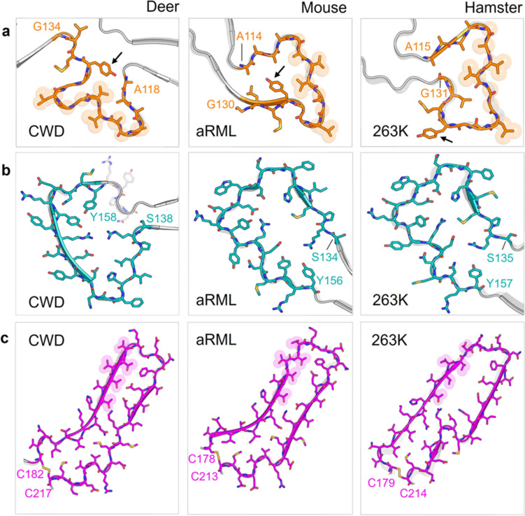
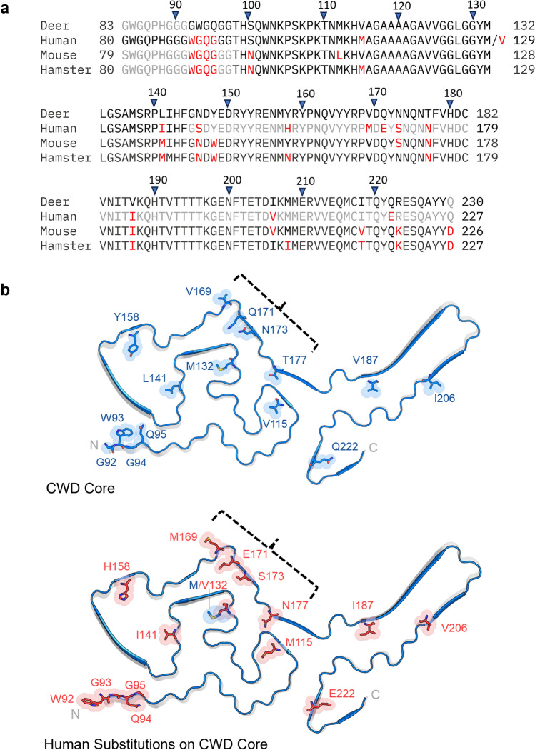
Chronic wasting disease (CWD) is a widely distributed prion disease of cervids with implications for wildlife conservation and also for human and livestock health. The structures of infectious prions that cause CWD and other natural prion diseases of mammalian hosts have been poorly understood. Here we report a 2.8 Å resolution cryogenic electron microscopy-based structure of CWD prion fibrils from the brain of a naturally infected white-tailed deer expressing the most common wild-type PrP sequence. Like recently solved rodent-adapted scrapie prion fibrils, our atomic model of CWD fibrils contains single stacks of PrP molecules forming parallel in-register intermolecular β-sheets and intervening loops comprising major N- and C-terminal lobes within the fibril cross-section. However, CWD fibrils from a natural cervid host differ markedly from the rodent structures in many other features, including a ~ 180° twist in the relative orientation of the lobes. This CWD structure suggests mechanisms underlying the apparent CWD transmission barrier to humans and should facilitate more rational approaches to the development of CWD vaccines and therapeutics.

Keywords: Amyloid; Chronic wasting disease; Cryo-electron microscopy; Deer; Prion; Structure.


Conflict of interest statement

The authors declare no competing interests.

Figures

Fig. 1
Fig. 1
PrP amino acid sequence, purity, seeding activity, and morphology of CWD PrPSc preparation. a PrP amino acid sequence determined for the white-tailed deer source of our CWD brain tissue before cellular processing to remove N-terminal signal peptide and C-terminal GPI anchor signal. b Silver-stained SDS-PAGE gel and western blot of CWD PrPSc. c End-point dilution RT-QuIC analysis of purified CWD prion preparation. Traces show mean ± SD of ThT fluorescence from quadruplicate RT-QuIC reactions seeded with serial tenfold dilutions of the preparation (as indicated) over time. Negative control reactions were seeded with normal brain homogenate (NBH) at 10–4 w/v tissue dilution. d Negative stain transmission EM of the CWD PrPSc preparation showing amyloid fibrils. Smaller circular structures with dense centers were also seen (arrows mark examples), which were presumably ferritin, a previously observed contaminant of PrPSc preparations [4] that would not be confused with amyloid fibrils in the cryo-EM analyses
Fig. 2
Fig. 2
Cryo-EM images and density maps of CWD prion fibrils. a Representative 2D cryo-EM image of unstained CWD fibrils in vitreous ice layer. Inset: fast Fourier transform of 2D image showing signals from regular 4.77 Å spacings (arrows). b Projection of density map of fibril cross-section derived from single-particle cryo-EM analysis. c Surface depictions of density map with colors showing local resolutions according to color bar. Orange arrowheads: peripheral densities not attributed to polypeptide in the subsequent modeling; open arrowheads: the sites of potential N-linked glycans; blue arrowhead: C-terminus where GPI anchor is attached. d Elongated projection of the fibril density map representing a 180° twist along the axis (i.e., the cross-over distance of 295 nm)
Fig. 3
Fig. 3
CWD prion model based on cryo-EM density map. a Extended fibril model as a ribbon diagram. b PrP residues 92–229 threaded through a cross-sectional density map. c Schematic depiction of fibril core residues showing sidechain orientations relative to the polypeptide backbone. d Ribbon cartoon of the fibril cross-section to identify structural elements as labeled. e Stacked hexameric segment of the fibril with β sheets. f Relative surface hydrophobicity as indicated in the color bar. g Coulombic charge representation. The analyses in f and g were performed using ChimeraX 1.4
Fig. 4
Fig. 4
Core elements of CWD PrPSc displayed on the backbone of the fibril (top). The letters in blue circles correspond to the individual panels. Residues N184 and N200 mark the N-linked glycosylation sites. An arrow points to hydrophobic residues in the neck of the hammer loop. An arrowhead marks N95, the stacked sidechains of which along the fibril axis form an amide zipper. a Cross-sectional view of central lysine cluster encompassing residues K104, K107, K109, and K113 (blue). b Side view of hydrogen bond in the interface of the N- and C-lobes formed by M112 and Y221. c Side view of stabilizing salt bridge formed between K113 and D181. d Cross-sectional view of hydrophobic residues (orange) in the E motif. e Side view of ambiguous cryo-EM density between positively charged residues R139 and R151. f Side view of hydrogen bond between Q220 and Y229 at the top of the C-arch
Fig. 5
Fig. 5
Comparison of CWD to rodent-adapted prion strains. a Ribbon diagrams of monomers within CWD (this paper), a22L [20], aRML [21], 263K [25], and GSS F198S [16] fibril core structures. The 2-protofilament subtype of the GSS fibril is depicted, with the second protofilament shown in grey. The red dashed line separates the N- and C-lobes of the CWD cross-section. b Lateral views of space-filling models of stacks of three monomers of CWD, a22L, aRML, and 263K EM models. The orientations of these views relative to the ribbon diagrams in a are as if looking at the latter from below. Thus, whereas the C-lobes of each model in b show the side with the exposed C-terminus, the orientation of the CWD N-lobe is swiveled 180° relative to that of the other N-lobes. Arrowheads indicate interface between the head of the N- and C-terminal lobes of a given monomer
Fig. 6
Fig. 6
Comparison of major structural motifs between deer CWD and rodent-adapted scrapie strains. a Residues ~114–134, which are part of the E motif in CWD and comprise the N-arch in both mouse (aRML) and hamster (263K) strains. Arrows mark tyrosine 131 (Y127 in aRML; Y128 in 263K). b Residues 135–157, which are part of the hammer loop in CWD and the middle arch in both mouse and hamster strains. c The disulfide arch, which begins at the corresponding disulfide bridges of the CWD, aRML, and 263K fibrils
Fig. 7
Fig. 7
Species-associated sequence variations in regions overlapping core structures of ex vivo PrPSc fibrils. a Sequence alignment with deer numbering on top and individual species numbering on the sides. Positions at which the sequence differs from deer PrP are highlighted in red; residues outside the respective ordered core regions (based on structures of deer CWD (this paper), human GSS F198S [16], mouse a22L [20], and hamster 263K [25]) are shown in gray; (Clustal Omega program was used for this analysis [30]). b Positions at which human PrP residues (bottom, red) differ from the corresponding deer sequence (top, blue) superimposed on the CWD core cross-section; (mutation of the deer sequence was performed in Pymol/2.6.0). Brackets mark four sequence variations between deer and humans that have been shown collectively to strongly contribute to the barrier to transmission of CWD to transgenic mice that overexpress human PrP [27]. M/V132 marks the position that corresponds to position 129 in human PrP, where either M (like deer) or V is found normally

References

    1. Afonine PV, Grosse-Kunstleve RW, Echols N, Headd JJ, Moriarty NW, Mustyakimov M et al (2012) Towards automated crystallographic structure refinement with phenix.refine. Acta Crystallogr D Biol Crystallogr 68:352–367. 10.1107/S0907444912001308 - PMC - PubMed
    1. Arifin MI, Hannaoui S, Chang SC, Thapa S, Schatzl HM, Gilch S (2021) Cervid prion protein polymorphisms: role in chronic wasting disease pathogenesis. Int J Mol Sci. 10.3390/ijms22052271 - PMC - PubMed
    1. Barad BA, Echols N, Wang RY, Cheng Y, DiMaio F, Adams PD et al (2015) EMRinger: side chain-directed model and map validation for 3D cryo-electron microscopy. Nat Methods 12:943–946. 10.1038/nmeth.3541 - PMC - PubMed
    1. Baron GS, Hughson AG, Raymond GJ, Offerdahl DK, Barton KA, Raymond LD et al (2011) Effect of glycans and the glycophosphatidylinositol anchor on strain dependent conformations of scrapie prion protein: improved purifications and infrared spectra. Biochemistry 50:4479–4490. 10.1021/bi2003907 - PMC - PubMed
    1. Barria MA, Libori A, Mitchell G, Head MW (2018) Susceptibility of human prion protein to conversion by chronic wasting disease prions. Emerg Infect Dis 24:1482–1489. 10.3201/eid2408.161888 - PMC - PubMed

Publication types

LinkOut - more resources