. 2024 Oct 24;15(1):9200.
doi: 10.1038/s41467-024-53106-7.

Supercharged fluorescent proteins detect lanthanides via direct antennae signaling

Affiliations

Supercharged fluorescent proteins detect lanthanides via direct antennae signaling

Kevin Y Huang et al. Nat Commun. .

Abstract

A sustainable operation for harvesting metals in the lanthanide series is needed to meet the rising demand for rare earth elements across diverse global industries. However, existing methods are limited in their capacity for detection and capture at environmentally and industrially relevant lanthanide concentrations. Supercharged fluorescent proteins have solvent-exposed, negatively charged residues that potentially create multiple direct chelation pockets for free lanthanide cations. Here, we demonstrate that negatively supercharged proteins can bind and quantitatively report concentrations of lanthanides via an underutilized lanthanide-to-chromophore pathway of energy transfer. The top-performing sensors detect lanthanides in the micromolar to millimolar range and remain unperturbed by environmentally significant concentrations of competing metals. As a demonstration of the versatility and adaptability of this energy transfer method, we show proximity and signal transmission between the lanthanides and a supramolecular assembly of supercharged proteins, paving the way for the detection of lanthanides via programmable protein oligomers and materials.


Conflict of interest statement

K.Y.H., A.D.E., and D.J.F.W. are co-inventors on the U.S. patent application pertaining to the use of supercharged proteins for lanthanide detection (Application No. 63/611,409). Applicant: Board of Regents, The University of Texas System. The remaining authors declare no competing interests.

Figures
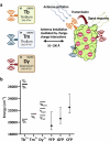
Fig. 1
Fig. 1. Schema for energy transfer.
a Schema of the posited lanthanide-based resonance energy transfer between the lanthanide antenna and the fluorescent protein (PDB: 2B3P). Antenna excitation gives rise to a detectable signal if the lanthanide and the biosensor are separated by 10–100 Å. Created in BioRender. Huang, K. (2023) BioRender.com/f23r502. b Spectral overlaps between terbium, thulium, dysprosium, and YFP, GFP, and CFP. The solid dashes represent select free-ion energy levels of trivalent terbium, thulium, and dysprosium. The vertical bars denote the excitation maxima +/− the spectral half-width of supercharged YFP, GFP and CFP. Source data are provided as a Source Data file.
Fig. 2
Fig. 2. Charge engineering introduces lanthanide chelation sites.
a Charge engineering schema for generating 18 protein biosensor variants of the base superfolder protein (PDB: 2B3P). b An illustration of potential lanthanide binding interactions and proximity to the protein chromophore (PDB: 2B3P). Solvent-exposed residues are mutated to increase the net charge while preserving the structure and function of the fluorescent protein. The charged surface enables lanthanide binding at various distances to the internal chromophore of the fluorescent protein. Created in BioRender. Huang, K. (2023) BioRender.com/f23r502.
Fig. 3
Fig. 3. Time-resolved spectroscopy reveals long-lived lanthanide luminescence signaling.
a Supernegative GFP−10 sensors fluoresce in the presence of 1 mM of Tb3+. Time-resolved luminescence responses measured at 0 µs, 100 µs, and 300 µs. GFP−10 concentration of 0.1 mg/mL (~3.7 µM) in 50 mM Tris-HCl at pH of 7. Green shading denotes the fraction of GFP−10 fluorescence attributable to Tb3+ placement. b Supernegative YFP−31 sensors fluoresce in the presence of 1 mM of Tb3+. Time-resolved luminescence responses measured at 0 µs, 100 µs, and 300 µs. YFP−31 concentration of 0.1 mg/mL (~3.7 µM) in 50 mM Tris-HCl at pH of 7. Yellow shading denotes the fraction of YFP−31 fluorescence attributable to Tb3+ placement. Source data are provided as a Source Data file.
Fig. 4
Fig. 4. Lanthanide sensitivities of supercharged protein biosensors.
a Ratiometric excitation responses of supernegative and superpositive protein sensors in the presence of 1 mM Tb3+, Tm3+, or Dy3+ in 50 mM Tris-HCl at pH 7. Fluorescent protein concentration of 0.1 mg/mL (~3.7 µM). Error bars denote S.D. +/− the mean. b GFP−10 biosensor ratiometric responses to Tb3+, Tm3+, Dy3+, Eu3+, Sm3+, and Yb3+ in the 1 nM–10 mM range. GFP−10 concentration of 0.1 mg/mL (~3.7 µM) in 50 mM Tris-HCl at pH of 7. c YFP−31 biosensor ratiometric responses to Tb3+, Tm3+, Dy3+, Eu3+, Sm3+, and Yb3+ in the 1 nM–10 mM range. YFP−31 concentration of 0.1 mg/mL (~3.7 µM) in 50 mM Tris-HCl at pH of 7. Experiments were performed in biological triplicate. Source data are provided as a Source Data file.
Fig. 5
Fig. 5. Responsivities of biosensors in the presence of interferents.
a Supernegative YFP series emission spectrum (ex. 280 nm) in the presence of equimolar (1 mM) amounts of Tb3+ or Al3+. Yellow and gray shading denote the fraction of YFP fluorescence attributable to Tb3+ placement and aluminum noise, respectively. b GFP−10 and YFP−31 biosensor responses to 1 mM of Tb3+ versus a 10-fold excess of contaminants (10 mM) found in mine drainage from the Virginia Canyon. 50 mM Tris-HCl at pH of 7. Error bars denote S.D. +/− the mean. Green and yellow shading represent the fraction of GFP and YFP fluorescence attributable to Tb3+ placement and aluminum noise, respectively. c Virginia Canyon (VC) and modified Virginia Canyon (mVC) interference of the GFP−10 ratiometric signal in the presence of 1 mM Tb3+. GFP−10 concentration of 0.1 mg/mL (~3.7 µM) in 50 mM Tris-HCl at pH of 7. d Virginia Canyon (VC) and modified Virginia Canyon (mVC) interference of the YFP−31 ratiometric signal in the presence of 1 mM Tb3+. YFP−31 concentration of 0.1 mg/mL (~3.7 µM) in 50 mM Tris-HCl at pH of 7. Experiments were performed in biological triplicate. Source data are provided as a Source Data file.
Fig. 6
Fig. 6. Supercharged protomer fluorescence in the presence of lanthanides.
a Schema of the posited LRET interaction between the lanthanide and the CFP+32/GFP−31 protomer (PDB: 6MDR). Created in BioRender. Huang, K. (2023) BioRender.com/f23r502. b Lanthanide and aluminum (1 mM) interference of the protomer signal (ex. 433 nm). An increased signal relative to the unperturbed protomer FRET is observed when exciting at 340 nm. Protomer concentration of 3.7 µM in 50 mM Tris-HCl at pH of 7. Created in BioRender. Huang, K. (2023) BioRender.com/f23r502. c The FRET signal is ‘restored’ in varying degrees when exciting at 340 nm versus 433 nm. Error bars denote S.D. +/− the mean. Experiments were performed in biological triplicate. Source data are provided as a Source Data file.

References

    1. Eliseeva, S. V. & Bünzli, J.-C. G. Lanthanide luminescence for functional materials and bio-sciences. Chem. Soc. Rev.39, 189–227 (2010). - PubMed
    1. Woodruff, D. N., Winpenny, R. E. P. & Layfield, R. A. Lanthanide single-molecule magnets. Chem. Rev.113, 5110–5148 (2013). - PubMed
    1. Edelmann, F. T. Lanthanide amidinates and guanidinates in catalysis and materials science: a continuing success story. Chem. Soc. Rev.41, 7657 (2012). - PubMed
    1. Wall, F. Rare earth elements. Encyclopedia of Geology 680–693 (Elsevier, 2021). 10.1016/B978-0-08-102908-4.00101-6.
    1. Lucas, J., Lucas, P., Le Mercier, T., Rollat, A. & Davenport, W. G. Rare Earths: Science, Technology, Production and Use. (Elsevier, 2015).

Publication types

MeSH terms

Substances

LinkOut - more resources