. 2024 Oct 24;15(1):9195.
doi: 10.1038/s41467-024-53412-0.

Stepwise phosphorylation and SUMOylation of PIDD1 drive PIDDosome assembly in response to DNA repair failure

Affiliations

Stepwise phosphorylation and SUMOylation of PIDD1 drive PIDDosome assembly in response to DNA repair failure

Richa B Shah et al. Nat Commun. .

Abstract

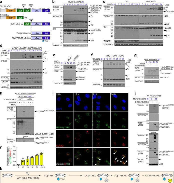
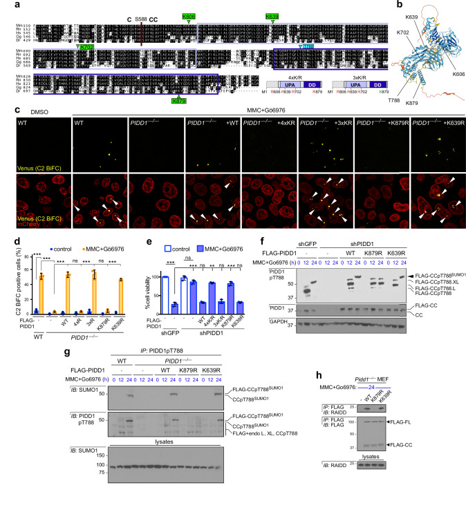
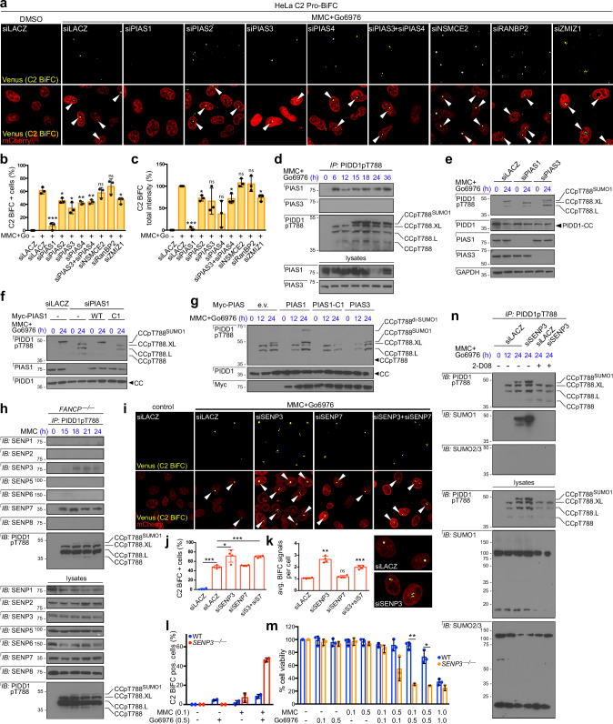
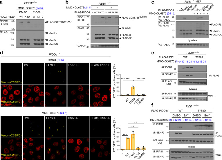
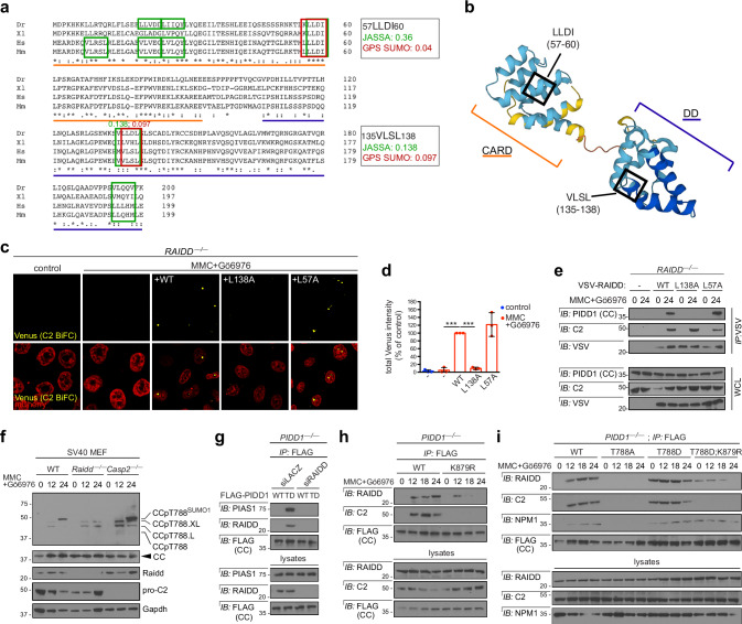
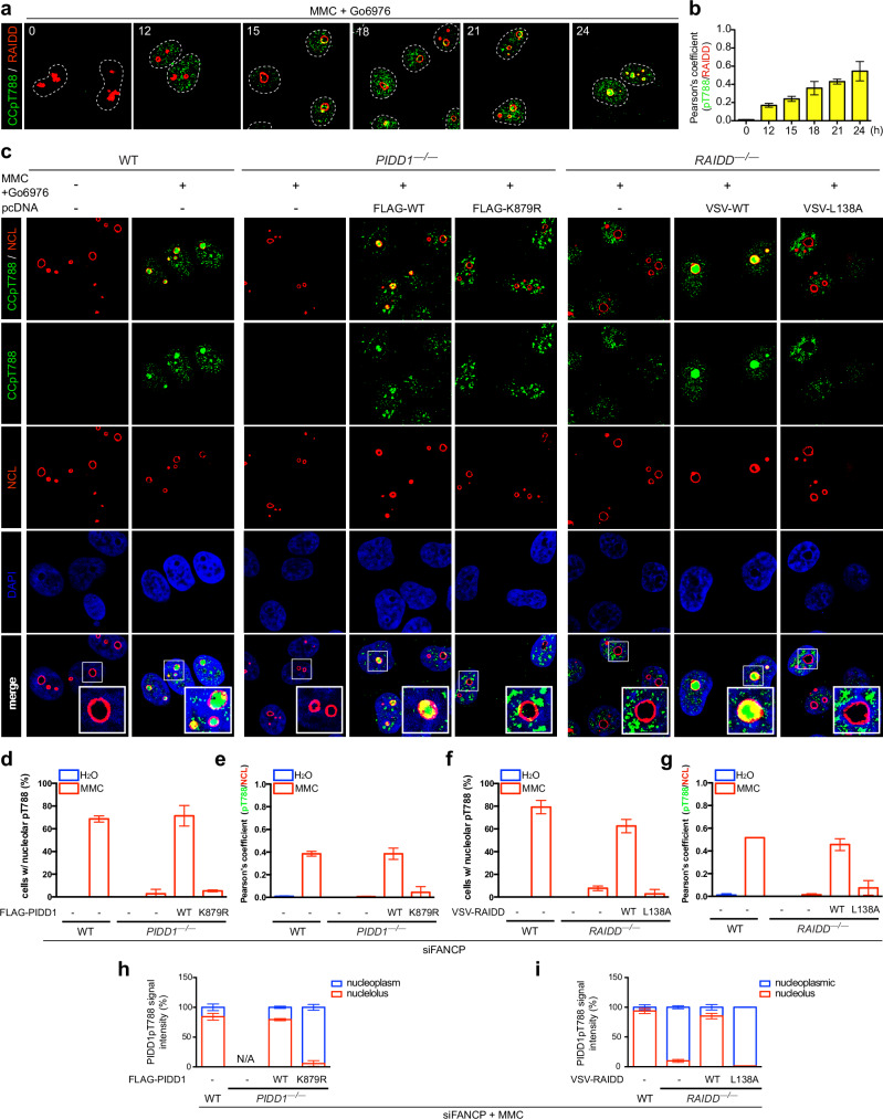
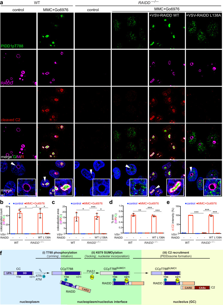
SUMOylation regulates numerous cellular stress responses, yet targets in the apoptotic machinery remain elusive. We show that a single, DNA damage-induced monoSUMOylation event controls PIDDosome (PIDD1/RAIDD/caspase-2) formation and apoptotic death in response to unresolved DNA interstrand crosslinks (ICLs). SUMO-1 conjugation occurs on conserved K879 in the PIDD1 death domain (DD); is catalyzed by PIAS1 and countered by SENP3; and is triggered by ATR phosphorylation of neighboring T788 in the PIDD1 DD, which enables PIAS1 docking. Phospho/SUMO-PIDD1 proteins are captured by nucleolar RAIDD monomers via a SUMO-interacting motif (SIM) in the RAIDD DD, thus compartmentalizing nascent PIDDosomes for caspase-2 recruitment. Denying SUMOylation or the SUMO-SIM interaction spares the onset of PIDDosome assembly but blocks its completion, thus eliminating the apoptotic response to ICL repair failure. Conversely, removal of SENP3 forces apoptosis, even in cells with tolerable ICL levels. SUMO-mediated PIDDosome control is also seen in response to DNA breaks but not supernumerary centrosomes. These results illuminate PIDDosome formation in space and time and identify a direct role for SUMOylation in the assembly of a major pro-apoptotic device.


Conflict of interest statement

The authors declare no competing interests.

Figures

Fig. 1
Fig. 1. PIDD1 is mono-SUMOylated in response to ICL repair failure.
a Domain structures of full-length (FL) PIDD1 and autocatalytic cleavage products. LRR, leucine-rich repeats; ZU5, domain present in Zona occludens, UNC5-like netrin receptors and Ankyrin; UPA, Uncharacterized protein domain in UNC5, PIDD and Ankyrin family; DD, death domain. bg HeLa cells of indicated genotypes (c), transfected with indicated siRNAs (d) or stably expressing indicated shRNAs (e) were treated with indicated drugs, harvested at indicated time points and analyzed on 10% bis-tris NuPAGE gels with indicated antibodies. MMC, mitomycin C; CPT, camptothecin; TOPO, topotecan; 2-D08, SUMO E2 ligase inhibitor; thalidomide, CRBN inhibitor. h HCT116 cells stably expressing WT or ΔGG (non-conjugatable) FLAG-SUMO-1 were treated with MMC and/or Go6976, harvested at 24 h, immunoprecipitated with anti-FLAG antibody and analyzed by western blot. i, i’ HeLa cells grown on cover slips were treated with MMC and Chk1 inhibitor Go6976, fixed at indicated time points (hour), stained with indicated antibodies and imaged by confocal microscopy. At least 35 cells per time point were scored in each of n = 3 independent experiments. Representative images (i) quantified in (i’), with data expressed as means +/− SD; **, p < 0.005; ***p < 0.001; ns, non-significant; two-tailed Student’s t-test. j HeLa cells treated with indicated drugs were immunoprecipitated with anti-PIDD1pT788 antibody at indicated time points and analyzed by western blot. k Cartoon summarizing the modified PIDD1pT788 species. All drugs were given at 1 μM. Cells in (bh, j) were TdR-synchronized as described in Supplementary Tables 1A (b, c, eh) and 1B (d). Source data are provided as a Source data file.
Fig. 2
Fig. 2. Identification of K879 as the PIDD1-CC SUMO-1 acceptor.
a, b ClustalW alignment of human (Hs), mouse (Mm), rat (Rn), chicken (Gg), and zebrafish (Dr) PIDD1 C termini. UPA and DD boxed in light blue and dark blue, respectively. Conserved lysine residues and ATM/ATR target T788 also indicated both in alignment (a) and AlphaFold2 model of full-length PIDD1 (b). c, d C2.Pro-BiFC HeLa cells of indicated genotypes were transfected with indicated FLAG-PIDD1 variants, treated with or without MMC and Go6976 (5 μM each) and imaged at 24 h. Arrowheads mark Venus signal, reflecting dimerization of C2 Pro-VN and C2 Pro-VC (see Supplementary Fig. 1h). At least 40 cells per sample were scored in each of n = 3 independent experiments. Representative images (c) were quantified in (d). e HeLa cells stably expressing the indicated shRNAs were transfected with indicated FLAG-PIDD1 variants, treated with or without MMC (0.1 μM) and Go6976 (1 μM) and stained with the vital dye alamarBlue at 5 days. f HeLa cells of indicated genotypes were transfected with indicated FLAG-PIDD1 variants, treated with or without MMC and Chk1 inhibitor Go6976 (1 μM each), harvested at indicated time points and analyzed by western blot. g HeLa cells of indicated genotypes were transfected with indicated FLAG-PIDD1 variants, treated with or without MMC and Go6976 (1 μM each), harvested at indicated time points, immunoprecipitated with anti-PIDD1pT788 antibodies and analyzed by western blot. h Pidd1—/— MEF transfected with indicated FLAG-PIDD1 variants and treated with MMC and Go6976 (1 μM each) were harvested at 24 h, immunoprecipitated with anti-FLAG antibody and analyzed by western blot. Cells in (fh) were TdR-synchronized as described in Supplementary Table 1B. Data in (d, e) expressed as means +/− SD of 3 independent experiments. **p < 0.005; ***p < 0.001; ns, non-significant; two-tailed Student’s t-test. Source data are provided as a Source data file.
Fig. 3
Fig. 3. PIDD1 SUMOylation is catalyzed by PIAS1 and reversed by SENP3.
ac C2.Pro-BiFC HeLa cells transfected with indicated siRNAs were treated with or without MMC and Go6976 (5 μM each) and analyzed by confocal microscopy at 24 h. At least 40 cells per sample were scored in each of n = 3 independent experiments. Representative images (a) were quantified for percentage of C2 BiFC positive cells (b) and overall signal intensity (c). d HeLa cells treated with MMC and Go6976 and harvested at indicated time points were immunoprecipitated with anti-PIDD1pT788 antibody and analyzed by western blot. e HeLa cells transfected with indicated siRNAs were treated with MMC and Go6976, harvested at indicated time points and analyzed by western blot. f HeLa cells transfected with indicated siRNAs, co-transfected with indicated Myc-PIAS1 constructs and treated and analyzed as in (e). C1, catalytically inactive. (g) HeLa cells transfected with indicated Myc-PIAS constructs and treated with MMC and Go6976 were analyzed as in (e). CCpT788di-SUMO1, di-SUMO-1ylated CCpT788. h FANCP—/— HeLa cells treated with MMC were harvested at indicated time points and PIDD1pT788 immunoprecipitates were analyzed by western blot. ik Cells treated and analyzed as in (a) were quantified for percentage of C2 BiFC positive cells (j) and average number of Venus puncta per cell (k). l Parent and SENP3—/— HeLa cells treated with MMC and Go6976 at indicated doses were analyzed as in (ik). At least 40 cells per sample were scored in each of n = 2 independent experiments (representative images shown in Supplementary Fig. 3c) (m) Cells as in (l) were stained with the vital dye alamarBlue at 5 days. n HeLa cells transfected with indicated siRNAs and treated with indicated drugs were immunoprecipitated with anti-PIDD1pT788 antibody and analyzed by western blot. Cells in (dh, n) were TdR-synchronized as described in Supplementary Tables 1A (d, h), 1B (eg), 1C (n). Unless otherwise indicated, drugs were given at 1μM. Data in (b, c, j, k, m) are expressed as means +/− SD, with *p < 0.05, **p < 0.005, ***p < 0.001, ns, non-significant, two-tailed Student’s t-test. Source data are provided as a Source data file.
Fig. 4
Fig. 4. DNA damage-induced PIDD1 phosphorylation triggers SUMOylation.
a, b PIDD1—/— HeLa cells transfected with indicated FLAG-PIDD1 variants, treated with MMC and Go6976 with or without SUMO E2 ligase inhibitor 2-D08 were harvested at 24 h and analyzed by western blot. c Pidd1—/— MEF transfected with indicated FLAG-PIDD1 variants and treated with or without MMC and Go6976 were harvested at 24 h, immunoprecipitated with anti-FLAG antibody and analyzed by western blot. d PIDD1—/— C2.Pro-BiFC HeLa cells were transfected with indicated FLAG-PIDD1 variants, treated with or without MMC and Go6976 (5 μM each) and imaged at 24 h. At least 40 cells per sample were scored in each of n = 3 independent experiments. Representative images were quantified (right), with data expressed as means +/− SD. **p < 0.005; ***p < 0.001; ns, non-significant, two-tailed Student’s t-test. e, f PIDD1—/— HeLa cells transfected with indicated FLAG-PIDD1 variants and treated with MMC and Go6976 (e) with or without ATR inhibitor BAY-1895344 (BAY, 0.75 mM) (f) were harvested at 24 h, immunoprecipitated with anti-PIDD1pT788 antibody and analyzed by western blot. Cells in (ac, e, f ) were TdR-synchronized as described in Supplementary Tables 1B and 1C. Unless otherwise indicated, drugs were given at 1 μM. Source data are provided as a Source data file.
Fig. 5
Fig. 5. SUMO-SIM interaction between PIDD1 and RAIDD DDs sustains nascent PIDD1/RAIDD complexes.
a, b ClustalW alignment of human (Hs), mouse (Mm), xenopus (Xl), and zebrafish (Dr) RAIDD homologs with GPS- SUMO and JASSA -predicted SUMO-interacting domains (SIM) boxed in red and green, respectively (probability scores also indicated) (a), with conserved putative SIMs boxed in black on AlphaFold2 RAIDD model (b). Caspase recruitment domain (CARD) and DD underlined in orange and blue, respectively. (c-d) RAIDD—/— C2.Pro-BiFC HeLa cells were transfected with indicated VSV-RAIDD variants, treated with or without MMC and Go6976 (5 μM each) and imaged at 24 h. At least 40 cells per sample were scored in each of n = 3 independent experiments. Representative images (c) were quantified (d), with data expressed as means +/− SD. ***p < 0.001, two-tailed Student’s t-test. e RAIDD—/— HeLa cells transfected with indicated VSV-RAIDD variants, and treated with or without MMC and Go6976 (1 μM each) were harvested at 24 h, immunoprecipitated with anti-VSV antibodies and analyzed by western blot. f SV40-transformed MEF of indicated genotypes treated with MMC and Go6976 (1 μM each) were harvested at indicated time points and analyzed by western blot. g PIDD1—/— HeLa cells transfected with indicated siRNA and cotransfected with indicated FLAG-PIDD1 variants were harvested at 24 h, immunoprecipitated with anti-FLAG antibody and analyzed by western blot. h, i PIDD1—/— HeLa cells transfected with indicated FLAG-PIDD1 variants, were treated MMC and Go6976 (1 μM each), harvested at indicated time points, immunoprecipitated with anti-FLAG antibody and analyzed by western blot. Cells in (ei) were TdR-synchronized as described in Supplementary Tables 1A (f) and 1B (e, gi). Source data are provided as a Source data file.
Fig. 6
Fig. 6. SUMO-SIM interaction between PIDD1 and RAIDD drives PIDD1 nucleolar incorporation.
ai HeLa cells of indicated genotypes and transfected with indicated FLAG-PIDD1 or VSV-RAIDD variants (ci) were treated with MMC and Go6976 (1 μM each), fixed at indicated time points (hours; (a, b)) and stained with indicated antibodies and DAPI to mark nuclei (contours marked by dashed white line in (a)). At least 40 cells per sample were scored in each of n = 2 independent experiments. Representative images (a, c) were analyzed for percentage of cells with nucleolar PIDD1pT788 signals (d, f), Pearson’s coefficient (b, e, g) and distribution of PIDD1pT788 signal intensity (h, i). Source data are provided as a Source data file.
Fig. 7
Fig. 7. SUMO-SIM interaction between PIDD1 and RAIDD is essential for endogenous PIDDosome formation.
ae Parent and RAIDD—/— HeLa cells transfected with indicated VSV-RAIDD variants were treated with or without MMC and Go6976 (1 μM each), fixed at 24 h and stained with indicated antibodies and DAPI. At least 20 cells per sample were scored in each of n = 3 independent experiments. Representative images (a) were analyzed for triple colocalization count (b), triple colocalization area (c), percent area of triple signal overlaps (d) and overall cleaved C2 signal intensity (e). Data expressed as means +/− SD. *p < 0.05; **p < 0.005; ***p < 0.001; ns, non-significant, two-tailed Student’s t-test. f Three-step model for PIDDosome formation (see text). Source data are provided as a Source data file.

Similar articles

Cited by

References

    1. Green, D. R. Inflammasomes and other caspase-activation platforms. Cold Spring Harb. Perspect Biol. 1410.1101/cshperspect.a041061 (2022). - PMC - PubMed
    1. Man, S. M. & Kanneganti, T. D. Converging roles of caspases in inflammasome activation, cell death and innate immunity. Nat. Rev. Immunol.16, 7–21 (2016). - PMC - PubMed
    1. Parrish, A. B., Freel, C. D. & Kornbluth, S. Cellular mechanisms controlling caspase activation and function. Cold Spring Harb. Perspect Biol.5, 10.1101/cshperspect.a008672 (2013). - PMC - PubMed
    1. Tinel, A. & Tschopp, J. The PIDDosome, a protein complex implicated in activation of caspase-2 in response to genotoxic stress. Science304, 843–846 (2004). - PubMed
    1. Ando, K. et al. PIDD death-domain phosphorylation by ATM controls prodeath versus prosurvival PIDDosome signaling. Mol. Cell47, 681–693 (2012). - PMC - PubMed

Publication types

MeSH terms

Substances

LinkOut - more resources