. 2024 Oct 24;14(1):25214.
doi: 10.1038/s41598-024-76414-w.

A refined TTC assay precisely detects cardiac injury and cellular viability in the infarcted mouse heart

Affiliations

A refined TTC assay precisely detects cardiac injury and cellular viability in the infarcted mouse heart

Zheheng Ding et al. Sci Rep. .

Abstract

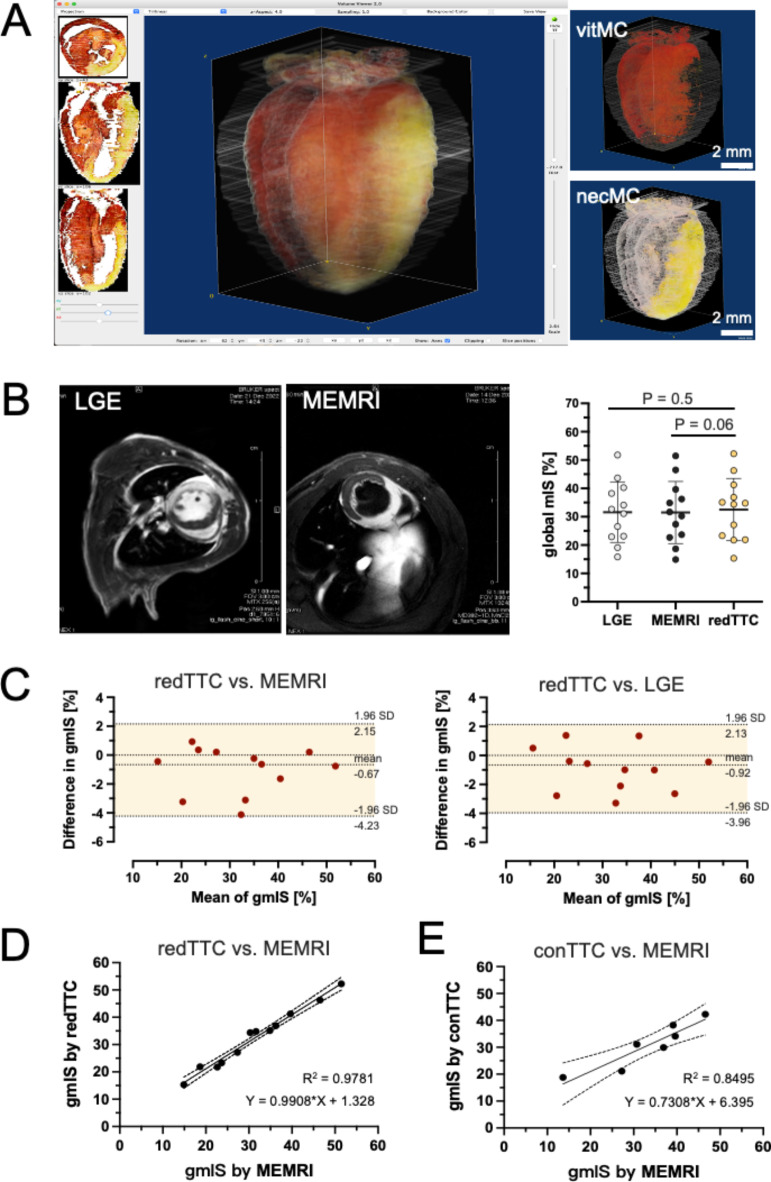
Histological analysis with 2,3,5-triphenyltetrazolium chloride (TTC) staining is the most frequently used tool to detect myocardial ischemia/reperfusion injury. However, its practicality is often challenged by poor image quality in gross histology, leading to an equivocal infarct-boundary delineation and potentially compromised measurement accuracy. Here, we introduce several crucial refinements in staining protocol and sample processing, which enable TTC images to be analyzed with light microscopy. The refined protocol involves a two-step TTC staining process (perfusion and immersion) and subsequent Zamboni fixation to differentiate myocardial viability and necrosis, and use of Coomassie brilliant blue to label area-at-risk. After the duo-staining steps were completed, the heart sample was embedded and sliced transversally by a cryostat into a series of thin sections (50 µm) for microscopic analysis. The refined TTC (redTTC) assay yielded remarkably high-quality images with striking color intensity and sharply defined boundaries, permitting unambiguous and reliable delineation of the infarct and area-at-risk. In the same animals, the redTTC assay showed good agreement with the in-vivo gold standard measurements (LGE and MEMRI). Meanwhile, redTTC imaging allows tracking of viable cardiomyocytes at cellular resolution, and with this enhanced capability, we convincingly demonstrated the pro-survival action of stem cells based-therapy. Therefore, the redTTC assay represents a significant technical advance that permits precise detection of the true extent of cardiac injury and cardiomyocyte viability. This approach is cost-effective and may be adapted for use in diverse applications, making it highly appealing to many laboratories performing ischemia/reperfusion injury experiments.

Keywords: Area-at-risk (AAR); CBB; Cardioprotection; Myocardial infarct size (mIS); Myocardial infarction; redTTC.


Conflict of interest statement

The authors declare no competing interests.

Figures

Fig. 1
Fig. 1
Microscopic TTC assay defines myocardial viability at cellular resolution. (A) Schematic of experimental protocol showing 2-step staining and cryosection for microscopic imaging. (B) A representative microscopic TTC image at mid-ventricular level of the 24-h MI heart, demonstrating two distinct areas with clear and unequivocal borderline: TTC-positive (deep red) indicates the vital myocardium (vitMC) and the picric acid-stained beige the necrotic myocardium (necMC). (C) At the cellular level, formazan precipitates were found in the cytoplasmic space of the viable cardiomyocytes figured out by WGA staining (green). (D) The formazan signal overlaps with viable cardiomyocyte, as defined by positive staining for cardiac troponin I (cTnI). (E) Quantitative analysis of segmental myocardial infarct size (mIS) in successive sections reveals dynamic variation in each slice, showing the greatest value in the apex and the lowest at the site of ligation (n = 5). The length of interventricular septum (IVS) correlated proportionally to the distance above the apex (n = 5), suggesting IVS may be considered as an internal landmark to reference the anatomical level of individual section.
Fig. 2
Fig. 2
Validation and comparison of redTTC assay to in-vivo and conventional methods. (A) With computer-aided volumetry, 3D reconstruction was generated by interactive surface plotting using ImageJ software. The bulk infarct (beige) from the entire heart was calculated as the global mIS. (B) The global mIS was compared in the same animals to the infarct size derived by in-vivo methods: namely, late gadolinium enhancement (LGE) and manganese‐enhanced MRI (MEMRI) (n = 12). Within a broad range of infarct size (15 – 52%), no significant difference was detected (paired t-test). (C) Bland–Altman analysis shows good agreement in the measurements of redTTC to MEMRI (left panel) or LGE images (right panel) methods. (D) The mIS derived by the redTTC assay was regressed against the values of MEMRI images (y = 0.99x + 1.33, n = 12) and both groups demonstrates a significant correlation (R2 = 0.98, p < 0.0001, n = 12). The dashed line indicates 95% confidence interval (CI). (E) The mIS derived by conventional TTC method (conTTC) also showed a significant correlation to the values of MEMRI images (y = 0.73x + 6.40, R2 = 0.85, n = 7, p < 0.01). When comparing both correlation coefficients, the redTTC assay shows significantly closer relation to MEMRI measurements, as compared to the conTTC method (z-test, p < 0.001).
Fig. 3
Fig. 3
Measurement of mIS:AAR ratio. (A) Schematic drawing of staining protocol for TTC and CBB double-staining. (B) Microscopic imaging of the double-staining section reveals that CBB markedly stains the remote myocardium (RM) blue. AAR is computed as the area distal of the RM, and the vital myocardium (vitMC in red) and the necrosis (necMC in beige) are separated into RGB single channels using a color-threshold tool. (C) The portions of RM, necMC and vitMC are calculated in the mid-ventricular section from 5 hearts (n = 5). Within the AAR, vitMC is found to predominantly locate in the epicardial, endocardial, anterior border zone (BZ) and posterior BZ regions, with the least found in the infarct zone (0.5%).
Fig. 4
Fig. 4
Detection of cardiomyocyte survival using redTTC assay. (A) Subtle islands of surviving cardiomyocytes are detected within the infarct zone. The vital myocardium (vitMC) is significantly more abundant in the hearts that received injections of active form of stem cells (ADSCWT1/gfp+, n = 5) compared to those injected with the dormant cells (ADSCWT1/gfp-, n = 6). Arrows indicate the injection site. (B) The vitMC survive either as single cells or in clusters within the infarct area. (C) TTC signal overlays precisely with cardiomyocytes, as identified by the typical striations in α-actinin staining (asterisk). Note that the necrotic cells also exhibit a high affinity to anti-α-actinin antibody. (D) TTC-positive cells retain cellular integrity, as shown by the intact membrane basement stained by laminin (green). (E) Positive casp-3 staining is primarily found in TTC-weak/negative cells, but not in TTC-dense cardiomyocytes, indicating an absence of apoptotic events in the surviving cardiomyocytes. ** indicates p < 0.01.

References

    1. Bolli, R. et al. Myocardial protection at a crossroads: The need for translation into clinical therapy. Circ. Res.95, 125–134 (2004). - PubMed
    1. Lindsey, M. L. et al. Guidelines for in vivo mouse models of myocardial infarction. Am. J. Physiol. Heart Circ. Physiol.321, H1056–H1073 (2021). - PMC - PubMed
    1. Redfors, B., Shao, Y. & Omerovic, E. Myocardial infarct size and area at risk assessment in mice. Exp. Clin. Cardiol.17, 268–272 (2012). - PMC - PubMed
    1. Rich, P. R., Mischis, L. A., Purton, S. & Wiskich, J. T. The sites of interaction of triphenyltetrazolium chloride with mitochondrial respiratory chains. FEMS Microbiol. Lett.202, 181–187 (2001). - PubMed
    1. Birnbaum, Y., Hale, S. L. & Kloner, R. A. Differences in reperfusion length following 30 minutes of ischemia in the rabbit influence infarct size, as measured by triphenyltetrazolium chloride staining. J. Mol. Cell. Cardiol.29, 657–666 (1997). - PubMed

LinkOut - more resources