. 2024 Oct 24;7(1):1385.
doi: 10.1038/s42003-024-07090-9.

Versatile gamma-tubulin complexes contribute to the dynamic organization of MTOCs during Drosophila spermatogenesis

Affiliations

Versatile gamma-tubulin complexes contribute to the dynamic organization of MTOCs during Drosophila spermatogenesis

Elham Alzyoud et al. Commun Biol. .

Abstract

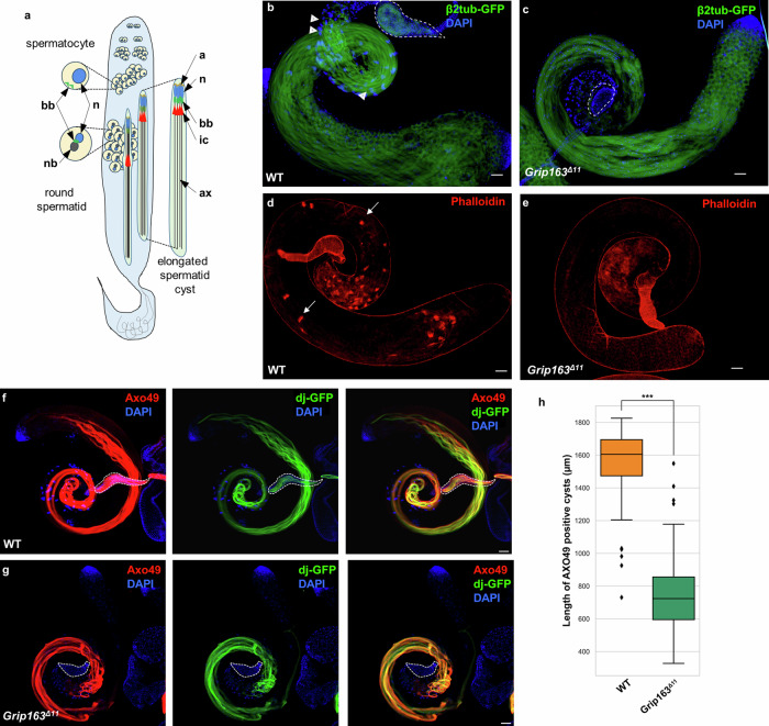
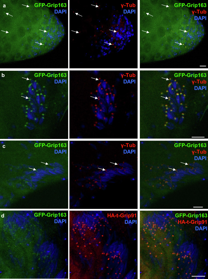
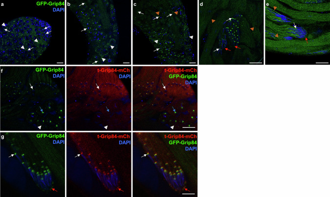
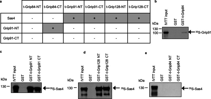
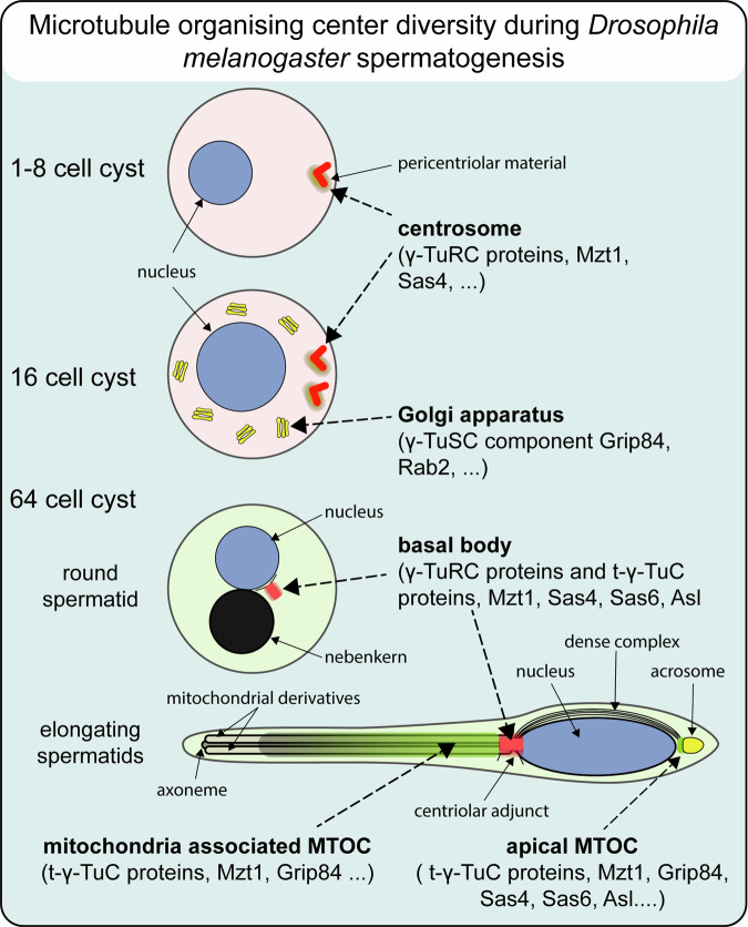
The initiation of microtubule formation is facilitated by γ-tubulin and γ-Tubulin Ring Complex (γ-TuRC) in various microtubule-organizing centers (MTOCs). While the heterogeneity of tissue-specific MTOCs and γ-TuRC in Drosophila testis has been described, their molecular composition and physiological significance are poorly understood. We investigated the testis-specific distribution and biochemical interaction of the canonical γ-TuRC proteins Grip163 and Grip84. We found that while Grip163 is present on the centrosome and basal body, Grip84 localizes to the centrosome and Golgi in spermatocytes and colocalizes with the testis-specific γ-Tubulin complexes (t-γ-TuC) at the basal body, apical nuclear tip, and near the elongated mitochondria after meiosis. We also showed the apical nuclear tip localization of some γ-TuRC interacting partners and proved their binding to t-γ-TuC proteins. These results highlight and prove the importance of the different γ-TuRCs in organizing the diverse MTOCs present during the extensive rearrangement of cell organelles during the spermatogenesis of Drosophila.


Conflict of interest statement

The authors declare no competing interests.

Figures

Fig. 1
Fig. 1. Mitochondrial and axonemal abnormalities lead to abnormal spermatid elongation in Grip163Δ11 mutant.
a Schematic representation of Drosophila spermatogenesis stages in the testis, highlighting the major cellular organelles (bb basal body, n nucleus, nb nebenkern, a acrosome, ax axoneme and cytoskeletal structures (ic investment cone) of the secondary spermatocyte, round spermatids and the elongated spermatid cyst. b, c Microtubules are visualized with the β2tub-GFP transgene (green), while the nuclei stained with DAPI (blue) in WT b and Grip163Δ11 mutant spermatids (c) Elongated cysts are present both in WT and Grip163Δ11, however, the seminal vesicle (dashed lines) of Grip163Δ11 mutant does not contain β2tub-GFP positive mature sperms. d, e The individualization complexes (IC) were stained with Phalloidin (red), ICs are formed normally (arrows) in WT, (c) and missing in Grip163Δ11 mutant (e). f, g Fully developed axonemes are visualized by Axo49 tubulin antibody (red) and elongated mitochondria are with DJ-GFP transgene (green) both in the testis and seminal vesicle (dashed line). Elongated polyglycylated axonemes and mitochondria were shorter in Grip163Δ11 mutant and the seminal vesicle was empty (dashed line) (g) compared to the WT (f). h Boxplot shows the length of AXO49 positive cysts in WT and Grip163Δ11 mutant. (WT ntestis = 12, Grip163Δ11 ntestis = 22, WT nmeasurement = 36, Grip163Δ11nmeasurement = 64) Statistical significance was tested by one-way ANOVA. (scale bars: 50 μm).
Fig. 2
Fig. 2. Abnormal mitochondrial and axonemal structures in the post-meiotic cysts of Grip163Δ11 mutant.
a, b Phase contrast images show a normal 1:1 (white outline), nucleus: nebenkern ratio in WT (a) and an abnormal ratio (red outline) in Grip163Δ11 mutant (b). c Violin plot showing the nucleus: nebenkern ratio in WT (n = 186) and Grip163Δ11 mutant round spermatids (n = 112). d, h Transmission electron micrograms of testis cross-sections, representing elongating cysts of WT (d, f) and Grip163Δ11 mutant (e, g, h). Disorganized cysts with axonemal and mitochondrial abnormalities are characteristic in Grip163Δ11 mutant. f WT elongating cyst shows normal axoneme structure with 9 + 2 tubulin dimers (highlighted in green and green arrowhead) and the attaching two mitochondrial derivatives (yellow and orange), with paracrystalline material formation in one of them (orange). g, h In Grip163Δ11 mutant the mitochondria and the axoneme detached from each other (black arrows), and multiple mitochondrial derivatives (highlighted in orange and yellow) attached to one axoneme (green and green arrowhead). h, h’ Mitochondria fused, enlarged (blue arrowheads) and detached from the axoneme (green) and doublet microtubules representing the disintegrated axonemes (black arrowheads) and the open axial membranes (red empty arrowheads) are characteristic of Grip163Δ11 mutant cyst. (scale bars: (ae) 2 μm, (fh’) 500 nm).
Fig. 3
Fig. 3. Mislocalization of centrosomal markers in Grip163Δ11mutant.
a, b GFP-PACT (green) and γ-Tubulin (red) are colocalizing on the centrosome (arrows) and basal body (arrowheads) in WT spermatocytes and spermatids (a, b). γ-tubulin, but not GFP-PACT centrosomal localization is affected in Grip163Δ11 mutant spermatocytes. In the round spermatids, GFP-PACT and γ-Tubulin are colocalised (c), but the post-meiotic elongating cysts are disorganized with scattered basal bodies (d). (scale bars: 20 μm).
Fig. 4
Fig. 4. Localization of GFP-Grip163 during spermatogenesis.
ac The endogenously tagged GFP-Grip163 colocalizes with γ-Tubulin at the (a) centrosome (arrows) and the (b, c) basal body (arrows) throughout spermatogenesis. d GFP-Grip163 has an overlapping localization with the t-γ-TuC represented by HA-t-Grip91 on the basal body/centriole adjunct in the elongating spermatids). (scale bars: 10 μm).
Fig. 5
Fig. 5. Localization of GFP-Grip84 during spermatogenesis.
ae In vivo tagged GFP-Grip84 localizes both to the centrosome (a) and the basal body (b, c) (white arrows) and in the post-meiotic elongating cyst GFP-Grip84 localizes also to the apical tip of the elongating nuclei (d, e) (red arrows). Additionally, it is localized surrounding the elongating mitochondria (ce) (orange arrowheads). Golgi-like localization of GFP-Grip84 was observed in spermatocytes (c) (white arrowheads). f, g GFP-Grip84 localizes to the centrosome before meiosis (blue arrows) and shows overlapping localization with t-Grip84-mCh at the basal body (white arrows) and the apical tip of the elongating nuclei (red arrows) in the post-meiotic spermatids (Scale bars: 20 μm).
Fig. 6
Fig. 6. γ-TuSC protein GFP-Grip84 is localizing to the Golgi in spermatocytes.
a GFP-Grip84 colocalizes with centrosomal protein Asl (anti-Asl, white arrow) and also to the Golgi apparatus (white arrowhead). b, c GFP-Grip84 colocalizes with antibody-stained cis-Golgi marker GM130 in the spermatocytes (arrowheads). d, e Rab2-YFP localizes to the Golgi (arrow) in spermatocytes visualized by the trans-Golgi marker GCC88 and also to the apical end of the elongating nuclei (arrowhead). (Scale bars: a, c, d 10 μm, B, E20 μm).
Fig. 7
Fig. 7. Sas4 is a component of the site, which has been suggested as a ncMTOC at the apical tip of the elongating nuclei.
a, b Antibody-stained Sas4 (red) colocalizes with the endogenous tag GFP-Grip84 at the centrosome (arrows) but not on the Golgi (arrowheads) in the spermatocytes. c Sas4 (red) is localizing to the basal body of the elongating spermatids (arrows) and additionally to the apical tip of the nuclei (arrowheads). d Antibody-stained Ana1 localizes only to the basal body (arrows) of the elongating spermatids, while the t-γ-TuC component HA-t-Grip91 localizes to the basal body(arrow) and the apical tip (arrowheads) of the elongating spermatids. (Scale bars: 20 μm).
Fig. 8
Fig. 8. Interactions of t-γ-TuC proteins with Sas4 and the canonical Grip91.
a Summary of Y2H analysis of the N- or C-terminal parts of t-γ-TuC proteins (as indicated in the table) with Sas4, and the N- or C-terminal parts of Grip91, respectively. be Autoradiographic images show the presence or absence of interaction between purified and immobilized GST-t-γ-TuC protein fragments and 35S-labeled Sas4 or Grip91, synthesized by in vitro transcription-translation (IVTT) reaction.
Fig. 9
Fig. 9. Summary of localization of γ-TuRC and t-γ-TuC (t-Grip84, t-Grip91, t-Grip128) containing MTOCs during different stages of spermatogenesis.
γ-TuRC and interactor proteins localize to the centrosome. γ-TuSC member Grip84 localizes to the Golgi apparatus of spermatocytes. Both γ-TuRC and t-γ-TuC present on the basal body and t-γ-TuC, Grip84, and Mzt1 also localize near the elongating spermatids’ mitochondria. t-γ-TuC, Grip84, Mzt1, Sas4, Sas6, and Asl enrich at the apical tip of the post-meiotic elongating nuclei.

References

    1. Tillery, M., Blake-Hedges, C., Zheng, Y., Buchwalter, R. & Megraw, T. Centrosomal and non-centrosomal microtubule-organizing centers (MTOCs) in Drosophila melanogaster. Cells7, 1–44 (2018). - PMC - PubMed
    1. Desai, A. & Mitchison, T. J. Microtubule polymerization dynamics. Annu. Rev. Cell Dev. Biol.13, 83–117 (1997). - PubMed
    1. Moritz, M., Braunfeld, M. B., Guénebaut, V., Heuser, J. & Agard, D. A. Structure of the γ-tubulin ring complex: a template for microtubule nucleation. Nat. Cell Biol.2, 365–370 (2000). - PubMed
    1. Zheng, Y., Wong, M. L., Alberts, B. & Mitchison, T. Nucleation of microtubule assembly by a gamma-tubulin-containing ring complex. Nature378, 578–583 (1995). - PubMed
    1. Fry, A. M., Sampson, J., Shak, C. & Shackleton, S. Recent advances in pericentriolar material organization: ordered layers and scaffolding gels. F1000Research6, 1622 (2017). - PMC - PubMed

LinkOut - more resources