. 2024 Oct 24;19(1):78.
doi: 10.1186/s13024-024-00766-0.

Gut-first Parkinson's disease is encoded by gut dysbiome

Affiliations

Gut-first Parkinson's disease is encoded by gut dysbiome

Mário F Munoz-Pinto et al. Mol Neurodegener. .

Abstract

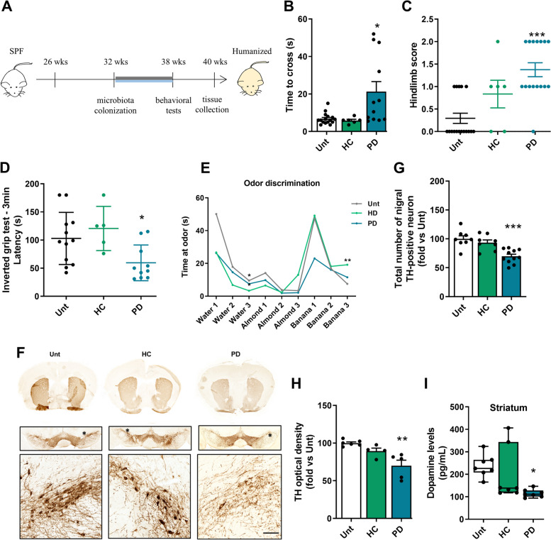
Background: In Parkinson's patients, intestinal dysbiosis can occur years before clinical diagnosis, implicating the gut and its microbiota in the disease. Recent evidence suggests the gut microbiota may trigger body-first Parkinson Disease (PD), yet the underlying mechanisms remain unclear. This study aims to elucidate how a dysbiotic microbiome through intestinal immune alterations triggers PD-related neurodegeneration.

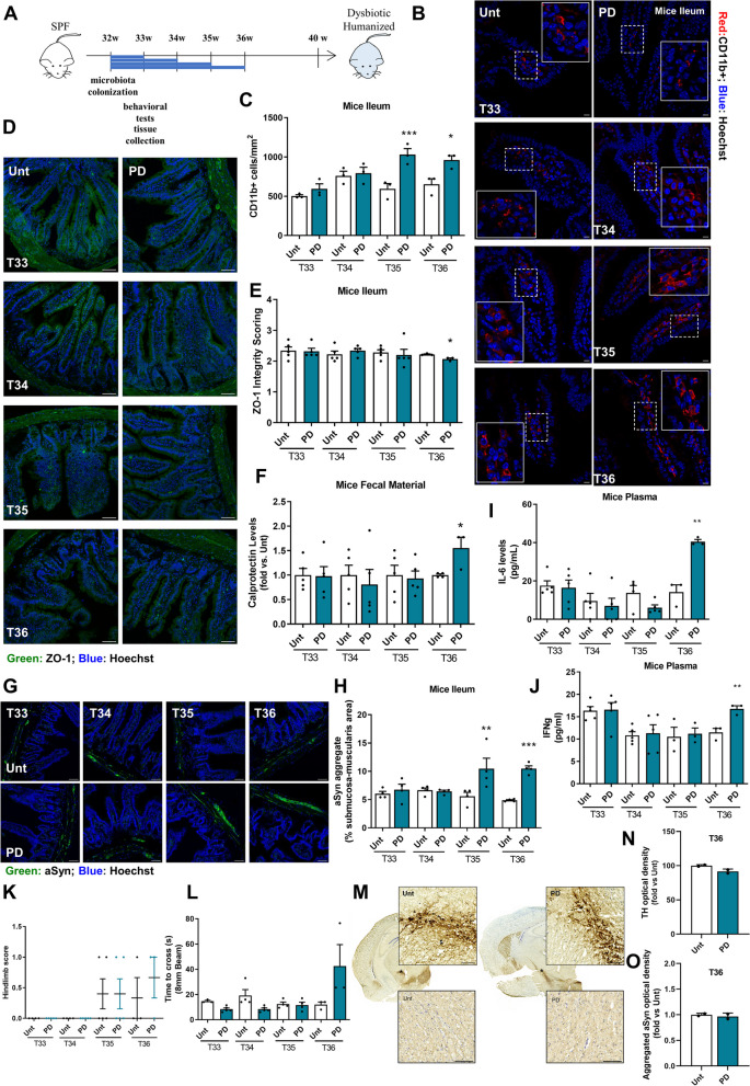
Methods: To determine the impact of gut dysbiosis on the development and progression of PD pathology, wild-type male C57BL/6 mice were transplanted with fecal material from PD patients and age-matched healthy donors to challenge the gut-immune-brain axis.

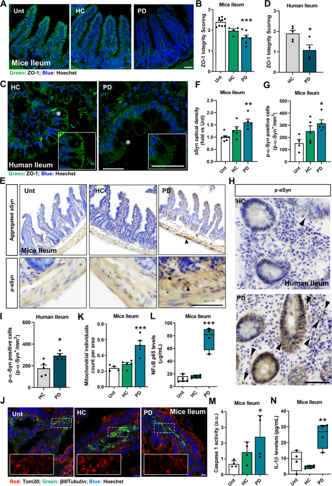
Results: This study demonstrates that patient-derived intestinal microbiota caused midbrain tyrosine hydroxylase positive (TH +) cell loss and motor dysfunction. Ileum-associated microbiota remodeling correlates with a decrease in Th17 homeostatic cells. This event led to an increase in gut inflammation and intestinal barrier disruption. In this regard, we found a decrease in CD4 + cells and an increase in pro-inflammatory cytokines in the blood of PD transplanted mice that could contribute to an increase in the permeabilization of the blood-brain-barrier, observed by an increase in mesencephalic Ig-G-positive microvascular leaks and by an increase of mesencephalic IL-17 levels, compatible with systemic inflammation. Furthermore, alpha-synuclein aggregates can spread caudo-rostrally, causing fragmentation of neuronal mitochondria. This mitochondrial damage subsequently activates innate immune responses in neurons and triggers microglial activation.

Conclusions: We propose that the dysbiotic gut microbiome (dysbiome) in PD can disrupt a healthy microbiome and Th17 homeostatic immunity in the ileum mucosa, leading to a cascade effect that propagates to the brain, ultimately contributing to PD pathophysiology. Our landmark study has successfully identified new peripheral biomarkers that could be used to develop highly effective strategies to prevent the progression of PD into the brain.

Keywords: Dopaminergic; Dorsal motor nucleus of the vagus; Dysbiome; Dysbiosis; Gut microbiome; Gut-brain axis; Inflammation; Innate immunity; Mitochondria; Parkinson’s disease; Substantia nigra.


Conflict of interest statement

All authors declare no competing interests.

Figures

Fig. 1
Fig. 1
Dopaminergic neurodegeneration and motor alterations in WT mice transplanted with fecal microbiota. A Schematic representation of experimental design (B) Balance and motor coordination performance were assessed with the beam walking test (n = 6–16 mice per group). C Hindlimb clasping reflex was monitored, as a quick phenotypic neurological scoring system to assess disease progression (n = 6–17 mice per group). D The inverted grip test was used to assess of limb muscle strength (n = 4–12 mice per group). E Odor discrimination and habituation was tested in time spent (s) non-social odors (n = 6–8 mice per group). F Representative photomicrographs of brain coronal sections immunostained for TH.+ in the striatum (STR) and substantia nigra (SN). G Total number of nigral TH-positive neurons in the SN assessed by stereological analysis (n = 8–11 mice per group). H OD analysis of the TH-positive fibres in the STR group normalized to the untreated group (n = 4–6 mice per group). I Striatal dopamine levels in pg/mL (n = 7 mice per group). *p < 0.05, **p < 0.01, ***p < 0.001, using one-way ANOVA with Dunnet´s test (G and H) or Kruskal–Wallis with Dunn´s test (B-D and I). Data are expressed as mean ± SEM. Scale bars are 100 µm (magnified inner square) and 1 mm. See also Tables S1-S2 and Figures S1-S4
Fig. 2
Fig. 2
Microbiota associated with the ileal mucosa of mice. A Alpha diversity measured using the Shannon index at the OTU level derived from 16S rDNA sequences obtained from mouse terminal ileum intestinal samples. (n values for Unt = 25; HC = 13; PD = 16). Unt vs. HC, by Kruskal–Wallis test, p = 0.982. Unt vs. PD, Kruskal–Wallis test, *p = 0.0335. HC vs. PD, Kruskal–Wallis test, *p = 0.0344). B Beta diversity evaluated by Principal Coordinate Analysis (PCoA) based on the Bray–Curtis index of OTUs derived from 16S rDNA sequencing of mouse terminal ileum intestinal samples. (n values for Unt = 25; HC = 13; PD = 16). Unt vs. HC, by PERMANOVA test, p = 0.3799. Unt vs. PD, PERMANOVA test, *p = 0.0126. HC vs. PD, PERMANOVA test, *p = 0.0126). C Pie charts showing the proportional taxonomic composition at the genus level of terminal ileum intestinal samples. D Differential abundance of segmented filamentous bacteria (SFB) and (E) Differential abundance of Enterobacteriaceae in mouse terminal ileum intestinal samples (n values for Unt = 25; HC = 13; PD = 16 analyzed by PERMANOVA with DESeq2 Wald test statistical analysis). Data are presented as mean ± SEM. Statistical significance was calculated using the Kruskal–Wallis test. ∗ p ≤ 0.05
Fig. 3
Fig. 3
Gut immunity remodeling in PD. A Representative immunofluorescence images of transverse mouse ileum sections stained with anti-CD11b. B Quantification of CD11b+ cells per mm2 in the ileum (n = 5–9 mice per group). C, D Measurement of specific inflammatory cytokines by ELISA. C TNF (n = 4–13 mice per group), (D) IL-6 (n = 4–5 mice per group). E Representative immunofluorescence images of human terminal ileum sections stained with anti-CD11b. F Quantification of CD11b+ cells per mm2 in the ileum (n = 4–5). G Representative photomicrographs images of transverse ileum sections stained with anti-CD4. H Quantification of CD4+ cells per mm2 in the ileum (n = 5–8 mice per group). I Representative immunofluorescence images of Th17 cells (CD4+/IL17+) in transverse sections of the ileum. J Quantification of Th17 cells (CD4+/IL17+) cells per mm2 in the ileum (n = 4–5 mice per group). K IL-17 levels (pg/mL) in the ileum (n = 6–15 mice per group) measured by ELISA. L Representative images of human terminal ileum sections stained with anti-IL-17 and CD4. M Quantification of CD4 + /IL-17 + cells per mm.2 in human ileum (n = 4–5). Data for histological analysis and IL-17 determination were obtained from different animal cohorts. *p < 0.05, **p < 0.01, ***p < 0.001, using one-way ANOVA with Dunnet´s test (B, D, H and J, K) or Kruskal–Wallis with Dunn´s test (C) and unpaired Student´s t-test (F and M). Data are expressed as mean ± SEM.. Scale bars are 50 µm. See also Tables S3-S4 and Figures S3
Fig. 4
Fig. 4
Gut microbiota of PD patients induces mouse ileal inflammation. A, B Assessment of intestinal barrier integrity. A Immunofluorescence staining for zonula occludens-1 (ZO-1). B ZO-1 integrity score (n = 4–9 mice per group). C Representative images of human ileum sections stained with anti-ZO-1. D ZO-1 integrity score (n = 4–5). E Photomicrographs showing histology for aSyn aggregates and p-aSyn (pS129) immunoreactivity in the ileum from fecal material-treated mice. F Quantitative analysis of OD for aSyn aggregates immunoreactivity in myenteric plexuses. Data were normalized to the untreated group (n = 5–7 mice per group). G Quantification of p-aSyn positive cells/mm2. (n = 4–5 mice per group). H Photomicrographs represent histology for p-aSyn (S129P) immunoreactivity in the terminal ileum of human subjects. I Quantification of p-aSyn positive cells/mm2 (n = 4–5). J Representative immunofluorescence images of the mitochondrial network (Tom20) in the myenteric plexus of untreated, HC mice and PD mice. K Quantification of mitochondrial individuals in βIII-tubulin-positive neurons per mm.2 in the ileum (n = 4–5 mice per group). L-N Proinflammatory markers measured by ELISA. L NFκB (n = 4–5 mice per group), (M) Caspase-1 activity (n = 4 mice per group) and (N) IL-1β (n = 4 mice per group). *p < 0.05, **p < 0.01, ***p < 0.001, by one-way ANOVA with Dunnet´s test (B, F, G, K-N) and unpaired Student´s t-test (D and I). Data are mean ± SEM. Scale bars are 50 µm in all images apart from J (10 µm). See also Tables S3 and Figure S3 and S5
Fig. 5
Fig. 5
Systemic inflammation and permeabilization of the blood–brain barrier. A Representative dot plots of CD45+CD3+CD4+ and CD45+CD3+CD8+ populations in serum samples by flow cytometry. B Quantification of the CD4/CD8 ratio (n = 7–9 mice per group). C-E Measurement of specific inflammatory cytokines in mouse plasma by ELISA. (C) IFNγ (n = 4–6 mice per group), (D) IL-6 levels (n = 4–6 mice per group) and (E) IL-17 levels (n = 3–8 mice per group). F Representative immunohistological images of SN coronal sections stained with IgG. (G) Quantification of IgG-positive microvascular leakage per mm2 in the SN (n = 5 mice per group). *p < 0.05, **p < 0.01, using one-way ANOVA with Dunnet´s test (C-E and G) or Kruskal–Wallis with Dunn´s test (B). Data are mean ± SEM. Scale bars are 50 µm and 500 µm (upper panel). See also Figure S6
Fig. 6
Fig. 6
aSyn pathology in the Dorsal Motor Nucleus of the Vagus and Substantia Nigra. A aSyn aggregates and p-aSyn (pS129) histological immunoreactivity in the dorsal motor nucleus of the vagus (DMV). B Quantitative analysis of OD for aSyn aggregates immunoreactivity in the DMV. Data are normalized to the untreated group (n = 6–8 mice per group). C Quantification of p-aSyn (S129P) positive cells/mm2 in DMV (n = 6–7 mice per group). D Photomicrographs showing histology for aSyn aggregates and p-aSyn (S129P) immunoreactivity in SN from fecal material-treated mice. E Quantitative analysis of OD for aSyn aggregates immunoreactivity in SN. Data are normalized to the untreated group (n = 10–13 mice per group). F Quantification of p-aSyn (S129P) positive cells/mm.2 immunoreactivity in SN (n = 5–8 mice per group). *p < 0.05, **p < 0.01, ***p < 0.001 by one-way ANOVA with Dunnet´s test (B, E and F) or Kruskal–Wallis with Dunn´s test (C). Data are mean ± SEM. Scale bars are 50 µm (magnified inner square) and 1 mm. See also Figure S7
Fig. 7
Fig. 7
Enhanced sensitivity of DMV TH + neurons. A Representative immunofluorescence photomicrographs of the localization of tyrosine hydroxylase (TH) and choline acetyltransferase (ChAT)-positive neurons in the DMV from untreated and HC and PD transplanted mice. B Quantification of the number of TH-positive neurons per mm2 in the DMV region (n = 7–9 mice per group). C Quantification of the number of ChAT-positive neurons per mm2 in the DMV region (n = 7–9 mice per group). D Representative immunofluorescence photomicrographs of Tom20 in the DMV from untreated and HC and PD transplanted mice. E Quantification of the mitochondrial individuals in TH-positive neurons per mm.2 in the DMV region (n = 3 mice per group). *p < 0.05 and **p < 0.01 using ANOVA one-way with Dunnet´s test. Data are represented as mean ± SEM. Scale bars are 500 µm except in inner box 50 µm (A) and 10 µm (D). See also Figure S7
Fig. 8
Fig. 8
Innate immunity and neuroinflammation activation in the PD mouse model. A Representative immunofluorescence micrographs of midbrain coronal sections stained with Tom20 and TH. B Quantification of the mitochondrial individuals in TH-positive neurons per mm.2 in the SN (n = 3 mice per group). C Representative immunoblot for TLR4 and pro-IL-1β levels. The blots were reprobed for βIII-tubulin to confirm equal protein loading. D Densitometric analysis of TLR4 levels normalized to βIII-tubulin (n = 5–7 mice per group) measured by WB. E NFκB levels in μg/mL (n = 4–5 mice per group) measured by ELISA. F Densitometric analysis of pro-IL-1β levels normalized to βIII-tubulin (n = 4–5 mice per group) measured by WB. G-J Proinflammatory markers measured by ELISA. G Caspase-1 activation (n = 5–8 mice per group). H IL-1β levels in pg/mL (n = 4–10 mice per group). I IL-8 levels in pg/mL (n = 4–5 mice per group). J IL-17 levels in pg/mL (n = 3–8 mice per group). K Representative images of coronal brain sections stained with Iba1 (microglial and macrophage-specific calcium-binding protein), Trem2 (Triggering Receptor Expressed on Myeloid Cells 2) and Hoechst 33,342 as a nuclear marker in SN. Enlarged boxes show the area of Trem2 included in the Iba1 signal. L Percentage of Trem2 area included in Iba1 expression (n = 4 mice per group). Data were obtained from different animal cohorts. *p < 0.05, **p < 0.01, ***p < 0.001, by one-way ANOVA with Dunnet´s test (B, D-G and I-L) or Kruskal–Wallis with Dunn´s test (H). Data are mean ± SEM. Scale bars are 10 µm (A) and 50 µm (K). See also Figure S8
Fig. 9
Fig. 9
Gut-First PD Progression. A Experimental design. B Representative immunofluorescence images of transversal mice ileum sections stained with anti-CD11b. C Quantification of CD11b+ cells per mm2 in the ileum (n = 3 mice per group). D Representative images of human ileum sections stained with anti-ZO-1. (E) ZO-1 integrity score (n = 3 mice per group). F Fecal material calprotectin levels (n = 4–5 mice per group) measured by ELISA. G Photomicrographs represent histology for aSyn aggregates immunoreactivity in the ileum from fecal material-treated mice. H Quantitative analysis of optical density (OD) for aSyn aggregates immunoreactivity in myenteric plexuses. (n = 4 mice per group). I-J Measurement of specific inflammatory cytokines in mice plasma by ELISA. I IL-6 and (J) IFNγ levels (n = 3–5 mice per group). K Hindlimb clasping reflex was monitored, as a quick phenotypic neurological scoring system for evaluating disease progression (n = 3–5 mice per group). L Balance and motor coordination performance was assessed with the beam walking test (n = 3–5 mice per group). M Representative photomicrographs of brain coronal sections immunostained for TH+ in the substantia nigra (SN). N OD analysis of the TH-positive fibres in the SN group normalized to the untreated group (n = 2 mice per group). O Quantification of p-aSyn (S129P) positive cells/mm2 immunoreactivity in SN (n = 2 mice per group). *p < 0.05, **p < 0.01, ***p < 0.001 by one-way ANOVA with Dunnet´s test (C, H-I) or unpaired Student´s t-test (EF, J). Data are mean ± SEM. Scale bars in M are 100 µm and 50 µm in the other images

References

    1. Sana TG, Lugo KA, Monack DM. T6SS: The bacterial “fight club” in the host gut. PLoS Pathog. 2017;13:e1006325-. Available from: 10.1371/journal.ppat.1006325. - PMC - PubMed
    1. Lin C-H, Chen C-C, Chiang H-L, Liou J-M, Chang C-M, Lu T-P, et al. Altered gut microbiota and inflammatory cytokine responses in patients with Parkinson’s disease. J Neuroinflammation. 2019;16:129. Available from: https://pubmed.ncbi.nlm.nih.gov/31248424. - PMC - PubMed
    1. Wallen ZD, Demirkan A, Twa G, Cohen G, Dean MN, Standaert DG, et al. Metagenomics of Parkinson’s disease implicates the gut microbiome in multiple disease mechanisms. Nat Commun. 2022;13:6958. Available from: 10.1038/s41467-022-34667-x. - PMC - PubMed
    1. Esteves AR, Munoz-Pinto MF, Nunes-Costa D, Candeias E, Silva DF, Magalhães JD, et al. Footprints of a microbial toxin from the gut microbiome to mesencephalic mitochondria. Gut. 2023;72:73. Available from: http://gut.bmj.com/content/72/1/73.abstract. - PMC - PubMed
    1. Braak H, Sastre M, Bohl JRE, de Vos RAI, Del Tredici K. Parkinson’s disease: lesions in dorsal horn layer I, involvement of parasympathetic and sympathetic pre- and postganglionic neurons. Acta Neuropathol. 2007;113:421–9. Available from: 10.1007/s00401-007-0193-x. - PubMed