

The lack of floater perception in eyes with asteroid hyalosis and its direct implications on laser vitreolysis
- PMID: 39449063
- PMCID: PMC11515566
- DOI: 10.1186/s40942-024-00601-0
The lack of floater perception in eyes with asteroid hyalosis and its direct implications on laser vitreolysis
Abstract
Purpose: To present a novel optical model explaining why the vast majority of patients with Asteroid Hyalosis (AH) do not perceive any floaters. This changes our understanding of floater perception and undermines the operation mode of YAG laser vitreolysis.
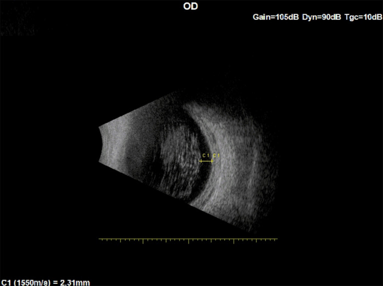
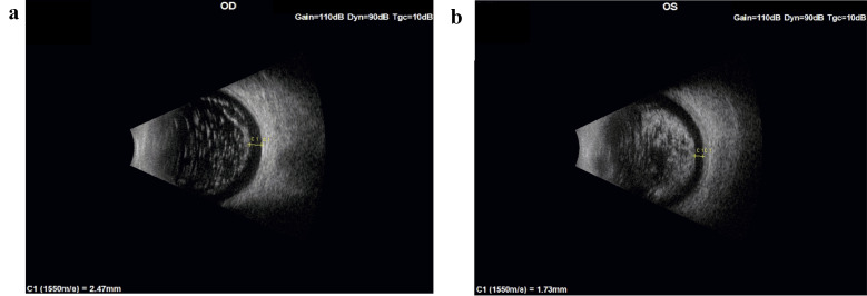
Methods: Relying on a previously published model of floater perception based on astronomical equations of a solar eclipse, and on ultrasound images of the vitreous in three eyes with AH, we explain why such patients do not perceive floaters in spite of opaque bodies filling their entire vitreous, to the point of, in severe cases of AH, obscuring the fundus view during ophthalmoscopy.
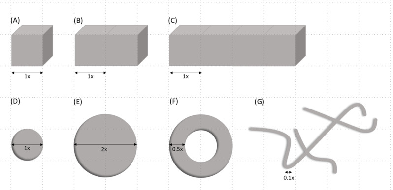
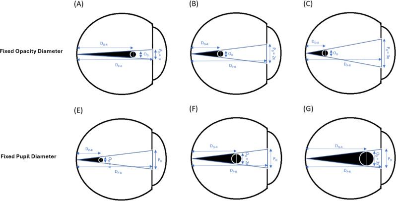
Main outcome measures: Developing an optical model of light rays that can quantify the maximal distance upon which a vitreous floater or opacity will cast a shadow on the retina.
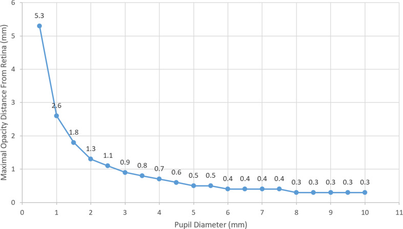
Results: Calculations using the proposed model demonstrated that with a 3 mm pupil, for a floater located between 1.5 mm and 2 mm from the retina, its shortest diameter must be > 215 microns and > 286 microns, respectively, to be perceived. Since AH floaters, based on ultrasound imaging, do not exist in the most peripheral 1.5 mm of the vitreous, it becomes understandable why these patients are asymptomatic.
Conclusions: Based on the proposed model and our findings, we deduced that even large, degenerative floaters whose width is usually narrower than a large retinal vein (125 microns), must be located very close to the retina and hence are not the floaters that are aimed at when performing YAG laser vitreolysis. We speculate that in successful cases, YAG vitreolysis works by a different mechanism, most likely a shock wave that displaces floaters further away from the retina. Hence, vitreolysis might not necessarily require the laser be aimed at the floaters, as symptomatic floaters may be located in the outer 1.5-2.0 mm of the vitreous body, a very risky zone for YAG laser shots.
Keywords: Asteroid hyalosis; Eclipse; Floaters; Myodesopsia; Umbra; Vitreolysis.
© 2024. The Author(s).
Conflict of interest statement
The authors declare no competing interests.
Figures
References
-
- Benson AH. Disease of the vitreous: a case of monocular asteroid hyalites. Trans Ophthalmol Soc UK. 1894;14:101–4.
LinkOut - more resources
Full Text Sources
Miscellaneous
