

Macrophage accumulation in dorsal root ganglion is associated with neuropathic pain in experimental autoimmune neuritis
- PMID: 39449726
- PMCID: PMC11500528
- DOI: 10.1515/tnsci-2022-0355
Macrophage accumulation in dorsal root ganglion is associated with neuropathic pain in experimental autoimmune neuritis
Abstract
Background: Neuropathic pain is a common symptom of Guillain-Barré syndrome (GBS). The infiltration of macrophages in the dorsal root ganglion (DRG) contributed to neuropathic pain in nerve injury. The underlying mechanisms of neuropathic pain in patients with GBS remain unknown. Experimental autoimmune neuritis (EAN) is a useful mice model of GBS. Our study aimed to explore whether the infiltration of macrophages in DRG is associated with neuropathic pain of EAN.
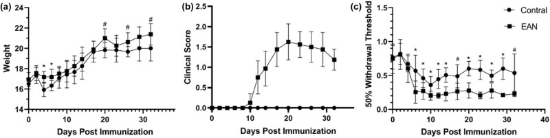
Methods: Male C57BL/6 mice were randomly divided into two groups, the EAN group (n = 12) and the control group (n = 12). Six mice in each group were sacrificed after anesthetization in the attack and remission phase, respectively. The 50% paw withdrawal threshold and clinical score were measured, and macrophages with its subtypes were detected in the spleen and DRG tissue.
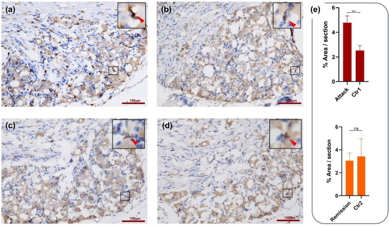
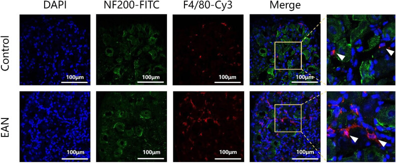
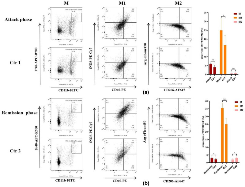
Results: More macrophages infiltrated the DRG of the EAN group in the attack phase and mostly surrounded neurons in the DRG. The proportion of macrophages and pro-inflammatory macrophages in the spleen of mice with EAN was significantly higher than the control group in the attack phase.
Conclusion: The infiltration of macrophages in DRG might be associated with neuropathic pain of EAN and pro-inflammatory macrophages may involve in neuropathic pain of EAN.
Keywords: dorsal root ganglion; experimental autoimmune neuritis; macrophages; neuropathic pain.
© 2024 the author(s), published by De Gruyter.
Conflict of interest statement
Conflict of interest: The authors state no conflict of interest.
Figures
Similar articles
-
Neuropathic pain in experimental autoimmune neuritis is associated with altered electrophysiological properties of nociceptive DRG neurons.Exp Neurol. 2017 Nov;297:25-35. doi: 10.1016/j.expneurol.2017.07.011. Epub 2017 Jul 19. Exp Neurol. 2017. PMID: 28734788
-
Mechanical allodynia and spinal up-regulation of P2X4 receptor in experimental autoimmune neuritis rats.Neuroscience. 2008 Mar 18;152(2):495-501. doi: 10.1016/j.neuroscience.2007.12.042. Epub 2008 Jan 12. Neuroscience. 2008. PMID: 18276080
-
Pain hypersensitivity in rats with experimental autoimmune neuritis, an animal model of human inflammatory demyelinating neuropathy.Brain Behav Immun. 2007 Jul;21(5):699-710. doi: 10.1016/j.bbi.2006.07.007. Epub 2006 Sep 26. Brain Behav Immun. 2007. PMID: 17005365
-
Beneficial or Harmful Role of Macrophages in Guillain-Barré Syndrome and Experimental Autoimmune Neuritis.Mediators Inflamm. 2018 Apr 26;2018:4286364. doi: 10.1155/2018/4286364. eCollection 2018. Mediators Inflamm. 2018. PMID: 29853789 Free PMC article. Review.
-
Th1/Th2/Th17/Treg cytokines in Guillain-Barré syndrome and experimental autoimmune neuritis.Cytokine Growth Factor Rev. 2013 Oct;24(5):443-53. doi: 10.1016/j.cytogfr.2013.05.005. Epub 2013 Jun 21. Cytokine Growth Factor Rev. 2013. PMID: 23791985 Review.
References
-
- Tomita S, Sekiguchi F, Kasanami Y, Naoe K, Tsubota M, Wake H, et al. Ca(v)3.2 overexpression in L4 dorsal root ganglion neurons after L5 spinal nerve cutting involves Egr-1, USP5 and HMGB1 in rats: An emerging signaling pathway for neuropathic pain. Eur J Pharmacol. 2020 Dec;888:173587. 10.1016/j.ejphar.2020.173587. - DOI - PubMed
-
- Sekiguchi F, Domoto R, Nakashima K, Yamasoba D, Yamanishi H, Tsubota M, et al. Paclitaxel-induced HMGB1 release from macrophages and its implication for peripheral neuropathy in mice: Evidence for a neuroimmune crosstalk. Neuropharmacology. 2018 Oct;141:201–13. 10.1016/j.neuropharm.2018.08.040. - DOI - PubMed
LinkOut - more resources
Full Text Sources