. 2024 Oct 24:7:0511.
doi: 10.34133/research.0511. eCollection 2024.

Diacylglycerol O-acyltransferase 2, a Novel Target of Flavivirus NS2B3 Protease, Promotes Zika Virus Replication by Regulating Lipid Droplet Formation

Affiliations

Diacylglycerol O-acyltransferase 2, a Novel Target of Flavivirus NS2B3 Protease, Promotes Zika Virus Replication by Regulating Lipid Droplet Formation

Xiaotong Luo et al. Research (Wash D C). .

Abstract

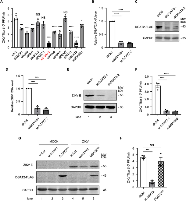
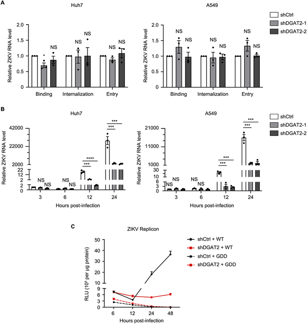
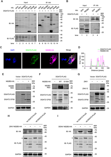
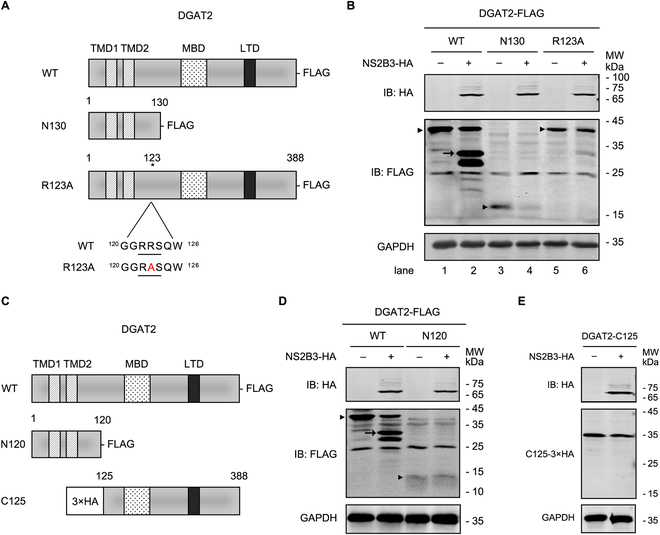
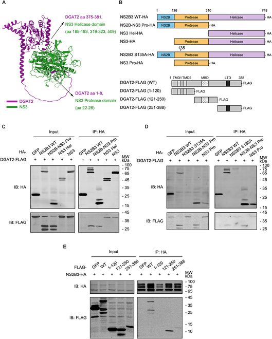
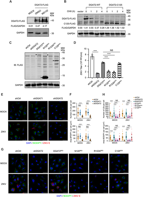
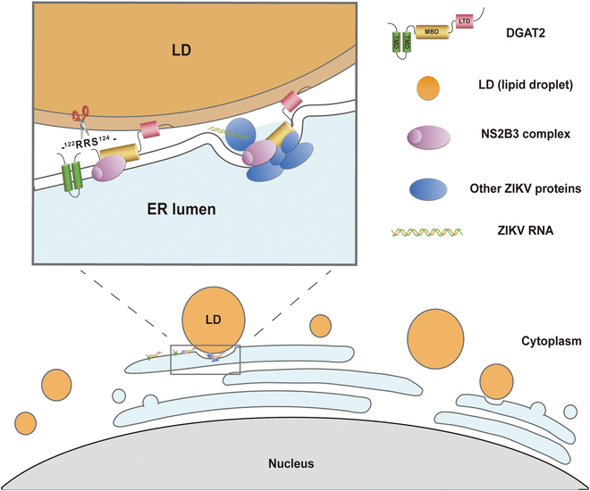
Various lipid metabolism-related factors are essential for Zika virus (ZIKV) replication. In this study, we revealed a crucial role of diacylglycerol O-acyltransferase 2 (DGAT2) in ZIKV replication using a short hairpin RNA-based gene knockdown technique. The replication of ZIKV was significantly inhibited by DGAT2 depletion in multiple cell lines and restored by trans-complementation with DGAT2. Mechanistically, DGAT2 is recruited in the viral replication complex by interacting with non-structural (NS) proteins. Among them, both human and murine DGAT2s can be cleaved by NS2B3 at the 122R-R-S124 site. Interestingly, the cleavage product of DGAT2 becomes more stable and is sufficient to promote the lipid droplet (LD) formation independent of its enzymatic activity. This work identifies DGAT2 as a novel target of the viral protease NS2B3 and elucidates that DGAT2 is recruited by viral proteins into the replication complex, thereby playing a proviral role by promoting LD formation, which advances our understanding of host-flavivirus interaction.


Conflict of interest statement

Competing interests: The authors declare that they have no competing interests.

Figures

Fig. 1.
Fig. 1.
The replication of ZIKV is impaired in DGAT2-knockdown cells. (A) Screening of lipid metabolism-related genes associated with ZIKV replication. The Huh7 cells were infected with ZIKV at an MOI of 1 after 48 h of transfection with the control siRNAs or lipid metabolism-related gene-specific siRNAs and then harvested at 24 h after infection for plaque assay. (B and C) Confirmation of DGAT2-knockdown efficiency. The total RNAs of the control, DGAT2-knockdown-1, and DGAT2-knockdown-2 Huh7 cells were extracted for qRT-PCR assay (B). Due to the lack of useful commercial antibodies to detect the endogenous cellular DGAT2 levels, FLAG-tagged DGAT2-expressing plasmid was alternatively cotransfected with the DGAT2-shRNA-expressing plasmid. Knockdown efficacy of DGAT2-FLAG was determined by Western blotting using anti-FLAG antibody (C). (D to F) Effect of DGAT2 knockdown on ZIKV replication levels. The control and DGAT2-knockdown cells were infected with ZIKV at an MOI of 1 and harvested at 24 h after infection for qRT-PCR (D) or Western blotting (E). The E protein levels in Western blotting were normalized against GAPDH (E). The supernatants were collected for plaque assay (F). (G and H) Detection of viral replication levels. The control, DGAT2KD, and DGAT2RES cells were infected with ZIKV (MOI = 1) and harvested at 24 h after infection for Western blotting (G). The supernatants were collected for plaque assay (H). Human GAPDH mRNA level was measured as an internal control for qRT-PCR. Data were shown as means ± SEM from at least 3 independent experiments. NS, no statistical significance; *P < 0.05; ***P < 0.001; ****P < 0.0001 (ANOVA with Dunnett’s multiple comparison test). GAPDH was probed as the loading control for Western blotting. Representative images of 3 independent experiments are shown.
Fig. 2.
Fig. 2.
DGAT2 functions at ZIKV RNA replication step. (A) Effect of DGAT2 knockdown on viral entry. The control and DGAT2-knockdown cells were inoculated with ZIKV at an MOI of 1, followed by incubation on ice for 45 min (virion binding) or at 37 °C for 30 min (virion internalization) or at 37 °C for 60 min (virion entry). Cells were harvested for qRT-PCR. (B) Effect of DGAT2 knockdown on ZIKV RNA replication. The control and DGATKD cells were infected with ZIKV (MOI = 1) and collected at 3, 6, 12, and 24 h after infection. The viral RNA levels were measured by qRT-PCR. Human GAPDH mRNA level was measured as an internal control. (C) Replicon assay. The control and DGAT2KD cells were transfected with ZIKV WT or GDD replicon RNAs, and harvested at indicated time points for luciferase assay. Data were shown as mean ± SEM of at least 3 independent experiments. *P < 0.05; ***P < 0.001; ****P < 0.0001 (ANOVA with Dunnett’s multiple comparison test).
Fig. 3.
Fig. 3.
DGAT2 interacts with ZIKV NS proteins and is cleaved by protease NS2B3. (A) Co-IP assay of DGAT2 and ZIKV replication complex components in 293T cells. The 293T cells were cotransfected with plasmids expressing DGAT2-FLAG and ZIKV NS-HA proteins, and then whole-cell extracts were prepared for co-IP assay using anti-HA agarose beads at 24 h after transfection. Samples were detected by Western blotting using anti-FLAG and anti-HA antibodies. Green fluorescent protein (GFP)-HA was probed as the NC. IB, immunoblot. (B) Co-IP assay of DGAT2 and ZIKV NS2B3 in Huh7 cells. The Huh7 cells were cotransfected with plasmids expressing DGAT2-FLAG and ZIKV NS2B3-HA proteins. At 24 h after transfection, the whole-cell lysates were harvested and then prepared for co-IP assay using anti-FLAG antibody. GFP-FLAG was probed as the NC. (C) Subcellular localization of DGAT2 and ZIKV NS2B3. The Huh7 cells were cotransfected with plasmids expressing DGAT2-FLAG and NS2B3-HA. Anti-FLAG and anti-HA antibodies were used to indicate the subcellular localization of DGAT2 (green) and NS2B3 (magenta). Nuclei were stained with DAPI (blue). Scale bar, 100 μm. (D) Colocalization analysis using ImageJ software. (E and F) Immunoblot of Huh7 cells coexpressing DGAT2 and NS2B3 protein. Plasmids expressing FLAG-tagged human DGAT2 and HA-tagged ZIKV NS2B3 (E) or S135A mutant (F) were cotransfected in Huh7 cells for 24 h and then detected by Western blotting analysis. (G) Immunoblot of Huh7 cells expressing DGAT2-FLAG protein in the context of ZIKV infection. The Huh7 cells were transfected with the plasmid expressing DGAT2-FLAG for 24 h and then infected with ZIKV (MOI = 1). Cells were collected for Western blotting at 24 h after infection using anti-FLAG and ZIKV E protein antibodies. (E to G) DGAT2 CP30, DGAT2 cleavage product of ~30 kDa. (H and I) Immunoblot of Huh7 cells cotransfected with flavivirus NS2B3 and human/rat DGAT2-expressing plasmids. The plasmid expressing HA-tagged ZIKV NS2B3 (H) or DENV NS2B3 (I) and FLAG-tagged human DGAT2 or rat DGAT2 were cotransfected in Huh7 cells for 24 h, and then the DGAT2-FLAG-expressing level was detected by Western blotting assay. The DGAT2 cleavage fragment was marked by arrowhead, and full-length DGAT2-FLAG band was identified by black triangles. Representative images of 3 independent experiments are shown.
Fig. 4.
Fig. 4.
The 122R-R-S124 motif of DGAT2 is the cleavage site of ZIKV protease NS2B3. (A) Schematic diagram of WT and mutant DGAT2 constructs. DGAT2-N130 contains the first 130 aa of the N-terminal sequence. The 123rd arginine site was mutated into alanine in DGAT2-R123A, and the location of the mutated site was depicted by asterisk. TMD, transmembrane domain; MBD, membrane binding domain; LTD, LD targeting domain. (B) Immunoblotof Huh7 cells coexpressing ZIKV NS2B3 with DGAT2 mutants. The plasmid expressing ZIKV NS2B3-HA and DGAT2-WT-FLAG/N130-FLAG/R123A-FLAG was cotransfected in Huh7 cells for 24 h and then detected by Western blotting analysis. (C) Schematic diagram of full-length and truncated DGAT2 constructs. DGAT2-N120 contains the N-terminal 120 aa of DGAT2. 3×HA-DGAT2-C125 contains 125 to 388 aa of DGAT2fused with 3×HA at its N-terminus. (D and E) Immunoblottingof Huh7 cells cotransfected with plasmids encoding ZIKV NS2B3 and DGAT2 truncates. The plasmids expressing ZIKV NS2B3-HA and DGAT2-WT-FLAG/N120-FLAG (D) or 3×HA-DGAT2-C125 (E) were cotransfected in Huh7 cells for 24 h and then detected by Western blotting analysis. GAPDH was probed as an internal control for Western blotting. The DGAT2 cleavage fragment was marked by arrowhead, and FLAG-expressing bands were identified by black triangles. Representative images of 3 independent experiments are shown.
Fig. 5.
Fig. 5.
The 121-250 aa fragment of DGAT2 is essential for DGAT2 to interact with NS2B3. (A) Structure prediction analysis of human DGAT2 and NS3. The structure prediction was analyzed by AlphaFold 3 software, and the putative interaction sites in the structure were denoted. (B) Schematic illustration of constructs expressing truncated NS2B3 and DGAT2. (C and D) Co-IP assay to map the DGAT2-interacting domain of NS2B3. The Huh7 cells were cotransfected with plasmids expressing DGAT2-FLAG and different truncated proteins of NS2B3-HA. At 24 h after transfection, whole-cell lysates were harvested and then prepared for co-IP assay using anti-HA agarose beads. The protein levels were measured by immunoblot with anti-HA and anti-FLAG antibodies. (E) Co-IP assay to map the NS2B3-interacting domain of DGAT2. The Huh7 cells were cotransfected with plasmids expressing NS2B3-HA and different truncated proteins of DGAT2-FLAG. Immunoprecipitation and immunoblot analysis were performed similarly as in (C) and (D). GFP-HA (C and D) and GFP-FLAG (E) were used as the NC. Representative images of 3 independent experiments are shown.
Fig. 6.
Fig. 6.
The cleavage product of DGAT2 (DGAT2-C125) is sufficient to promote ZIKV replication by regulating LD formation. (A) Inhibition of DGAT2 degradation by MG132. Huh7 cells expressing DGAT2-FLAG were treated with DMSO or MG132 for 10 h. The cell extracts were subjected to Western blotting analysis with anti-FLAG antibody. (B) Stability of DGAT2-C125 protein. The plasmid expressing DGAT2-FLAG or DGAT2-C125-FLAG was transfected into Huh7 cells for 24 h and then treated with the translation inhibitor CHX for the indicated times (0, 1, 2, and 4 h). The proteins were detected by Western blotting with anti-FLAG antibody. (C) Expression levels of DGAT2 truncated proteins. The Huh7 cells were transfected with the DGAT2-N120-FLAG-, DGAT2-R123A-FLAG-, or DGAT2-C125-FLAG-expressing plasmid. The cells were collected at 24 h after transfection and then detected by Western blotting with anti-FLAG antibody. The black triangles indicate the DGAT2 truncated protein bands. Representative images of 3 independent experiments are shown. GAPDH was analyzed as a loading control in (A) to (C). (D) Viral titers in DGAT2 truncated protein-expressing cells. The control, DGAT2KD, DGAT2RES, DGAT2-N120RES, DGAT2-R123ARES, and DGAT2-C125RES Huh7 cells were infected with ZIKV (MOI = 1) and harvested at 24 h after infection for plaque assay. (E to H) LDs levels in cells. The control, DGAT1KD, DGAT2KD, DGAT2RES, DGAT2-N120RES, DGAT2-R123ARES, and DGAT2-C125RES Huh7 cells were infected with ZIKV (MOI = 1) and then fixed at 24 h after infection (E and G). BODIPY staining was used to detect the formation of LDs (green). ZIKV E protein was stained by fluorescent antibody (red), and DAPI (blue) was used to indicate the nucleus (scale bars, 50 μm). The numbers and areas of LDs (at least 30 cells per sample) were analyzed by using ImageJ analysis software (F and H). Representative images of 3 independent experiments are shown. Data are shown as means ± SEM from at least 3 independent experiments. ****P < 0.0001 (ANOVA with Dunnett’s multiple comparison test).
Fig. 7.
Fig. 7.
A proposed model to illustrate the mechanism of DGAT2 promoting ZIKV replication. In response to ZIKV infection, DGAT2 is cleaved by ZIKV protease NS2B3 and becomes more stable. Cleaved DGAT2 is subsequently recruited by NS proteins to the viral replication complex. DGAT2 may function as a tethering bridge between ER and LD, regulating LD accumulation to supply energy for ZIKV replication.

Similar articles

Cited by

References

    1. Rasmussen SA, Jamieson DJ, Honein MA, Petersen LR. Zika virus and birth defects--Reviewing the evidence for causality. N Engl J Med. 2016;374(20):1981–1987. - PubMed
    1. Lazear HM, Diamond MS. Zika virus: New clinical syndromes and its emergence in the Western Hemisphere. J Virol. 2016;90(10):4864–4875. - PMC - PubMed
    1. Saiz J-C, Vázquez-Calvo Á, Blázquez AB, Merino-Ramos T, Escribano-Romero E, Martín-Acebes MA. Zika virus: The latest newcomer. Front Microbiol. 2016;7:496. - PMC - PubMed
    1. Heaton NS, Perera R, Berger KL, Khadka S, Lacount DJ, Kuhn RJ, Randall G. Dengue virus nonstructural protein 3 redistributes fatty acid synthase to sites of viral replication and increases cellular fatty acid synthesis. Proc Natl Acad Sci USA. 2010;107(40):17345–17350. - PMC - PubMed
    1. Huang Y, Lin Q, Huo Z, Chen C, Zhou S, Ma X, Gao H, Lin Y, Li X, He J, et al. . Inositol-requiring enzyme 1α promotes Zika virus infection through regulation of stearoyl coenzyme a desaturase 1-mediated lipid metabolism. J Virol. 2020;94(23):e01229-20. - PMC - PubMed

LinkOut - more resources