

Dynamic micro-optical coherence tomography enables structural and metabolic imaging of the mammalian cochlea
- PMID: 39449964
- PMCID: PMC11499234
- DOI: 10.3389/fnmol.2024.1436837
Dynamic micro-optical coherence tomography enables structural and metabolic imaging of the mammalian cochlea
Abstract
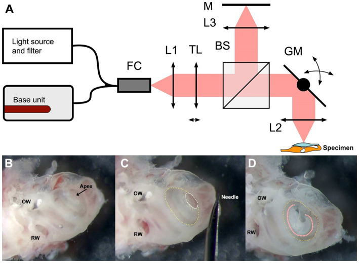
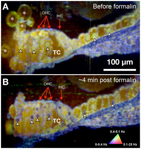
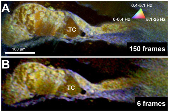
Sensorineural hearing loss (SNHL) is caused by damage to the mechanosensory hair cells and auditory neurons of the cochlea. The development of imaging tools that can directly visualize or provide functional information about a patient's cochlear cells is critical to identify the pathobiological defect and determine the cells' receptiveness to emerging SNHL treatments. However, the cochlea's small size, embedded location within dense bone, and sensitivity to perturbation have historically precluded high-resolution clinical imaging. Previously, we developed micro-optical coherence tomography (μOCT) as a platform for otologic imaging in animal models and human cochleae. Here we report on advancing μOCT technology to obtain simultaneously acquired and co-localized images of cell viability/metabolic activity through dynamic μOCT (DμOCT) imaging of intracellular motion. DμOCT obtains cross-sectional images of ATP-dependent movement of intracellular organelles and cytoskeletal polymerization by acquiring sequential μOCT images and computing intensity fluctuation frequency metrics on a pixel-wise basis. Using a customized benchtop DμOCT system, we demonstrate the detailed resolution of anatomical and metabolic features of cells within the organ of Corti, via an apical cochleostomy, in freshly-excised adult mouse cochleae. Further, we show that DμOCT is capable of capturing rapid changes in cochlear cell metabolism following an ototoxic insult to induce cell death and actin stabilization. Notably, as few as 6 frames can be used to reconstruct cochlear DμOCT images with sufficient detail to discern individual cells and their metabolic state. Taken together, these results motivate future development of a DμOCT imaging probe for cellular and metabolic diagnosis of SNHL in humans.
Keywords: cochlea; hair cell; metabolic imaging; micro-optical coherence tomography; organ of Corti; sensorineural hearing loss.
Copyright © 2024 Schulz-Hildebrandt, Spasic, Hou, Ting, Batts, Tearney and Stankovic.
Conflict of interest statement
Guillermo Tearney and Konstantina Stankovic have received sponsored research funding from WayVector. Guillermo Tearney, Konstantina Stankovic, Hinnerk Schulz-Hildebrandt, and Fang Hou are inventors on patents pertaining to μOCT and DμOCT technology. The remaining authors declare that the research was conducted in the absence of any commercial or financial relationships that could be construed as a potential conflict of interest. The author(s) declared that they were an editorial board member of Frontiers, at the time of submission. This had no impact on the peer review process and the final decision.
Figures

Similar articles
-
Novel application of metabolic imaging of early embryos using a light-sheet on-a-chip device: a proof-of-concept study.Hum Reprod. 2025 Jan 1;40(1):41-55. doi: 10.1093/humrep/deae249. Hum Reprod. 2025. PMID: 39521726 Free PMC article.
-
Organ of Corti macrophages: a distinct group of cochlear macrophages with potential roles in supporting cell degeneration and survival.Front Immunol. 2025 Jun 25;16:1617146. doi: 10.3389/fimmu.2025.1617146. eCollection 2025. Front Immunol. 2025. PMID: 40636110 Free PMC article.
-
Systemic Inflammatory Response Syndrome.2025 Jun 20. In: StatPearls [Internet]. Treasure Island (FL): StatPearls Publishing; 2025 Jan–. 2025 Jun 20. In: StatPearls [Internet]. Treasure Island (FL): StatPearls Publishing; 2025 Jan–. PMID: 31613449 Free Books & Documents.
-
Management of urinary stones by experts in stone disease (ESD 2025).Arch Ital Urol Androl. 2025 Jun 30;97(2):14085. doi: 10.4081/aiua.2025.14085. Epub 2025 Jun 30. Arch Ital Urol Androl. 2025. PMID: 40583613 Review.
-
Cost-effectiveness of using prognostic information to select women with breast cancer for adjuvant systemic therapy.Health Technol Assess. 2006 Sep;10(34):iii-iv, ix-xi, 1-204. doi: 10.3310/hta10340. Health Technol Assess. 2006. PMID: 16959170
Cited by
-
The State of High-Resolution Imaging of the Human Inner Ear: A Look Into the Black Box.Adv Sci (Weinh). 2025 Jul;12(28):e00556. doi: 10.1002/advs.202500556. Epub 2025 Jun 5. Adv Sci (Weinh). 2025. PMID: 40470704 Free PMC article. Review.
-
Intracochlear Imaging Using IVUS and OFDI: A Cadaveric Feasibility Study.Laryngoscope Investig Otolaryngol. 2025 Aug 5;10(4):e70224. doi: 10.1002/lio2.70224. eCollection 2025 Aug. Laryngoscope Investig Otolaryngol. 2025. PMID: 40766102 Free PMC article.
References
-
- Arnold B., Jäger L., Grevers G. (1996). Visualization of inner ear structures by three-dimensional high-resolution magnetic resonance imaging. Otol. Neurotol. 17, 480–485. - PubMed
Grants and funding
LinkOut - more resources
Full Text Sources