

Evaluation of Random and Aligned Polycaprolactone Nanofibrous Electrospun Scaffold for Human Periodontal Ligament Engineering in Biohybrid Titanium Implants
- PMID: 39450145
- PMCID: PMC11502134
- DOI: 10.1155/2024/2571976
Evaluation of Random and Aligned Polycaprolactone Nanofibrous Electrospun Scaffold for Human Periodontal Ligament Engineering in Biohybrid Titanium Implants
Abstract
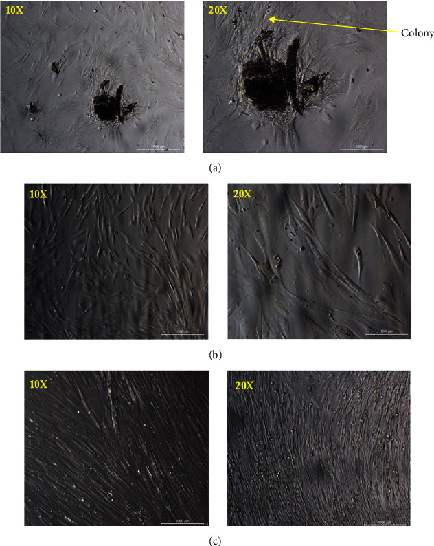
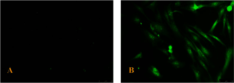
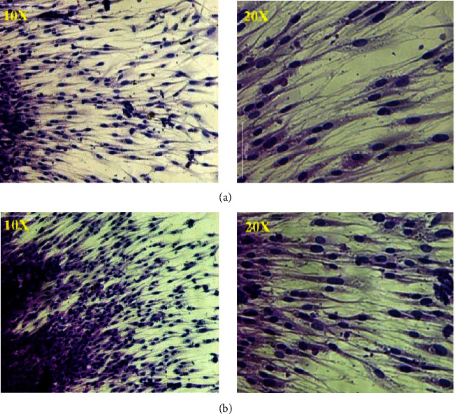
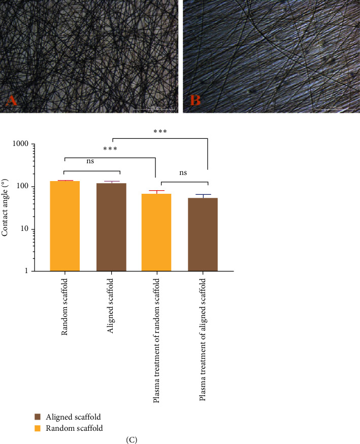
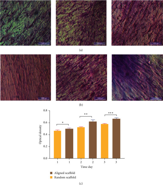
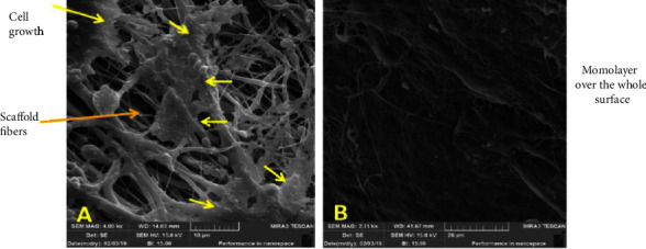
Background: Stem cells are introduced to regenerate some living tissue to restore function and longevity. The study aims to isolate in vitro human periodontal ligament stem cells (hPDLSCs) and investigate their proliferation rate on plasma-treated aligned and random polycaprolactone (PCL) nanofibrous scaffolds made via an electrospinning technique to attempt periodontal-like tissue in dental implants. Materials and Methods: hPDLSCs were isolated from extracted human premolars and cultured on plasma-treated or untreated PCL-aligned and random scaffolds to enhance adhesion of periodontal ligament (PDL) cells as well as interaction and proliferation. Cell morphology, adhesion, and proliferation rate were evaluated using field emission scanning electron microscopy (FESEM) and the methyl tetrazolium (MTT) assay. The wettability of PCL scaffolds was tested using a goniometer. Results: The hydrophilicity of plasma-treated scaffolds was significantly increased (p ≤ 0.05) in both aligned and random nanofibers compared to the nontreated nanofibrous scaffold. Cells arranged in different directions on the random nanofiber scaffold, while for aligned scaffold nanofibers, the cells were arranged in a pattern that followed the direction of the aligned electrospun nanofibres. The rate of hPDLSC proliferation on an aligned PCL nanofiber scaffold was significantly higher than on a random PCL nanofibrous scaffold with a continuous, well-arranged monolayer of cells, as shown in FESEM. Conclusion: The aligned PCL nanofiber scaffold is superior to random PCL when used as an artificial scaffold for hPDLSC regeneration in PDL tissue engineering applications.
Keywords: dental implant; electrospinning technique; periodontal ligament; poly(caprolactone) nanofibrous; stem cells.
Copyright © 2024 Ihab N. Safi et al.
Conflict of interest statement
The authors declare no conflicts of interest.
Figures
Similar articles
-
Preparing polycaprolactone scaffolds using electrospinning technique for construction of artificial periodontal ligament tissue.J Taibah Univ Med Sci. 2020 Aug 17;15(5):363-373. doi: 10.1016/j.jtumed.2020.07.007. eCollection 2020 Oct. J Taibah Univ Med Sci. 2020. PMID: 33132808 Free PMC article.
-
Incorporation of aligned PCL-PEG nanofibers into porous chitosan scaffolds improved the orientation of collagen fibers in regenerated periodontium.Acta Biomater. 2015 Oct;25:240-52. doi: 10.1016/j.actbio.2015.07.023. Epub 2015 Jul 15. Acta Biomater. 2015. PMID: 26188325
-
Comparison of random and aligned PCL nanofibrous electrospun scaffolds on cardiomyocyte differentiation of human adipose-derived stem cells.Iran J Basic Med Sci. 2014 Nov;17(11):903-11. Iran J Basic Med Sci. 2014. PMID: 25691933 Free PMC article.
-
The Effects of Plasma Treated Electrospun Nanofibrous Poly (ε-caprolactone) Scaffolds with Different Orientations on Mouse Embryonic Stem Cell Proliferation.Cell J. 2014 Fall;16(3):245-54. Epub 2014 Oct 4. Cell J. 2014. PMID: 24611137 Free PMC article.
-
Advances in Electrospun Poly(ε-caprolactone)-Based Nanofibrous Scaffolds for Tissue Engineering.Polymers (Basel). 2024 Oct 10;16(20):2853. doi: 10.3390/polym16202853. Polymers (Basel). 2024. PMID: 39458681 Free PMC article. Review.
References
-
- Ramazanli V. N. Effect of PH and Temperature on the Synthesis of Silver Nano Particles Extracted From Olive Leaf. Advances in Biology & Earth Sciences . 2021;6(2):169–173.
LinkOut - more resources
Full Text Sources
